Eccrine hidradenoma is a rare, benign cutaneous tumor that arises from eccrine sweat glands distributed throughout the body. It is also known as eccrine acrospiroma, clear cell hidradenoma, nodular hidradenoma, or eccrine sweat gland adenoma (
1,
3).
The incidence of eccrine hidradenoma peaks in the 4th to 5th decades and appears to be higher in women than in men (
4). It can occur anywhere on the body, and commonly reported locations are the face, scalp, ear lobe, axilla, trunk, and upper extremity (
5). Eccrine hidradenoma of the breast is very rare, and only 14 cases have been reported in English literature (
1,
3,
5-
14). Its features somewhat different from similar lesions found in other parts of the body. The majority of eccrine hidradenomas in the breast present as painless lumps with a slow growth rate. Patients may have pain, nipple discharge, or skin ulceration (
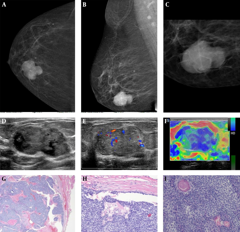
3). Eccrine hidradenoma of the breast commonly arises in the nipple and subareolar areas, but it can also originate in the deeper breast parenchyma like our case (
11).
On histopathology, eccrine hidradenoma appears as either a well-circumscribed or lobulated mass in the dermis, with both solid and cystic components. The cystic component varies in size and is faintly eosinophilic and homogeneous (
12). The solid component contains 2 types of cells. The dominant type is round and has a clear cytoplasm. The other is polyhedral, with basophilic cytoplasm. The tumor may also display focal cystic changes. Malignant transformation is extremely rare but possible. However, neither clinical presentation nor histologic appearance can predict malignant transformation. Malignant lesions tend to invade local tissue, in contrast with benign lesions (
4).
The imaging findings of eccrine hidradenoma have not been well described because of its rarity and insufficient imaging evaluation.
The mammographic findings of eccrine hidradenoma are nonspecific. It can appear as a well-circumscribed, hyperdense mass on mammography (
13). The most commonly reported features on ultrasonography are the presence of a circumscribed, complex, solid and cystic mass with vascularity on color Doppler study (
6,
13,
15). The solid component may contain calcifications. The cystic component can show variable complexity in the presence of hemorrhage (
14,
15). It can also appear as a circumscribed, solid, hypoechoic mass (
16,
17). On magnetic resonance imaging, it may be seen as a circumscribed, solid, or cystic mass, usually with an intermediate-to-low signal on T1-weighted images and an intermediate-to-high signal on T2-weighted images. The solid component of the mass shows enhancement after intravenous contrast administration (
16,
18).
The treatment of eccrine hidradenoma is complete wide excision. It is important to achieve clear resection margins (
15). Recurrence has been reported and is associated with incomplete excision (
15).
Although rare, malignant transformation can arise primarily or secondarily from a preexisting benign eccrine hidradenoma (
4). Malignant eccrine hidradenomas, also known as hidradenocarcinomas or malignant clear cell hidradenomas, can metastasize through either lymphatic or hematogenous dissemination (
4). Aggressive surgical treatment is necessary for these lesions, although the optimal treatment method has not been established. These tumors are not susceptible to radiotherapy and chemotherapy and are associated with a poor prognosis (
19).
Radiologists should consider eccrine hidradenoma when a mixed solid and cystic mass is observed in the breast on ultrasonography, as it may mimic primary malignancy.