1. Introduction
Postoperative pulmonary complications are the most frequent and significant contributors to morbidity, mortality, and healthcare costs (1). These complications have been reported to be associated with several perioperative factors, including older age, history of pulmonary disease and smoking, previous cardiac surgery, application and duration of cardiopulmonary bypass (CPB), median sternotomy, administration of anesthetics, neuromuscular relaxants, and sedatives, fluid imbalance, delayed tracheal extubation, systemic inflammatory response, intraoperative phrenic nerve injury, and postoperative acute kidney injury (2, 3). Acute hypoxemic respiratory failure (AHRF) is a significant pulmonary complication following cardiac surgical procedures (4). Although cardiac pulmonary edema, atelectasis, acute lung injury, exacerbation of chronic obstructive pulmonary disease, pneumonia, pneumothorax, pleural effusions, pulmonary embolism, and phrenic nerve injury are the main causes of AHRF after cardiac surgery (4-6), this condition with a partial pressure of oxygen (PaO2) < 60 mmHg on a fraction of inspired oxygen (FiO2) of 0.5 (PaO2/FiO2 ≤ 120) can occur during mechanical ventilatory support in up to 10% of the patients undergoing surgery on CPB. This usually results from a severe perioperative cardiopulmonary insult, such as long duration of CPB and postoperative low cardiac output syndrome (2).
In the present case report, we describe a man who developed AHRF with an extremely low PaO2 during mechanical ventilation under intravenous sedation, an infrequent complication after an elective cardiac surgical procedure (2, 3).
2. Case Presentation
A 68-year old man was admitted to the cardiac surgery intensive care unit (ICU) of our tertiary hospital in Athens, Greece after an elective Bentall procedure due to ascending aortic aneurysm in conjunction with a malfunctioning aortic valve. The patient had undergone aortic valve replacement 10 years ago and his medical history revealed no lung diseases. Besides, he was not an active smoker during the last 10 years.
The duration of Bentall surgical procedure and CPB was 440 and 227 minutes, respectively. In addition, aortic cross clamping time was 154 minutes. A total of six units of red blood cells, 15 units of platelets, and 2.5 liter of crystalloids were given intraoperatively. As aforementioned, after completion of the surgical procedure, the patient was admitted to the cardiac surgery ICU of our center and was managed postoperatively with the intensive care protocol of our ICU. Specifically, the patient was placed on a volume-cycled respirator for total ventilatory support under intravenous sedation with propofol (25 μg/kg/min) with the following settings: FiO2: 1.0, tidal volume: 650 ml, respiratory rate: 12 breaths/min, and 5 cmH2O of positive end expiratory pressure (PEEP). Also, the following signs were evaluated at his ICU arrival: arterial blood pressure (systolic/diastolic/mean): 100/40/60 mmHg, central venous pressure: 5 mmHg, heart rate: 110 beats/min, percutaneous oxygen saturation (SpO2): 99%, and body temperature: 34.6°C. During the early postoperative period, despite his inotropic (dobutamine, 4μg/kg/min), vasopressor (norepinephrine, 1.1 μg/kg/min), and antiarrhytmic (amiodarone, 150 mg over the first 10 min followed by 360 mg over the next 6 hours) support, the patient was hemodynamically unstable with low mean arterial pressure, supraventricular tachycardia, and copious urine output, not responsive to intravenously administered diuretics (furosemide) and fluids. Moreover, we attempted to place Swan-Ganz catheter, but this effort was deemed ineffective due to ventricular arrhythmia.
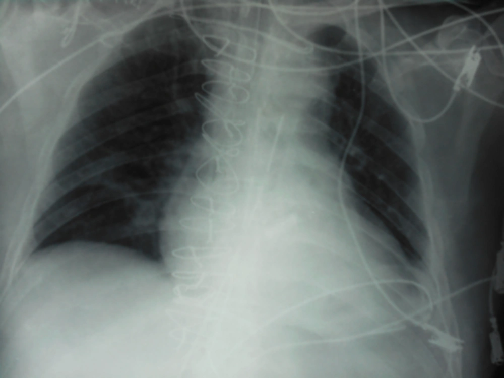
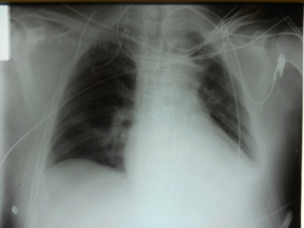
In addition, a baseline chest X-ray was performed, which was clear (Figure 1). At almost six hours after his ICU admission, the patient suddenly presented severe oxygen desaturation (SpO2 75%) during mechanical ventilatory support on 0.7 FiO2 with the following findings from the arterial blood gas test: PaO2: 39.1 mmHg, partial pressure of carbon dioxide (PaCO2): 36.6 mmHg, and pH: 7.39. We immediately applied measures for the acute respiratory insufficiency management, including increasing FiO2 to 1.0 on ventilator, ensuring of adequate intravenous sedation, physical examination, and chest X-ray. In addition, we confirmed the proper position of the endotracheal tube and ensured adequate ventilator function and settings. Physical examination of both respiratory and cardiovascular systems in conjunction with chest X-ray (Figure 2) revealed no pathological findings indicative of pneumothorax, atelectasis, pleural effusion, acute pulmonary edema, pneumonia, pulmonary embolism, and acute lung injury, including acute respiratory distress syndrome and cardiac tamponade. Simultaneously, we tried to stabilize the patient’s heart rhythm and rate by administration of esmolol (initial dose of 500 μg/kg/min over 1 min followed by maintenance dose of 50 μg/kg/min) through continuous intravenous infusion, replacing amiodarone. Also, in order to increase the arterial blood pressure and maintain satisfactory cardiac output and tissue perfusion, we increased the intravenously administered dose of norepinephrine and fluids (colloids and crystalloids). Finally, the patient presented a significantly slow oxygenation improvement with PaO2 > 80 mmHg on 1.0 FiO2 seven hours after the above-mentioned acute respiratory insufficiency episode.
3. Discussion
Although AHRF is a frequent complication in patients undergoing surgery on CPB, presence of severe hypoxemia during mechanical ventilation is a fairly uncommon condition during the early postoperative period. Prolonged duration of CPB and postoperative low cardiac output syndrome are the main risk factors of this poor respiratory outcome (2, 3).
Our patient was at a high risk of respiratory complications postoperatively. Specifically, he had a significantly long CPB duration due to his high operative severity and he was hemodynamically unstable during the early postoperative period with accompanying low cardiac output. Nevertheless, it is worth mentioning that our patient had a clear X-ray despite his severe and persistent hypoxemia on high FiO2 levels during his mechanical ventilatory support. He might have developed type IV respiratory failure, which occurs in the patients who are in shock or hypoperfusion states without the associated pulmonary problems (5). Our claim was strengthened by the PaO2 improvement after achievement of hemodynamic stabilization. Generally, response to hypoxemia depends on patients’ ability to recognize the hypoxemic state. Then, in order to increase cardiac output and minute ventilation to improve the situation, peripheral chemoreceptors located in the arch of aorta and at the bifurcation of carotid artery send different signals to the brain (7).
We ought to differentiate our case from the patients who develop acute respiratory failure after an operation for type A aortic dissection. These patients undergo an emergency surgery, which is associated with significantly increased probability of occurrence of postoperative pulmonary complications (1). In addition, acute respiratory failure is the clinical consequence of an increase in permeability of the alveolar capillary membrane and might be related to the diffuse inflammatory response observed in acute type A dissection patients (8).
3.1. Conclusion
In conclusion, AHRF on mechanical ventilatory support with clear chest X-ray and without pulmonary pathological findings could present due to low cardiac output and tissue hypo-perfusion during the early postoperative period of a major surgery, such as operation on aorta using CPB. Clinicians, including intensivists and critical care nurses, may improve tissue oxygenation and treat the underlying cause of respiratory failure through early identification of this life-threatening condition and taking measures for ensuring normal hemodynamic parameters and adequate tissue perfusion.

