Evaluation of Drugs and Strategies for Treating Coronary Artery Ectasia: Update and Future Perspective

authors:

avatar Mohammad Javad Taghizadeh 1 , avatar Shima Khodadadi 2 , avatar Somayeh Zamanifard 3 , *

Atherosclerosis Research Center, Ahvaz Jundishapur University of Medical Sciences, Ahvaz, Iran
Department of Internal Medicine, School of Medicine, Iran University of Medical Sciences,Tehran,Iran
Departement of Cardiology, Shariati Hospital, Tehran University of Medical Sciences, Tehran, Iran

how to cite: Taghizadeh M J, Khodadadi S, Zamanifard S. Evaluation of Drugs and Strategies for Treating Coronary Artery Ectasia: Update and Future Perspective. Jundishapur J Chronic Dis Care. 2022;11(3):e123301. https://doi.org/10.5812/jjcdc-123301.

Abstract

Context:

Although the mechanisms involved in the pathogenesis of coronary artery ectasia (CAE) and its treatment methods are not known for certainty, increased inflammatory and coagulation responses can be responsible for the formation of ectasia due to vascular disorders.

Evidence Acquisition:

The content used in this paper was obtained from English language articles (2005 - 2020) retrieved from the PubMed database and Google scholar search engine using “coronary artery ectasia”, “treatment”, “drug”, and “aneurysm” keywords.

Results:

The proven effect of inflammation and coagulation in CAE has posed a significant challenge for disease management. Therefore, anti-inflammatory and anticoagulation drugs can be treatment options for these patients. Increased inflammatory responses and some coagulation factors in CAE patients is undeniable. The study of these two systems in CAE patients and the evaluation of drugs affecting these mechanisms to achieve a definitive conclusion requires further and more extensive studies.

Conclusions:

We evaluated the hypothesis that anti-inflammatory and anticoagulation drugs with improved vascular endothelial function may accelerate the healing process of CAE patients; thus, they may be treatment options. Finally, it can be said that identifying molecular pathways related to drugs can improve their effectiveness in treating patients and increasing their survival. In addition, identifying upstream and downstream pathways can help diagnose the disease pathogenesis in addition to treating patients.

1. Context

Coronary artery ectasia (CAE) or aneurysm is a type of heart disorder characterized by localized or diffuse non-obstructive arteries. Chronic disease is one of the prominent features of the disease that can affect the treatment and clinical course of patients (1). Besides, CAE is an angiographic finding in 1 - 5% of patients with coronary artery disease (CAD), which is more common in men (2, 3). Although the exact pathophysiology of CAE is unclear, atherosclerosis is thought to be responsible for more than 50% of CAE cases; it is the most common cause, followed by congenital and acquired factors (4, 5). Also, CAE may occur during atherosclerosis development. There are other theories that CAE may also occur in the coronary and other vascular systems, so it may be independent of atherosclerosis (6). Moreover, CAE can be associated with many factors such as increased inflammatory responses, impaired immune function, and endothelial dysfunction (7). An imbalance in the production of inflammatory factors and increased secretion of cytokines are associated with coagulation development and impaired immune cells function, both of which are considered essential factors in the pathogenesis of vascular disease (8).

Anti-inflammatory drugs may help control the coagulation system and the clinical condition of CAE patients. In the present study, we aimed to investigate the possible responses of patients to anti-inflammatory and anticoagulation drugs and various theories about the inflammatory and coagulation mechanisms in CAE patients.

2. Classification of Ectasia

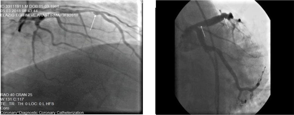
Ectasia is divided into several groups based on the number of vessels involved. In the first type, ectasia usually involves two or three blood vessels. In the second type, ectasia is usually diffuse in one vessel and localized in the other. In other types of ectasia, diffuse or localized ectasia is usually seen alone (8).

3. Anti-inflammatory Drugs

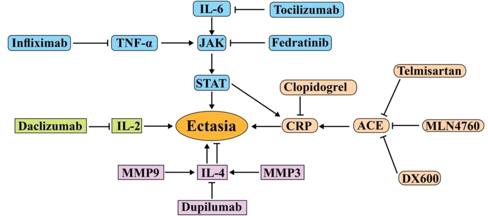
Inflammation is a vital process for CAD and atherosclerosis (9). Increased inflammatory mediators such as interleukins, C-reactive protein (CRP), and erythrocyte sedimentation rate (ESR) are considered risk factors for CAD, which are associated with increased morbidity and mortality (10, 11). The increasing number of neutrophils, monocytes, and lymphocytes as cells producing inflammatory factors in CAE patients and markers derived from dendritic cells in ectasia patients with CAD indicates the role of inflammation in CAE patients (Table 1) (2, 12). IL-6 secreted by monocytes can impair the function of the coagulation system and inflammation by increasing procoagulant factors and decreasing anticoagulants (13). IL-6 not only can play a role in inflammatory responses alone but also can be a potent stimulus for the production of other inflammatory mediators such as CRP and tumor necrosis factor-α (TNF-α) (14). According to these findings, the administration of IL-6 and JAK2-STAT3 inhibitors, along with cardiac drugs, can help CAE patients recover (Figure 1). Tocilizumab and sarilumab are IL-6 inhibitors that bind to IL-6 receptors to block its inflammatory activity and effects (15, 16). Tocilizumab acts like a double-edged sword, as it can lead to the development of aneurysms (17). It increases adverse blood fats and cholesterol, which are important risk factors for heart disease (Figure 1) (18).

Table 1.

Effects of Some Anti-inflammatory Drugs on Clinical Parameters of Coronary Artery Ectasia Patients

MedicinesMechanismOutcomeReferences
Tocilizumab and sarilumabAnti-IL-6 receptor antibodyIncreasing undesirable fats and cholesterol and coronary artery aneurysm development; directly affecting the inflammatory condition and, therefore, indirectly controlling the clotting system(16, 19)
Infliximab and etanerceptTNF-α inhibitorReducing CRP, ESR, fibrinogen, and arterial aneurysms; decreasing platelet count and reactivity; increasing MPV(20, 21)
FedratinibJAK2 dedicated inhibitorInhibiting JAK2, a critical mediator in signaling inflammation, and thus reducing the effects of increased inflammatory cytokines(22, 23)
Daclizumab and basiliximabAnti-IL-2 receptorImproving vascular endothelial function in postoperative stent placement(24)
DupilumabIL-4 receptor inhibitor monoclonal antibodyReducing the risk of atherosclerosis by preventing inflammatory risk factors; inhibiting IL-4 as an anti-inflammatory cytokine and thus leading to thrombosis and coagulation promotion(25, 26)
Secukinumab IL-17 inhibitorImproving endothelial function(27)
AnakinraIL-1 receptor antagonistDecreasing the levels of ferritin, fibrinogen, and CRP; preventing macrophage activation syndrome and reducing atherosclerosis immune-inflammatory response(28)
LipoxinsBinding to its receptors (ALX and GPR32) on human endothelial cellsStimulating the endothelial production of vasoprotective and antithrombotic mediators; genetic deletion of lipoxin receptor causes aortic dilation induced by angiotensin II infusion, decreases vascular collagen, and increases inflammation.(29)
Molecular pathways involved in the pathogenesis of ACE and the drugs used to treat them. Tocilizumab and fedratinib prevent inflammation and progression of ACE by targeting IL-6 and JAK, respectively. MLN4760 and DX600 also prevent the progression of ACE by inhibiting the angiotensin converter. Dupilumab can be effective in treating ectasia by targeting IL-4. Daclizumab can also be effective in treating ectasia by targeting IL-2. Abbreviation: JAK, Janus kinase; ACE, angiotensin-converting enzyme; CRP, C-reactive protein; TNF-α, tumor necrosis factor-α.

Given that CAE is six times more prevalent in patients with familial hypercholesterolemia than in healthy individuals, it can be hypothesized that tocilizumab use may be associated with increased cholesterol in CAE patients (30). However, it can control IL-6-related inflammatory responses and their destructive effects on the coagulation system by directly affecting inflammatory conditions; thus, it effectively reduces the risk of CAE. Side treatment of CAE patients with tocilizumab is an important and controversial issue; its use does not seem effective in this group of patients (Table 1). To control the effects of tocilizumab, it can be used along with lipid-lowering drugs. Alirocumab is a human monoclonal antibody against proprotein convertase subtilisin-kexin type 9 (PCSK9) that helps the liver reduce the level of bad cholesterol in the blood (31). It is used to treat atherosclerosis and prevent fat deposition in the arteries’ walls (32). Thus, it can be hypothesized that CAE patients with high blood lipids, which need anti-inflammatory drugs to control the coagulation system, can use lipid-controlling drugs along with cytokine inhibitors.

Fedratinib, as a specific inhibitor of JAK2, can block the activity of many inflammatory factors that are affected by this receptor or activated by IL-6, IL-17, and IL-22 (22). Also, IL-6 activates STAT3 by activating JAK2 and JAK1, which, in turn, induce Th17 differentiation and the secretion of inflammatory cytokines from this cell (33). Therefore, JAK inhibitors such as fedratinib can lead to the disconnection of IL-6 signaling with Th17 and the coagulation system. It seems that JAK2 inhibition, as one of the main factors involved in the inflammatory process, can prevent an overactive coagulation system (Figure 1).

Production of TNF-α from monocytes, macrophages, and lymphocytes, which are enlarged cells in CAE patients, has destructive effects on endothelial cells (ECs) (34). Besides, TNF-α plays a vital role in the pathogenesis of CAE by increasing the expression of adhesion molecules and altering vascular permeability (35). Coronary circulation involvement occurs following vascular injuries in CAE patients, which slows and disrupts blood circulation in these patients (36). In fact, the dysfunction of ECs by inflammatory mediators causes more inflammatory responses and worsens the patient’s clinical condition (30). As a result, blocking TNF-α and using antibodies against it may effectively control inflammation in CAE patients.

Infliximab and etanercept as TNF-α inhibitors can reduce CRP, ESR, and fibrinogen levels and prevent the coagulation system from becoming overactive in CAE patients (37, 38) (Table 1). Studies have shown that etanercept can cause hyperlipidemia (39), so its administration to CAE patients is controversial, and more studies are needed to reach a definitive conclusion about the effect of this drug on CAE patients’ blood lipids. Infliximab has a positive effect on the clinical condition of patients with arterial aneurysms, and its injection has reduced CRP (37). As high blood fats are a prognostic factor in coronary heart disease and the accumulation of lipoproteins triggers inflammatory immune responses and atherosclerotic plaques, it is possible to prevent etanercept-induced hyperlipidemia by ezetimibe. It is a potent inhibitor of sterol uptake and selectively inhibits the uptake of bile cholesterol and dietary cholesterol in the small intestine (Figure 2) (40, 41).

Ectasia involving vessels as diffuse and localized (42)

Based on the evidence, ezetimibe combined with statins is more effective in reducing inflammation. In addition to inhibiting 3-hydroxy-3-methylglutaryl coenzyme A (HMG-CoA) reductase, statins can lower low-density lipoprotein (LDL) cholesterol. It prevents fatty acid-related disorders and improves cardiovascular disease by reducing inflammation and resolving endothelial disorders (43-45).

Angiogenesis is a significant regulator of plaque growth, and statins can also inhibit angiogenesis. Statins also reduce the expression of cyclooxygenase-2 (CoX-2) and matrix metalloproteinase-9 (MMP-9) in vascular endothelium (46). As known, CoX is a pro-inflammatory enzyme with pro-angiogenic effects, and there is a functional relationship between CoX-2 activity and MMPs secretion (47, 48). Due to the properties of statins, it seems that prescribing this drug for CAE patients with hyperlipidemia can be effective in improving vascular endothelial function by inhibiting MMPs.

The imbalance between T-helper (Th) cells may lead to ectasia and atherosclerosis (49). Th1 cells are involved in the pathophysiology of heart disorders by secreting IL-2. Studies have shown that CAE patients have higher IL-2 levels than healthy individuals (50, 51). Another study found that IL-2 and IL-4 were significantly lower in CAE patients than in healthy individuals (52, 53). Secreted IL-4 by Th2 can have protective effects against vascular damage and be involved in vascular remodeling by inhibiting the production of inflammatory cytokines (54). Due to the protective and anti-inflammatory role of IL-4, its reduction in CAE patients is associated with a poor prognosis. The induction of anti-inflammatory pathways by IL-4 and suppression of monocytes indicate the atheroprotective effect of IL-4 (55, 56).

On the other hand, an increase in IL-10, an anti-inflammatory factor, has been reported in CAE patients (57). It seems that this increase in IL-10, along with an increase in inflammatory factors, could indicate a regulated immune response to the progression of inflammation, which can inhibit the activity of inflammatory cytokines by over-production of IL-10. An imbalance between the inflammatory factors produced by Th1 and the anti-inflammatory factors secreted by Th2 contributes to CAE and impaired vascular function.

Drugs such as daclizumab and basiliximab, which bind to IL-2 receptors on the surface of T cells, can inhibit IL-2 dysfunction. They eliminate the imbalance between Th1 and Th2 by increasing recombinant IL-10 and IL-4 or increasing Th2 expression (56, 58). No study has been performed on the effects of daclizumab and basiliximab on CAE patients. Further studies about these drugs and anti-inflammatory cytokines may speed up the treatment procedure for CAE patients.

Stimulated inflammatory responses may reflect the increased activity of MMPs. Destruction of the extracellular matrix by MMPs weakens the connective tissues of the vessel wall and makes them thinner and eventually dilated (59, 60). Studies have shown that MMP3 and MMP9 levels are high in CAE patients, and aneurysm development may be associated with increased MMP3 and MMP9. Besides, MMP3 hydrolyzes extracellular matrix compounds such as proteoglycans and collagens and can also activate MMP1 and MMP9 (61, 62).

One study showed that reducing MMP9 could prevent atherosclerotic damage and ectasia (63). Inactivation of MMP3 also reduces aneurysms (64). However, an increase in IL-4 is associated with an increase in MMP3 and MMP9 (48), so blocking the IL-4 receptor can reduce the risk of CAE (Table 1). In response to inflammation, IL-4 level increasedby the anti-inflammatory effects; on the other hand, its increase is associated with MMP3 and MMP9 elevation (65, 66). These two effects can be considered risk factors in CAE; it seems that the different functions of these cytokines are essential in CAE patients.

Dupilumab is a human monoclonal antibody that inhibits its signaling by binding to the IL-4 receptor and down-regulates inflammatory synthesis. It reduces the risk of atherosclerosis by inhibiting MMP12, IL-6, and Th2-related factors (25). On the other hand, by inhibiting IL-4 as an anti-inflammatory cytokine, this drug can lead to the development of thrombosis, coagulation, and even ischemic stroke (67). One study showed that IL-4 deficiency could lead to size reduction of vascular lesions in atherosclerosis (68). The multiple roles of IL-4 in the inflammatory and coagulation process and different outcomes with the use of its inhibitors are essential issues; further investigation and research are required to diagnose and treat CAE.

In general, reducing and controlling the level of MMPs can prevent damage to the extracellular matrix. Tissue inhibitor of MMPs (TIMPs) regulates the activity of MMPs, which inhibits and suppresses TIMPs by increasing inflammatory interleukins and MMPs (69). Thus, to improve the function of TIMPs in CAE patients and prevent vascular injury, reducing and inhibiting the production of mediators and inflammatory cells could be a way to treat and improve the clinical condition of CAE patients.

Inflammation is recognized as a significant component in the process of atherogenesis. Consequently, anti-inflammatory therapies can prevent the accumulation of inflammatory cells and vascular disorders in CAE patients. It should be noted that the administration of anti-inflammatory drugs to CAE patients requires the measurement of inflammatory cytokines in patients, followed by the use of an enhanced cytokine-associated inhibitor. The study of CAE patients regarding inflammatory factors and their treatment with anti-inflammatory drugs has not been discussed enough to provide a definitive theory in CAE patients. Future studies should pay attention to the role of anti-inflammatory drugs in these patients.

4. Anti-thrombotic Drugs

The imbalance between fibrinolytic systems and coagulation is associated with thrombosis and vascular disorders in CAE patients. The formation of thrombosis in ectatic vessels is one of the most important causes of mortality in patients with CAE (70, 71). It is not clear why CAE increases the risk of thrombosis, but studies have shown that fibrolytic system function is not normal in ectatic patients. The levels of α1-antitrypsin, α2-macroglobulin, and α2 plasmin inhibitor as plasmin inhibitors (one of the main factors in fibrinolysis) are increased in CAE patients (72). Increased plasmin inhibitors reduce the efficiency of the fibrinolysis system in CAE patients, which is associated with an increased risk of clinical coronary events (Table 2).

Table 2.

Effect of Some Antithrombotic Drugs on Clinical Parameters of Coronary Artery Ectasia Patients

MedicinesMechanismOutcomeReferences
Urokinase and streptokinaseActivation of plasminogen to plasminDecreasing fibrinogen levels; activating fibrinolysis to degenerate thrombosis (Excessive use can lead to bleeding)(73, 74)
rt-PAS (alteplase, reteplase and tenecteplase)Catalyzing the conversion of plasminogen to plasminPatency of blood vessels from coagulation lesions (Its short half-life may lead to the recurrence of blood clots)(75, 76)
AspirinIrreversibly inhibiting cyclooxygenaseElimination of thrombosis and improvement of patients with chronic treatment; slowing down the blood clotting process (High platelet aggregation with aspirin consumption increases myocardial infarction)(77, 78)
Clopidogrel and ticlopidinePlatelet ADP receptor (P2Y12) antagonistsIncreasing thrombolysis induced by streptokinase; causing anti-inflammatory effects; reducing the risk of thromboembolic attack (Individual platelet responses depend on the genetic, cellular, clinical, and environmental factors)(79, 80)
Tirofiban and eptifibatidegpIIb/IIIa receptor blockersImproving endothelial dysfunction in different clinical situations. Reversal of endothelial dysfunction induced after PCI(81, 82)
DabigatranDirect thrombin inhibitorAttenuating endothelial dysfunction; decreasing expression of inflammatory molecules; preventing the development of atherosclerosis; preventing systemic thromboembolic events in patients with atrial fibrillation(83)

Urokinase and streptokinase, two anticoagulant enzymes, have been shown to reduce the risk of heart attack and mortality (73). By saturating the α2-antiplasmin binding site, these two enzymes cause hyperplasminemia and increase fibrinogen degradation (74). It should be noted that these two intracoronary thrombolytic drugs increase the risk of postoperative bleeding in patients with fibrinogen levels less than 100 mg/dL (84). A study compared the urokinase and streptokinase and showed that postoperative bleeding was higher in patients receiving streptokinase than in those receiving urokinase. Unlike streptokinase, urokinase is not antigenic, and antibody-mediated resistance does not occur with this enzyme.

Like urokinase, recombinant tissue plasminogen activator (rt-PA) has no antigenic or pyrogenic properties and produces much less systemic fibrinolysis than streptokinase (85, 86). However, the short half-life of rt-PA may lead to recurrence and early clotting of arteries. Therefore, it seems that urokinase could be a better anticoagulation drug for treating vascular lesions due to increased coagulation in CAE patients. It is recommended that CAE patients be treated with anticoagulants or antiplatelet agents (87).

Ticlopidine is a drug used to reduce the risk of thromboembolic attack in patients with a history of thrombotic lesions or high-risk patients. It is a platelet aggregation inhibitor that blocks the binding of platelets to fibrinogen by blocking ADP receptors on platelet membranes (79, 88). The combined use of ticlopidine, aspirin, and heparin effectively controls coagulation activation in patients with coronary dilation (77).

Aspirin inhibits the production of prostaglandin and prothrombotic thromboxane by the acetylation of COX-1 (78). It can also control the inflammatory conditions in CAE patients due to its anti-inflammatory effects (89). Dipyridamole, like aspirin, inhibits thromboxane formation and increases the extracellular concentration of adenosine by inhibiting adenosine reuptake. In addition, dipyridamole increases the concentration of platelet AMP by increasing adenylate cyclase activity and inhibiting the enzyme phosphodiesterase (90, 91). It has been suggested that the combination of aspirin with statins is more effective in controlling inflammation (92). In addition to the antithrombotic and lipid-lowering properties of aspirin and statins, respectively, they have anti-inflammatory properties; all two are essential factors in CAE development, and it is possible to simultaneously prescribe these two drugs to prevent internal vascular lesions.

Compared to aspirin, heparin can lead to platelet consumption and thrombocytopenia (93). Thrombocytopenia leads to the production of more active platelets and the formation of more coagulation clots in the arteries. As a result, aspirin seems to be better than heparin for CAE patients; however, both can control inflammation and reduce platelet adhesion and aggregation.

Clopidogrel is also an ADP-selective agent, and its anti-aggregation property is several times higher than that of ticlopidine (88, 94). Clopidogrel injection significantly increases streptokinase-induced thrombolysis, so the concomitant use of clopidogrel during streptokinase treatment may facilitate clot lysis (95). Antiplatelet therapies such as clopidogrel have shown clinical efficacy in many vascular diseases (96-98). Previous studies have reported that clopidogrel removal has been associated with proinflammatory effects such as increased levels of P-selectin and CRP (99, 100). As a result, it can be hypothesized that clopidogrel may also have anti-inflammatory effects in addition to preventing coagulation. However, the most effective dose and timing of its use are still debated, and the critical point is that increasing the dose of clopidogrel does not always prevent thrombotic events and individual platelet response to antiplatelet therapy. Individual platelet response depends on antiplatelet therapy, genetic, cellular, clinical, and environmental factors (78, 101, 102). For example, patients with older age and more coronary lesions show increased platelet activity and are more resistant to aspirin after treatment with clopidogrel (103). Also, in these individuals, after dual antiplatelet therapy via aspirin and clopidogrel, platelet reactivity increases, leading to cardiovascular events increment (104, 105). This variation in response to clopidogrel in patients with CAD may be associated with endothelial dysfunction. As a result, initiating the treatment of CAE patients with clopidogrel requires a detailed examination of the patient’s clinical characteristics; initiating the treatment without considering the patient and disease characteristics can worsen the condition.

CAE patients have more serum P-selectin, β-thromboglobulin, and platelet factor 4 than healthy individuals, indicating an increase in platelet activity and size (106). Larger platelets are more metabolically and enzymatically active than smaller ones and have faster aggregation (107). This increase in platelet size may be due to intravascular thrombosis in the ectatic segment, which reduces platelet consumption. Platelet depletion eventually leads to stress in megakaryocytes and the production of larger platelets (108, 109).

According to the above, failure to treat intravascular thrombosis can worsen patients’ clinical conditions; it also increases the platelet count. Platelets not only are the main axis of the coagulation system but also are involved in the inflammatory processes. Therefore, the continuous clot formation in CAE patients’ arteries leads to more platelet production, worsening inflammatory and coagulation conditions.

A study has shown that the injection of active platelets aggravates atherosclerotic lesions (110). Highly active platelets also cause more damage to endothelial function (111, 112). The vascular endothelium is one of the main factors to inhibit platelet aggregation and adhesion. The secretion of vasoactive mediators by active platelets may cause endothelial dysfunction (113), which may increase thrombotic events by inadequate production of nitric oxide and prostacyclin. They counteract platelet aggregation by increasing platelet activity (114). As a result, antiplatelet drugs can control the risk of increased platelet activity and vascular dysfunction.

Intravenous gamma globulin (IVGG) is a drug that can reduce the incidence of coronary aneurysms (115). It can also prevent abnormal coagulation by reducing fever and CRP. One study found that IVGG with aspirin was more effective than use it alone for coronary lesion. The action mechanism of this drug in improving vascular function is not yet known, but several studies have shown its effect on improving thrombosis and platelet aggregation (115). This drug can indeed have a positive effect on the coagulation control of CAE patients, but high doses of IVGG can increase platelet count (116). The dosage of this drug in CAE patients who have prone vessels to thrombosis is an important and controversial issue.

It is not clear why CAE increases the risk of thrombotic events. Endothelial dysfunction, slow blood flow, and the imbalance between coagulation and fibrinolysis can be the causes of this complication (117, 118). Inflammation and coagulation are two strongly related processes, and the produced factors in both mechanisms affect each other (119). For example, IL-6 and IL-3 increments can generate more reactive and larger platelets (120).

Inflammatory factors have a devastating effect on the coagulation system and endothelial cells function, which seems to cause thrombosis in CAE patients.

Given that platelets are the common center of inflammation and thrombosis (108), it can be argued that the platelet activity-related factors in CAE patients can have a prognostic role in the formation of thrombosis in these individuals. Evaluation of inflammatory parameters in these patients can also indicate the amount of coagulation activity; it also determines how to control the function of this system in CAE patients.

5. Angiotensin-Converting Enzyme and Beta-Blockers

Angiotensin-converting enzyme (ACE) and ACE2 are two involved enzymes in the pathogenesis of heart disease, and their functions are controversial during vascular disease. ACE converts angiotensin I to angiotensin II, and ACE2 cleaves angiotensin II (vasoconstrictor peptide) to produce angiotensin 1-7 (vasodilator) (33, 121). Angiotensin II is an effective peptide for vascular biology and inflammation that increases vascular permeability by stimulating the production of prostaglandins and vascular endothelial cell growth factor (VEGF) (122).

Monocytes/macrophages and dendritic cells increase in CAE patients and produce angiotensin II and express its receptor (123). Angiotensin II is a potent vasoconstrictor (especially in arteries) that can increase the expression of inflammatory cytokines and CRP genes in arteries and cardiac fibroblasts (124, 125). In contrast, angiotensin 1-7 has a wide range of anti-inflammatory and antioxidant effects. The balance between angiotensin II and angiotensin 1-7 is essential in vascular disease (126). Measurement of angiotensin II can indicate the CAE patients’ vascular conditions; in patients with an elevated plasma level of angiotensin II, the use of suppressive drugs can be fruitful.

Activation of angiotensin receptors leads to the production of vasodilators and nitric oxide. Cardiovascular therapy is based on methods that limit the production or binding of angiotensin II to its receptor. Valsartan is an angiotensin II receptor antagonist. It can reduce the fibrinogen level and improve endothelial function (127). However, studies have shown that valsartan can increase D-dimer, which is a risk factor for heart disease (128).

Telmisartan is also an angiotensin II receptor antagonist with a higher half-life and affinity than valsartan (129). Telmisartan can weaken the coagulation system by reducing endothelial and platelet markers and fibrinogen (130). The effects of both drugs on the coagulation and vascular systems are similar, but it seems that due to the longer half-life of telmisartan and the lack of increased D-dimer, it could be a better treatment option than valsartan (131).

The ACE inhibitors have beneficial effects in patients with heart problems, and their inhibition improves vascular endothelial function, possibly due to the lack of production of angiotensin II (132). Quinapril and ramipril are two ACE inhibitors that block the production of angiotensin II by ACE. These two drugs reduce the coagulation factors that are associated with the development of coagulation and clot formation in blood vessels; they also improve heart failure, heart regeneration, and recurrence of ischemic heart disease (133, 134).

One study found that lisinopril as an ACE inhibitor increased ACE2, which could benefit CAE patients. Studies have shown that the inactivation of ACE2 in mice causes severe cardiac dysfunction and increases the oxidative stress mediated by angiotensin II (135). Acquired or genetic deficiency of ACE2 leads to an increase in circulating angiotensin II or tissue angiotensin II.

Interestingly, ACE2 expression decreases atherosclerotic plaques, which can be associated with increased angiotensin II over time and worsening the patient’s clinical condition (136). Increased angiotensin II decreases ACE2 activity in the cardiac myocytes and ACE2 mRNA in cardiac fibroblasts (125). In conclusion, the inhibition of ACE2 and the adverse effects of its inhibition on cardiac function may be due to the persistence of angiotensin II and its lack of conversion. This suggests that angiotensin II downregulates ACE2. In contrast, an increase in ACE2 is associated with an increase in angiotensin II and angiotensin 1-7, which indicates that angiotensin II and angiotensin 1-7 can be considered the regulators of ACE2 in cardiovascular disease (137).

On the other hand, increased ACE2 expression is associated with increased angiotensin 1-7 and ventricular cardiac disorders (138). Angiotensin 1-7 is a vasodilator, and its increased level may be associated with ischemic cardiomyopathy. As a result, it can be hypothesized that an imbalance in ACE2 expression and activity is not conducive to cardiac function. Its inactivity is associated with the effects of angiotensin II, and its increased activity is associated with the adverse effects of angiotensin 1-7. Further and broader studies are suggested to reach definitive conclusions about the role of ACE2 in CAE patients.

Since ACE2 is expressed in the heart and its reduction is associated with a decrease in the heart’s pumping ability, the recombinant human enzyme ACE2 (rhACE2) is thought to be a new treatment for patients with vascular disorders (139). Evidence suggests that the treatment of coronary artery mice with losartan or olmesartan (angiotensin receptor antagonist) increases cardiac ACE2 mRNA and its activity (140). Although there is no convincing evidence for using this treatment in CAE patients, studies have shown that this enzyme improves systolic and diastolic right ventricular function (141). We hypothesize that using this enzyme during vascular occlusion surgeries or prescribing it to patients with vascular disorders can improve the blood circulation and coagulation system and play a cardioprotective role.

Treatment with lisinopril or losartan increases angiotensin 1-7 levels by increasing production or preventing its breakdown (142). The and DX600 are newly discovered ACE2 inhibitors. MLN4760 is more selective than DX600 for ACE2. Studies examining the effects of these drugs on heart disease have concluded that the inhibition of ACE2 worsens the disease (143). These drugs have not been studied in CAE patients.

Further studies on CAE patients are needed to reach a definitive conclusion about the effect of angiotensin-inhibiting drugs on vascular conditions. Genetic testing of ACE2 and ACE in CAE patients and measurement of angiotensin levels are considered essential measures which can be done to determine disease prognosis and the coagulation system. Therefore, achieving new and targeted drug therapies for CAE patients requires extensive clinical trials.

6. Surgical Procedures

When medical treatment is not enough to improve heart health, different surgeries are recommended for the patient. Percutaneous transluminal coronary angioplasty (PTCA) is usually performed in CAE patients. The doctor removes the coronary aneurysm in the PTCA procedure and improves vascular flow using a balloon-tipped catheter by pushing it into a blocked artery (144, 145). In many cases, after the artery has been opened, an expandable mesh stent is inserted into the artery to prevent future narrowing and re-occlusion of the arteries (146). In addition to PTCA, coronary artery bypass surgery is performed rarely. In this type of surgery, using a blood vessel that is removed from another part of the body, a blood vessel is placed in the form of a bypass at the site of the clogged artery (147).

Unlike coronary artery bypass surgery, stent placement is minimally invasive because it does not require incisions or injuries. This is often done under local or mild anesthesia and usually takes about an hour but may take longer if multiple stents are placed (148). Patients undergoing stent placement have much less pain and discomfort and a much shorter recovery time than patients undergoing coronary artery bypass surgery. Polytetrafluoroethylene-coated stents also appear to be more effective (149). However, angioplasty does not apply to everyone, and coronary artery bypass surgery may be a better option than angioplasty when the main artery that carries blood to the left of the heart is blocked, the heart muscle is weak, or multiple blood vessels are defective (150). For patients with diabetes or multiple clogged arteries, coronary artery bypass surgery may be a better option, too (151).

Although angioplasty is a less invasive procedure for opening blocked arteries than bypass surgery, it has some risks. The most common and significant risk of angioplasty is narrowing the artery (re-contraction). If the operation involves only angioplasty without stent placement, narrowing the artery occurs again in about 30% of the cases (152). Stents are designed to reduce the risk of recurrent artery occlusion. Uncoated metal stents reduce the chance of re-occlusion by up to 20% and drug-coated stents by less than 5% (153). Today, most angioplasty operations are performed with third-generation drug stents with a restenosis rate below 3%. Even after this operation, blood clots are possibly forming inside the stents. These clots can block the artery again and cause a heart attack. Therefore, anticoagulants or anti-inflammatory drugs are essential to reduce the risk of clot formation inside the stent. Also, measuring the patient’s angiotensin level before surgery can be a prognostic factor for the patient’s recovery process and vascular function; it can be prevented by using postoperative complications inhibitors.

7. Future Perspective

Inflammation is a pathological process that plays a vital role in developing multiple cardiovascular diseases. The association between inflammation and coagulation system and vascular endothelial function has also been established. According to the above, the incidence of CAE may improve due to a previous allergy or viral infection; they harm coagulation and Endothelial cell by stimulating the immune system and upsetting the balance between the production of inflammatory cytokines.

Therefore, anticoagulants and antithrombotic drugs can certainly be a way to prevent clotting events and improve endothelial function in CAE patients. In this study, we summarized some of the coagulation and inflammatory drugs to determine the effect of co-administration of these drugs on the recovery process acceleration of CAR patients. Also, identifying the pathogenesis of the disease can be effective in designing appropriate treatment methods to improve the patients’ response to treatment. In addition, the use of drugs along with surgery can be more effective in treating patients.
References

  • 1.

    Giannoglou GD, Antoniadis AP, Chatzizisis YS, Damvopoulou E, Parcharidis GE, Louridas GE. Prevalence of ectasia in human coronary arteries in patients in northern Greece referred for coronary angiography. Am J Cardiol. 2006;98(3):314-8. [PubMed ID: 16860015]. https://doi.org/10.1016/j.amjcard.2006.02.034.

  • 2.

    Satran A, Bart BA, Henry CR, Murad MB, Talukdar S, Satran D, et al. Increased prevalence of coronary artery aneurysms among cocaine users. Circulation. 2005;111(19):2424-9. [PubMed ID: 15883217]. https://doi.org/10.1161/01.CIR.0000165121.50527.DE.

  • 3.

    Swaye PS, Fisher LD, Litwin P, Vignola PA, Judkins MP, Kemp HG, et al. Aneurysmal coronary artery disease. Circulation. 1983;67(1):134-8. [PubMed ID: 6847792]. https://doi.org/10.1161/01.cir.67.1.134.

  • 4.

    Li JJ, Nie SP, Qian XW, Zeng HS, Zhang CY. Chronic inflammatory status in patients with coronary artery ectasia. Cytokine. 2009;46(1):61-4. [PubMed ID: 19232498]. https://doi.org/10.1016/j.cyto.2008.12.012.

  • 5.

    Roberts WC. Natural history, clinical consequences, and morphologic features of coronary arterial aneurysms in adults. Am J Cardiol. 2011;108(6):814-21. [PubMed ID: 21791334]. https://doi.org/10.1016/j.amjcard.2011.05.009.

  • 6.

    Pasterkamp G, Schoneveld AH, van der Wal AC, Haudenschild CC, Clarijs RJ, Becker AE, et al. Relation of arterial geometry to luminal narrowing and histologic markers for plaque vulnerability: the remodeling paradox. J Am Coll Cardiol. 1998;32(3):655-62. [PubMed ID: 9741507]. https://doi.org/10.1016/s0735-1097(98)00304-0.

  • 7.

    Devabhaktuni S, Mercedes A, Diep J, Ahsan C. Coronary Artery Ectasia-A Review of Current Literature. Curr Cardiol Rev. 2016;12(4):318-23. [PubMed ID: 27142049]. [PubMed Central ID: PMC5304254]. https://doi.org/10.2174/1573403x12666160504100159.

  • 8.

    Fujimaru T, Ito S, Masuda H, Oana S, Kamei K, Ishiguro A, et al. Decreased levels of inflammatory cytokines in immunoglobulin-resistant Kawasaki disease after plasma exchange. Cytokine. 2014;70(2):156-60. [PubMed ID: 25082649]. https://doi.org/10.1016/j.cyto.2014.07.003.

  • 9.

    Li JJ. Inflammation: an important mechanism for different clinical entities of coronary artery diseases. Chin Med J (Engl). 2005;118(21):1817-26. [PubMed ID: 16336821].

  • 10.

    Turhan H, Erbay AR, Yasar AS, Balci M, Bicer A, Yetkin E. Comparison of C-reactive protein levels in patients with coronary artery ectasia versus patients with obstructive coronary artery disease. Am J Cardiol. 2004;94(10):1303-6. [PubMed ID: 15541253]. https://doi.org/10.1016/j.amjcard.2004.07.120.

  • 11.

    Tokgozoglu L, Ergene O, Kinay O, Nazli C, Hascelik G, Hoscan Y. Plasma interleukin-6 levels are increased in coronary artery ectasia. Acta Cardiol. 2004;59(5):515-9. [PubMed ID: 15529557]. https://doi.org/10.2143/AC.59.5.2005226.

  • 12.

    Kornowski R, Mintz GS, Lansky AJ, Hong MK, Kent KM, Pichard AD, et al. Paradoxic decreases in atherosclerotic plaque mass in insulin-treated diabetic patients. Am J Cardiol. 1998;81(11):1298-304. [PubMed ID: 9631966]. https://doi.org/10.1016/s0002-9149(98)00157-x.

  • 13.

    Mutlu GM, Green D, Bellmeyer A, Baker CM, Burgess Z, Rajamannan N, et al. Ambient particulate matter accelerates coagulation via an IL-6-dependent pathway. J Clin Invest. 2007;117(10):2952-61. [PubMed ID: 17885684]. [PubMed Central ID: PMC1978421]. https://doi.org/10.1172/JCI30639.

  • 14.

    Carty CL, Heagerty P, Heckbert SR, Jarvik GP, Lange LA, Cushman M, et al. Interaction between fibrinogen and IL-6 genetic variants and associations with cardiovascular disease risk in the Cardiovascular Health Study. Ann Hum Genet. 2010;74(1):1-10. [PubMed ID: 20059469]. [PubMed Central ID: PMC2946374]. https://doi.org/10.1111/j.1469-1809.2009.00551.x.

  • 15.

    Gualtierotti R, Ingegnoli F, Boscolo M, Griffini S, Grovetti E, Cugno M. Tocilizumab Effects on Coagulation Factor XIII in Patients with Rheumatoid Arthritis. Adv Ther. 2019;36(12):3494-502. [PubMed ID: 31654331]. [PubMed Central ID: PMC6860466]. https://doi.org/10.1007/s12325-019-01118-x.

  • 16.

    Huizinga TW, Fleischmann RM, Jasson M, Radin AR, van Adelsberg J, Fiore S, et al. Sarilumab, a fully human monoclonal antibody against IL-6Ralpha in patients with rheumatoid arthritis and an inadequate response to methotrexate: efficacy and safety results from the randomised SARIL-RA-MOBILITY Part A trial. Ann Rheum Dis. 2014;73(9):1626-34. [PubMed ID: 24297381]. [PubMed Central ID: PMC4145418]. https://doi.org/10.1136/annrheumdis-2013-204405.

  • 17.

    Jung JY, Kim MY, Suh CH, Kim HA. Off-label use of tocilizumab to treat non-juvenile idiopathic arthritis in pediatric rheumatic patients: a literature review. Pediatr Rheumatol Online J. 2018;16(1):79. [PubMed ID: 30547812]. [PubMed Central ID: PMC6295005]. https://doi.org/10.1186/s12969-018-0296-z.

  • 18.

    Gabay C, Riek M, Hetland ML, Hauge EM, Pavelka K, Tomsic M, et al. Effectiveness of tocilizumab with and without synthetic disease-modifying antirheumatic drugs in rheumatoid arthritis: results from a European collaborative study. Ann Rheum Dis. 2016;75(7):1336-42. [PubMed ID: 26374404]. [PubMed Central ID: PMC4941183]. https://doi.org/10.1136/annrheumdis-2015-207760.

  • 19.

    Venkiteshwaran A. Tocilizumab. MAbs. 2009;1(5):432-8. [PubMed ID: 20065633]. [PubMed Central ID: PMC2759492]. https://doi.org/10.4161/mabs.1.5.9497.

  • 20.

    Sicotte NL, Voskuhl RR. Onset of multiple sclerosis associated with anti-TNF therapy. Neurology. 2001;57(10):1885-8. [PubMed ID: 11723281]. https://doi.org/10.1212/wnl.57.10.1885.

  • 21.

    Wang X, Wang G, Wang J, Liu S, Zhou R, Chen L, et al. Coagulation state in patients with Crohn's disease: the effect of infliximab therapy. Eur J Gastroenterol Hepatol. 2014;26(9):955-63. [PubMed ID: 25072381]. https://doi.org/10.1097/MEG.0000000000000133.

  • 22.

    Wu D, Yang XO. TH17 responses in cytokine storm of COVID-19: An emerging target of JAK2 inhibitor Fedratinib. J Microbiol Immunol Infect. 2020;53(3):368-70. [PubMed ID: 32205092]. [PubMed Central ID: PMC7156211]. https://doi.org/10.1016/j.jmii.2020.03.005.

  • 23.

    Bright JJ, Du C, Sriram S. Tyrphostin B42 inhibits IL-12-induced tyrosine phosphorylation and activation of Janus kinase-2 and prevents experimental allergic encephalomyelitis. J Immunol. 1999;162(10):6255-62. [PubMed ID: 10229872].

  • 24.

    Martin ST, Kato TS, Farr M, McKeen JT, Cheema F, Ji M, et al. Similar survival in patients following heart transplantation receiving induction therapy using daclizumab vs. basiliximab. Circ J. 2015;79(2):368-74. [PubMed ID: 25501951]. [PubMed Central ID: PMC4967552]. https://doi.org/10.1253/circj.CJ-14-0718.

  • 25.

    He H, Olesen CM, Pavel AB, Clausen ML, Wu J, Estrada Y, et al. Tape-Strip Proteomic Profiling of Atopic Dermatitis on Dupilumab Identifies Minimally Invasive Biomarkers. Front Immunol. 2020;11:1768. [PubMed ID: 32849633]. [PubMed Central ID: PMC7423990]. https://doi.org/10.3389/fimmu.2020.01768.

  • 26.

    Attaway A, Ayache M, Velani S, McKell J. Insights into Asthma Therapies, Cardiovascular Effects, and Mechanisms from Recent Clinical Trials. Am J Respir Crit Care Med. 2017;196(7):920-2. [PubMed ID: 28812907]. https://doi.org/10.1164/rccm.201702-0428RR.

  • 27.

    von Stebut E, Reich K, Thaci D, Koenig W, Pinter A, Korber A, et al. Impact of Secukinumab on Endothelial Dysfunction and Other Cardiovascular Disease Parameters in Psoriasis Patients over 52 Weeks. J Invest Dermatol. 2019;139(5):1054-62. [PubMed ID: 30508547]. https://doi.org/10.1016/j.jid.2018.10.042.

  • 28.

    Shafferman A, Birmingham JD, Cron RQ. High dose Anakinra for treatment of severe neonatal Kawasaki disease: a case report. Pediatr Rheumatol Online J. 2014;12:26. [PubMed ID: 25045337]. [PubMed Central ID: PMC4103976]. https://doi.org/10.1186/1546-0096-12-26.

  • 29.

    Petri MH, Thul S, Andonova T, Lindquist-Liljeqvist M, Jin H, Skenteris NT, et al. Resolution of Inflammation Through the Lipoxin and ALX/FPR2 Receptor Pathway Protects Against Abdominal Aortic Aneurysms. JACC Basic Transl Sci. 2018;3(6):719-27. [PubMed ID: 30623131]. [PubMed Central ID: PMC6314955]. https://doi.org/10.1016/j.jacbts.2018.08.005.

  • 30.

    Sudhir K, Ports TA, Amidon TM, Goldberger JJ, Bhushan V, Kane JP, et al. Increased prevalence of coronary ectasia in heterozygous familial hypercholesterolemia. Circulation. 1995;91(5):1375-80. [PubMed ID: 7867176]. https://doi.org/10.1161/01.cir.91.5.1375.

  • 31.

    Jones PH, Bays HE, Chaudhari U, Pordy R, Lorenzato C, Miller K, et al. Safety of Alirocumab (A PCSK9 Monoclonal Antibody) from 14 Randomized Trials. Am J Cardiol. 2016;118(12):1805-11. [PubMed ID: 27729106]. https://doi.org/10.1016/j.amjcard.2016.08.072.

  • 32.

    Kuhnast S, van der Hoorn JW, Pieterman EJ, van den Hoek AM, Sasiela WJ, Gusarova V, et al. Alirocumab inhibits atherosclerosis, improves the plaque morphology, and enhances the effects of a statin. J Lipid Res. 2014;55(10):2103-12. [PubMed ID: 25139399]. [PubMed Central ID: PMC4174003]. https://doi.org/10.1194/jlr.M051326.

  • 33.

    Najafi S, Rajaei E, Moallemian R, Nokhostin F. The potential similarities of COVID-19 and autoimmune disease pathogenesis and therapeutic options: new insights approach. Clin Rheumatol. 2020;39(11):3223-35. [PubMed ID: 32885345]. [PubMed Central ID: PMC7471540]. https://doi.org/10.1007/s10067-020-05376-x.

  • 34.

    Vilcek J, Lee TH. Tumor necrosis factor. New insights into the molecular mechanisms of its multiple actions. J Biol Chem. 1991;266(12):7313-6. [PubMed ID: 1850405].

  • 35.

    Page MJ, Bester J, Pretorius E. The inflammatory effects of TNF-alpha and complement component 3 on coagulation. Sci Rep. 2018;8(1):1812. [PubMed ID: 29379088]. [PubMed Central ID: PMC5789054]. https://doi.org/10.1038/s41598-018-20220-8.

  • 36.

    Sen N, Tavil Y, Yazici HU, Hizal F, Acikgoz SK, Abaci A, et al. Mean platelet volume in patients with coronary artery ectasia. Med Sci Monit. 2007;13(8):CR356-9. [PubMed ID: 17660725].

  • 37.

    Burns JC, Mason WH, Hauger SB, Janai H, Bastian JF, Wohrley JD, et al. Infliximab treatment for refractory Kawasaki syndrome. J Pediatr. 2005;146(5):662-7. [PubMed ID: 15870671]. https://doi.org/10.1016/j.jpeds.2004.12.022.

  • 38.

    Abedin M, Scheurich D, Reimold SC, Reimold AM. Acute coronary syndrome after infliximab infusion. Cardiol Rev. 2006;14(1):50-2. [PubMed ID: 16371767]. https://doi.org/10.1097/01.crd.0000178320.51474.ac.

  • 39.

    Lahdenne P, Vahasalo P, Honkanen V. Infliximab or etanercept in the treatment of children with refractory juvenile idiopathic arthritis: an open label study. Ann Rheum Dis. 2003;62(3):245-7. [PubMed ID: 12594111]. [PubMed Central ID: PMC1754468]. https://doi.org/10.1136/ard.62.3.245.

  • 40.

    Tabas I. Macrophage death and defective inflammation resolution in atherosclerosis. Nat Rev Immunol. 2010;10(1):36-46. [PubMed ID: 19960040]. [PubMed Central ID: PMC2854623]. https://doi.org/10.1038/nri2675.

  • 41.

    Davis HR, Lowe RS, Neff DR. Effects of ezetimibe on atherosclerosis in preclinical models. Atherosclerosis. 2011;215(2):266-78. [PubMed ID: 21397230]. https://doi.org/10.1016/j.atherosclerosis.2011.02.010.

  • 42.

    Yılmaz M, Nail Bilen M. An angiographic curiosity: Coronary artery ectasia. A review of possible aetiological factors, clinical and histopathological features and treatment. Cardiovascular Disorders and Medicine. 2018;3(4):1-6. https://doi.org/10.15761/cdm.1000178.

  • 43.

    Al Badarin FJ, Kullo IJ, Kopecky SL, Thomas RJ. Impact of ezetimibe on atherosclerosis: is the jury still out? Mayo Clin Proc. 2009;84(4):353-61. [PubMed ID: 19339654]. [PubMed Central ID: PMC2665981]. https://doi.org/10.1016/S0025-6196(11)60545-4.

  • 44.

    Albert MA, Danielson E, Rifai N, Ridker PM, Prince Investigators. Effect of statin therapy on C-reactive protein levels: the pravastatin inflammation/CRP evaluation (PRINCE): a randomized trial and cohort study. JAMA. 2001;286(1):64-70. [PubMed ID: 11434828]. https://doi.org/10.1001/jama.286.1.64.

  • 45.

    Egashira K, Hirooka Y, Kai H, Sugimachi M, Suzuki S, Inou T, et al. Reduction in serum cholesterol with pravastatin improves endothelium-dependent coronary vasomotion in patients with hypercholesterolemia. Circulation. 1994;89(6):2519-24. [PubMed ID: 8205659]. https://doi.org/10.1161/01.cir.89.6.2519.

  • 46.

    Massaro M, Zampolli A, Scoditti E, Carluccio MA, Storelli C, Distante A, et al. Statins inhibit cyclooxygenase-2 and matrix metalloproteinase-9 in human endothelial cells: anti-angiogenic actions possibly contributing to plaque stability. Cardiovasc Res. 2010;86(2):311-20. [PubMed ID: 19946014]. https://doi.org/10.1093/cvr/cvp375.

  • 47.

    Gately S, Li WW. Multiple roles of COX-2 in tumor angiogenesis: a target for antiangiogenic therapy. Semin Oncol. 2004;31(2 Suppl 7):2-11. [PubMed ID: 15179620]. https://doi.org/10.1053/j.seminoncol.2004.03.040.

  • 48.

    Callejas NA, Casado M, Diaz-Guerra MJ, Bosca L, Martin-Sanz P. Expression of cyclooxygenase-2 promotes the release of matrix metalloproteinase-2 and -9 in fetal rat hepatocytes. Hepatology. 2001;33(4):860-7. [PubMed ID: 11283850]. https://doi.org/10.1053/jhep.2001.23002.

  • 49.

    Triantafyllis AS, Kalogeropoulos AS, Rigopoulos AG, Sakadakis EA, Toumpoulis IK, Tsikrikas S, et al. Coronary artery ectasia and inflammatory cytokines: link with a predominant Th-2 immune response? Cytokine. 2013;64(1):427-32. [PubMed ID: 23742784]. https://doi.org/10.1016/j.cyto.2013.05.003.

  • 50.

    Miller JD, Clabaugh SE, Smith DR, Stevens RB, Wrenshall LE. Interleukin-2 is present in human blood vessels and released in biologically active form by heparanase. Immunol Cell Biol. 2012;90(2):159-67. [PubMed ID: 21606942]. [PubMed Central ID: PMC3162067]. https://doi.org/10.1038/icb.2011.45.

  • 51.

    Rizos I, Tsiodras S, Rigopoulos AG, Dragomanovits S, Kalogeropoulos AS, Papathanasiou S, et al. Interleukin-2 serum levels variations in recent onset atrial fibrillation are related with cardioversion outcome. Cytokine. 2007;40(3):157-64. [PubMed ID: 17923414]. https://doi.org/10.1016/j.cyto.2007.08.013.

  • 52.

    Mach F, Schonbeck U, Sukhova GK, Bourcier T, Bonnefoy JY, Pober JS, et al. Functional CD40 ligand is expressed on human vascular endothelial cells, smooth muscle cells, and macrophages: implications for CD40-CD40 ligand signaling in atherosclerosis. Proc Natl Acad Sci U S A. 1997;94(5):1931-6. [PubMed ID: 9050882]. [PubMed Central ID: PMC20020]. https://doi.org/10.1073/pnas.94.5.1931.

  • 53.

    Amento EP, Ehsani N, Palmer H, Libby P. Cytokines and growth factors positively and negatively regulate interstitial collagen gene expression in human vascular smooth muscle cells. Arterioscler Thromb. 1991;11(5):1223-30. [PubMed ID: 1911708]. https://doi.org/10.1161/01.atv.11.5.1223.

  • 54.

    Upadhya S, Mooteri S, Pai R. Role of interleukins in atherogenesis. International Journal of Angiology. 2011;10(4):227-36. https://doi.org/10.1007/bf01637039.

  • 55.

    Vasse M, Paysant J, Soria J, Collet JP, Vannier JP, Soria C. Regulation of fibrinogen biosynthesis by cytokines, consequences on the vascular risk. Haemostasis. 1996;26 Suppl 4:331-9. [PubMed ID: 8979138]. https://doi.org/10.1159/000217313.

  • 56.

    Nassar GM, Morrow JD, Roberts L2, Lakkis FG, Badr KF. Induction of 15-lipoxygenase by interleukin-13 in human blood monocytes. J Biol Chem. 1994;269(44):27631-4. [PubMed ID: 7961680].

  • 57.

    Schonbeck U, Sukhova GK, Gerdes N, Libby P. T(H)2 predominant immune responses prevail in human abdominal aortic aneurysm. Am J Pathol. 2002;161(2):499-506. [PubMed ID: 12163375]. [PubMed Central ID: PMC1850720]. https://doi.org/10.1016/S0002-9440(10)64206-X.

  • 58.

    Sigal E, Sloane DL, Conrad DJ. Human 15-lipoxygenase: induction by interleukin-4 and insights into positional specificity. J Lipid Mediat. 1993;6(1-3):75-88. [PubMed ID: 8358018].

  • 59.

    van Laake LW, Vainas T, Dammers R, Kitslaar PJ, Hoeks AP, Schurink GW. Systemic dilation diathesis in patients with abdominal aortic aneurysms: a role for matrix metalloproteinase-9? Eur J Vasc Endovasc Surg. 2005;29(4):371-7. [PubMed ID: 15749037]. https://doi.org/10.1016/j.ejvs.2005.01.009.

  • 60.

    Carrell TW, Burnand KG, Wells GM, Clements JM, Smith A. Stromelysin-1 (matrix metalloproteinase-3) and tissue inhibitor of metalloproteinase-3 are overexpressed in the wall of abdominal aortic aneurysms. Circulation. 2002;105(4):477-82. [PubMed ID: 11815431]. https://doi.org/10.1161/hc0402.102621.

  • 61.

    Dollery CM, McEwan JR, Henney AM. Matrix metalloproteinases and cardiovascular disease. Circ Res. 1995;77(5):863-8. [PubMed ID: 7554139]. https://doi.org/10.1161/01.res.77.5.863.

  • 62.

    Dogan A, Tuzun N, Turker Y, Akcay S, Kaya S, Ozaydin M. Matrix metalloproteinases and inflammatory markers in coronary artery ectasia: their relationship to severity of coronary artery ectasia. Coron Artery Dis. 2008;19(8):559-63. [PubMed ID: 19005290]. https://doi.org/10.1097/MCA.0b013e3283109079.

  • 63.

    Luttun A, Lutgens E, Manderveld A, Maris K, Collen D, Carmeliet P, et al. Loss of matrix metalloproteinase-9 or matrix metalloproteinase-12 protects apolipoprotein E-deficient mice against atherosclerotic media destruction but differentially affects plaque growth. Circulation. 2004;109(11):1408-14. [PubMed ID: 14993123]. https://doi.org/10.1161/01.CIR.0000121728.14930.DE.

  • 64.

    Silence J, Lupu F, Collen D, Lijnen HR. Persistence of atherosclerotic plaque but reduced aneurysm formation in mice with stromelysin-1 (MMP-3) gene inactivation. Arterioscler Thromb Vasc Biol. 2001;21(9):1440-5. [PubMed ID: 11557669]. https://doi.org/10.1161/hq0901.097004.

  • 65.

    Sasaguri T, Arima N, Tanimoto A, Shimajiri S, Hamada T, Sasaguri Y. A role for interleukin 4 in production of matrix metalloproteinase 1 by human aortic smooth muscle cells. Atherosclerosis. 1998;138(2):247-53. [PubMed ID: 9690907]. https://doi.org/10.1016/s0021-9150(97)00296-7.

  • 66.

    Chizzolini C, Rezzonico R, De Luca C, Burger D, Dayer JM. Th2 cell membrane factors in association with IL-4 enhance matrix metalloproteinase-1 (MMP-1) while decreasing MMP-9 production by granulocyte-macrophage colony-stimulating factor-differentiated human monocytes. J Immunol. 2000;164(11):5952-60. [PubMed ID: 10820278]. https://doi.org/10.4049/jimmunol.164.11.5952.

  • 67.

    Iwase R, Ishiguro T, Fujita K, Ishibashi S, Yokota T. Dupilumab for Atopic Dermatitis, a Possible Risk Factor of Juvenile Ischemic Stroke: A Case Report. J Stroke Cerebrovasc Dis. 2020;29(6):104763. [PubMed ID: 32265139]. https://doi.org/10.1016/j.jstrokecerebrovasdis.2020.104763.

  • 68.

    Frostegard J, Ulfgren AK, Nyberg P, Hedin U, Swedenborg J, Andersson U, et al. Cytokine expression in advanced human atherosclerotic plaques: dominance of pro-inflammatory (Th1) and macrophage-stimulating cytokines. Atherosclerosis. 1999;145(1):33-43. [PubMed ID: 10428293]. https://doi.org/10.1016/s0021-9150(99)00011-8.

  • 69.

    Newman KM, Ogata Y, Malon AM, Irizarry E, Gandhi RH, Nagase H, et al. Identification of matrix metalloproteinases 3 (stromelysin-1) and 9 (gelatinase B) in abdominal aortic aneurysm. Arterioscler Thromb. 1994;14(8):1315-20. [PubMed ID: 8049193]. https://doi.org/10.1161/01.atv.14.8.1315.

  • 70.

    Swanton RH, Thomas ML, Coltart DJ, Jenkins BS, Webb-Peploe MM, Williams BT. Coronary artery ectasia--a variant of occlusive coronary arteriosclerosis. Br Heart J. 1978;40(4):393-400. [PubMed ID: 646906]. [PubMed Central ID: PMC482810]. https://doi.org/10.1136/hrt.40.4.393.

  • 71.

    Erden I, Erden EC, Ozhan H, Karabulut A, Ordu S, Yazici M. Outcome of primary percutaneous intervention in patients with infarct-related coronary artery ectasia. Angiology. 2010;61(6):574-9. [PubMed ID: 20395236]. https://doi.org/10.1177/0003319709361197.

  • 72.

    Wu W, Liu R, Chen L, Chen H, Zhang S. Disequilibrium of Blood Coagulation and Fibrinolytic System in Patients With Coronary Artery Ectasia. Medicine (Baltimore). 2016;95(8). e2779. [PubMed ID: 26937905]. [PubMed Central ID: PMC4779002]. https://doi.org/10.1097/MD.0000000000002779.

  • 73.

    Tennant SN, Dixon J, Venable TC, Page HL, Roach A, Kaiser AB, et al. Intracoronary thrombolysis in patients with acute myocardial infarction: comparison of the efficacy of urokinase with streptokinase. Circulation. 1984;69(4):756-60. [PubMed ID: 6607784]. https://doi.org/10.1161/01.cir.69.4.756.

  • 74.

    van Breda A, Katzen BT, Deutsch AS. Urokinase versus streptokinase in local thrombolysis. Radiology. 1987;165(1):109-11. [PubMed ID: 3628756]. https://doi.org/10.1148/radiology.165.1.3628756.

  • 75.

    Chester KW, Corrigan M, Schoeffler JM, Shah M, Toy F, Purdon B, et al. Making a case for the right '-ase' in acute ischemic stroke: alteplase, tenecteplase, and reteplase. Expert Opin Drug Saf. 2019;18(2):87-96. [PubMed ID: 30712409]. https://doi.org/10.1080/14740338.2019.1573985.

  • 76.

    Gurbel PA, Hayes K, Bliden KP, Yoho J, Tantry US. The platelet-related effects of tenecteplase versus alteplase versus reteplase. Blood Coagul Fibrinolysis. 2005;16(1):1-7. [PubMed ID: 15650539]. https://doi.org/10.1097/00001721-200501000-00001.

  • 77.

    Gregorini L, Marco J, Fajadet J, Bernies M, Cassagneau B, Brunel P, et al. Ticlopidine and aspirin pretreatment reduces coagulation and platelet activation during coronary dilation procedures. J Am Coll Cardiol. 1997;29(1):13-20. [PubMed ID: 8996289]. https://doi.org/10.1016/s0735-1097(96)00428-7.

  • 78.

    Ansari N, Najafi S, Shahrabi S, Saki N. PEAR1 polymorphisms as a prognostic factor in hemostasis and cardiovascular diseases. J Thromb Thrombolysis. 2021;51(1):89-95. [PubMed ID: 32445063]. https://doi.org/10.1007/s11239-020-02149-w.

  • 79.

    Muller C, Buttner HJ, Petersen J, Roskamm H. A randomized comparison of clopidogrel and aspirin versus ticlopidine and aspirin after the placement of coronary-artery stents. Circulation. 2000;101(6):590-3. [PubMed ID: 10673248]. https://doi.org/10.1161/01.cir.101.6.590.

  • 80.

    Cheng Z, Liu Y, Zhang S, Wu W, Shen Z, Fan Z, et al. Clinical characteristics and coronary features of coronary ectasia and aneurysm in China. World J Cardiovasc Dis. 2013;3(1):18-21. https://doi.org/10.4236/wjcd.2013.31005.

  • 81.

    Warnholtz A, Ostad MA, Heitzer T, Goldmann BU, Nowak G, Munzel T. Effect of tirofiban on percutaneous coronary intervention-induced endothelial dysfunction in patients with stable coronary artery disease. Am J Cardiol. 2005;95(1):20-3. [PubMed ID: 15619388]. https://doi.org/10.1016/j.amjcard.2004.08.057.

  • 82.

    Heitzer T, Ollmann I, Koke K, Meinertz T, Munzel T. Platelet glycoprotein IIb/IIIa receptor blockade improves vascular nitric oxide bioavailability in patients with coronary artery disease. Circulation. 2003;108(5):536-41. [PubMed ID: 12874186]. https://doi.org/10.1161/01.CIR.0000081774.31064.62.

  • 83.

    Rahadian A, Fukuda D, Salim HM, Yagi S, Kusunose K, Yamada H, et al. Thrombin inhibition by dabigatran attenuates endothelial dysfunction in diabetic mice. Vascul Pharmacol. 2020;124:106632. [PubMed ID: 31759113]. https://doi.org/10.1016/j.vph.2019.106632.

  • 84.

    Marder VJ. The use of thrombolytic agents: choice of patient, drug administration, laboratory monitoring. Ann Intern Med. 1979;90(5):802-8. [PubMed ID: 434689]. https://doi.org/10.7326/0003-4819-90-5-802.

  • 85.

    Verstraete M, Bernard R, Bory M, Brower RW, Collen D, de Bono DP, et al. Randomised trial of intravenous recombinant tissue-type plasminogen activator versus intravenous streptokinase in acute myocardial infarction. Report from the European Cooperative Study Group for Recombinant Tissue-type Plasminogen Activator. Lancet. 1985;1(8433):842-7. [PubMed ID: 2858711]. https://doi.org/10.1016/s0140-6736(85)92208-1.

  • 86.

    Fletcher AP, Alkjaersig N, Sherry S, Genton E, Hirsh J, Bachmann F. The Development of Urokinase as a Thrombolytic Agent. Maintenance of a Sustained Thrombolytic State in Man by Its Intravenous Infusion. J Lab Clin Med. 1965;65:713-31. [PubMed ID: 14281368].

  • 87.

    Bove AA, Vlietstra RE. Spasm in ectatic coronary arteries. Mayo Clin Proc. 1985;60(12):822-6. [PubMed ID: 4068760]. https://doi.org/10.1016/s0025-6196(12)64787-9.

  • 88.

    Quinn MJ, Fitzgerald DJ. Ticlopidine and clopidogrel. Circulation. 1999;100(15):1667-72. [PubMed ID: 10517740]. https://doi.org/10.1161/01.cir.100.15.1667.

  • 89.

    Morris T, Stables M, Hobbs A, de Souza P, Colville-Nash P, Warner T, et al. Effects of low-dose aspirin on acute inflammatory responses in humans. J Immunol. 2009;183(3):2089-96. [PubMed ID: 19597002]. https://doi.org/10.4049/jimmunol.0900477.

  • 90.

    Best LC, McGuire MB, Jones PB, Holland TK, Martin TJ, Preston FE, et al. Mode of action of dipyridamole on human platelets. Thromb Res. 1979;16(3-4):367-79. [PubMed ID: 229583]. https://doi.org/10.1016/0049-3848(79)90084-7.

  • 91.

    Fukawa K, Saitoh K, Irino O, Ohkubo K, Hashimoto S. Inhibitory mechanism of dipyridamole on platelet aggregation ex vivo. Thromb Res. 1982;27(3):333-40. [PubMed ID: 6291192]. https://doi.org/10.1016/0049-3848(82)90080-9.

  • 92.

    Pignone M, Earnshaw S, Tice JA, Pletcher MJ. Aspirin, statins, or both drugs for the primary prevention of coronary heart disease events in men: a cost-utility analysis. Ann Intern Med. 2006;144(5):326-36. [PubMed ID: 16520473]. https://doi.org/10.7326/0003-4819-144-5-200603070-00007.

  • 93.

    Giromini M, Bouvier CA, Dami R, Denizot M, Jeannet M. Effect of dipyridamole and aspirin in thrombotic microangiopathy. Br Med J. 1972;1(5799):545-6. [PubMed ID: 5062699]. [PubMed Central ID: PMC1787406]. https://doi.org/10.1136/bmj.1.5799.545.

  • 94.

    Herbert JM. Clopidogrel and antiplatelet therapy. Expert Opin Investig Drugs. 2008;3(5):449-55. https://doi.org/10.1517/13543784.3.5.449.

  • 95.

    Yao S, Mcnatt J, Anderson H, Cui K, Maffrand J, Buja L, et al. Concomitant Administration of Clopidogrel with Tissue-Type Plasminogen-Activator Delays Reocclusion After Thrombolysis in Canine Coronary-Arteries. Arteriosclerosis. Dallas, Tx, USA: American Heart Association; 1990. p. 75231-4596.

  • 96.

    Caprie Steering Committee. A randomised, blinded, trial of clopidogrel versus aspirin in patients at risk of ischaemic events (CAPRIE). CAPRIE Steering Committee. Lancet. 1996;348(9038):1329-39. [PubMed ID: 8918275]. https://doi.org/10.1016/s0140-6736(96)09457-3.

  • 97.

    Yusuf S, Zhao F, Mehta SR, Chrolavicius S, Tognoni G, Fox KK, et al. Effects of clopidogrel in addition to aspirin in patients with acute coronary syndromes without ST-segment elevation. N Engl J Med. 2001;345(7):494-502. [PubMed ID: 11519503]. https://doi.org/10.1056/NEJMoa010746.

  • 98.

    Mehta SR, Yusuf S, Peters RJ, Bertrand ME, Lewis BS, Natarajan MK, et al. Effects of pretreatment with clopidogrel and aspirin followed by long-term therapy in patients undergoing percutaneous coronary intervention: the PCI-CURE study. Lancet. 2001;358(9281):527-33. [PubMed ID: 11520521]. https://doi.org/10.1016/s0140-6736(01)05701-4.

  • 99.

    Malek LA, Grabowski M, Spiewak M, Filipiak KJ, Szpotanska M, Imiela T, et al. Relation between impaired antiplatelet response to clopidogrel and possible pleiotropic effects. J Thromb Thrombolysis. 2007;24(3):301-5. [PubMed ID: 17404690]. https://doi.org/10.1007/s11239-007-0026-8.

  • 100.

    Woo JS, Kim W, Jang HH, Kim JB, Kim WS, Kim KS. Effect of platelet reactivity, endothelial function, and inflammatory status on outcomes in patients with stable angina pectoris on clopidogrel therapy. Am J Cardiol. 2014;113(5):786-92. [PubMed ID: 24388620]. https://doi.org/10.1016/j.amjcard.2013.11.025.

  • 101.

    Price MJ, Berger PB, Teirstein PS, Tanguay JF, Angiolillo DJ, Spriggs D, et al. Standard- vs high-dose clopidogrel based on platelet function testing after percutaneous coronary intervention: the GRAVITAS randomized trial. JAMA. 2011;305(11):1097-105. [PubMed ID: 21406646]. https://doi.org/10.1001/jama.2011.290.

  • 102.

    Siasos G, Tousoulis D, Stefanadis C. CYP2C19 genotype and cardiovascular events. JAMA. 2012;307(14):1483-4. author reply 1484-5. [PubMed ID: 22496257]. https://doi.org/10.1001/jama.2012.445.

  • 103.

    Vaturi M, Vaduganathan M, Bental T, Solodky A, Kornowski R, Lev EI. Relation of aspirin response to age in patients with stable coronary artery disease. Am J Cardiol. 2013;112(2):212-6. [PubMed ID: 23566542]. https://doi.org/10.1016/j.amjcard.2013.03.022.

  • 104.

    Patti G, Nusca A, Mangiacapra F, Gatto L, D'Ambrosio A, Di Sciascio G. Point-of-care measurement of clopidogrel responsiveness predicts clinical outcome in patients undergoing percutaneous coronary intervention results of the ARMYDA-PRO (Antiplatelet therapy for Reduction of MYocardial Damage during Angioplasty-Platelet Reactivity Predicts Outcome) study. J Am Coll Cardiol. 2008;52(14):1128-33. [PubMed ID: 18804738]. https://doi.org/10.1016/j.jacc.2008.06.038.

  • 105.

    Parodi G, Marcucci R, Valenti R, Gori AM, Migliorini A, Giusti B, et al. High residual platelet reactivity after clopidogrel loading and long-term cardiovascular events among patients with acute coronary syndromes undergoing PCI. JAMA. 2011;306(11):1215-23. [PubMed ID: 21934054]. https://doi.org/10.1001/jama.2011.1332.

  • 106.

    Yasar AS, Erbay AR, Ayaz S, Turhan H, Metin F, Ilkay E, et al. Increased platelet activity in patients with isolated coronary artery ectasia. Coron Artery Dis. 2007;18(6):451-4. [PubMed ID: 17700216]. https://doi.org/10.1097/MCA.0b013e3282a30665.

  • 107.

    Martin JF. Platelet Heterogeneity in Vascular Disease. Platelet Heterogeneity. 1990. p. 205-26. https://doi.org/10.1007/978-1-4471-1763-6_11.

  • 108.

    Hartnell GG, Parnell BM, Pridie RB. Coronary artery ectasia. Its prevalence and clinical significance in 4993 patients. Br Heart J. 1985;54(4):392-5. [PubMed ID: 4052280]. [PubMed Central ID: PMC481917]. https://doi.org/10.1136/hrt.54.4.392.

  • 109.

    Rath S, Har-Zahav Y, Battler A, Agranat O, Rotstein Z, Rabinowitz B, et al. Fate of nonobstructive aneurysmatic coronary artery disease: angiographic and clinical follow-up report. Am Heart J. 1985;109(4):785-91. [PubMed ID: 3984833]. https://doi.org/10.1016/0002-8703(85)90639-8.

  • 110.

    Huo Y, Schober A, Forlow SB, Smith DF, Hyman MC, Jung S, et al. Circulating activated platelets exacerbate atherosclerosis in mice deficient in apolipoprotein E. Nat Med. 2003;9(1):61-7. [PubMed ID: 12483207]. https://doi.org/10.1038/nm810.

  • 111.

    Kaul S, Waack BJ, Padgett RC, Brooks RM, Heistad DD. Altered vascular responses to platelets from hypercholesterolemic humans. Circ Res. 1993;72(4):737-43. [PubMed ID: 8443865]. https://doi.org/10.1161/01.res.72.4.737.

  • 112.

    Sachais BS. Platelet-endothelial interactions in atherosclerosis. Curr Atheroscler Rep. 2001;3(5):412-6. [PubMed ID: 11487452]. https://doi.org/10.1007/s11883-001-0080-1.

  • 113.

    Warnholtz A, Ostad MA, Velich N, Trautmann C, Schinzel R, Walter U, et al. A single loading dose of clopidogrel causes dose-dependent improvement of endothelial dysfunction in patients with stable coronary artery disease: results of a double-blind, randomized study. Atherosclerosis. 2008;196(2):689-95. [PubMed ID: 17214996]. https://doi.org/10.1016/j.atherosclerosis.2006.12.009.

  • 114.

    Davi G, Patrono C. Platelet activation and atherothrombosis. N Engl J Med. 2007;357(24):2482-94. [PubMed ID: 18077812]. https://doi.org/10.1056/NEJMra071014.

  • 115.

    Furusho K, Kamiya T, Nakano H, Kiyosawa N, Shinomiya K, Hayashidera T, et al. High-dose intravenous gammaglobulin for Kawasaki disease. Lancet. 1984;2(8411):1055-8. [PubMed ID: 6209513]. https://doi.org/10.1016/s0140-6736(84)91504-6.

  • 116.

    Imbach P, Barandun S, d'Apuzzo V, Baumgartner C, Hirt A, Morell A, et al. High-dose intravenous gammaglobulin for idiopathic thrombocytopenic purpura in childhood. Lancet. 1981;1(8232):1228-31. [PubMed ID: 6112565]. https://doi.org/10.1016/s0140-6736(81)92400-4.

  • 117.

    Brunetti ND, Salvemini G, Cuculo A, Ruggiero A, De Gennaro L, Gaglione A, et al. Coronary artery ectasia is related to coronary slow flow and inflammatory activation. Atherosclerosis. 2014;233(2):636-40. [PubMed ID: 24553454]. https://doi.org/10.1016/j.atherosclerosis.2014.01.018.

  • 118.

    Kim JY, Yoon J, Yoo BS, Lee SH, Choe KH. Vascular endothelial function and carotid intima-media thickness in patients with isolated coronary artery ectasia and exercise-induced angina pectoris. Int J Cardiol. 2010;145(3):568-70. [PubMed ID: 20550975]. https://doi.org/10.1016/j.ijcard.2010.05.039.

  • 119.

    Wagner DD, Burger PC. Platelets in inflammation and thrombosis. Arterioscler Thromb Vasc Biol. 2003;23(12):2131-7. [PubMed ID: 14500287]. https://doi.org/10.1161/01.ATV.0000095974.95122.EC.

  • 120.

    Debili N, Masse JM, Katz A, Guichard J, Breton-Gorius J, Vainchenker W. Effects of the recombinant hematopoietic growth factors interleukin-3, interleukin-6, stem cell factor, and leukemia inhibitory factor on the megakaryocytic differentiation of CD34+ cells. Blood. 1993;82(1):84-95. [PubMed ID: 7686791].

  • 121.

    Dagenais GR, Pogue J, Fox K, Simoons ML, Yusuf S. Angiotensin-converting-enzyme inhibitors in stable vascular disease without left ventricular systolic dysfunction or heart failure: a combined analysis of three trials. Lancet. 2006;368(9535):581-8. [PubMed ID: 16905022]. https://doi.org/10.1016/S0140-6736(06)69201-5.

  • 122.

    Cheetham C, O'Driscoll G, Stanton K, Taylor R, Green D. Losartan, an angiotensin type I receptor antagonist, improves conduit vessel endothelial function in Type II diabetes. Clin Sci (Lond). 2001;100(1):13-7. [PubMed ID: 11115412].

  • 123.

    Kitazono T, Padgett RC, Armstrong ML, Tompkins PK, Heistad DD. Evidence that angiotensin II is present in human monocytes. Circulation. 1995;91(4):1129-34. [PubMed ID: 7850951]. https://doi.org/10.1161/01.cir.91.4.1129.

  • 124.

    Flammer AJ, Sudano I, Hermann F, Gay S, Forster A, Neidhart M, et al. Angiotensin-converting enzyme inhibition improves vascular function in rheumatoid arthritis. Circulation. 2008;117(17):2262-9. [PubMed ID: 18427133]. https://doi.org/10.1161/CIRCULATIONAHA.107.734384.

  • 125.

    Nakamura A, Johns EJ, Imaizumi A, Yanagawa Y, Kohsaka T. Effect of beta(2)-adrenoceptor activation and angiotensin II on tumour necrosis factor and interleukin 6 gene transcription in the rat renal resident macrophage cells. Cytokine. 1999;11(10):759-65. [PubMed ID: 10525314]. https://doi.org/10.1006/cyto.1999.0488.

  • 126.

    El-Saka MH, Madi NM, Ibrahim RR, Alghazaly GM, Elshwaikh S, El-Bermawy M. The ameliorative effect of angiotensin 1-7 on experimentally induced-preeclampsia in rats: Targeting the role of peroxisome proliferator-activated receptors gamma expression & asymmetric dimethylarginine. Arch Biochem Biophys. 2019;671:123-9. [PubMed ID: 31295432]. https://doi.org/10.1016/j.abb.2019.07.006.

  • 127.

    Sironi L, Calvio AM, Arnaboldi L, Corsini A, Parolari A, de Gasparo M, et al. Effect of valsartan on angiotensin II-induced plasminogen activator inhibitor-1 biosynthesis in arterial smooth muscle cells. Hypertension. 2001;37(3):961-6. [PubMed ID: 11244025]. https://doi.org/10.1161/01.hyp.37.3.961.

  • 128.

    Oubina MP, de Las Heras N, Vazquez-Perez S, Cediel E, Sanz-Rosa D, Ruilope LM, et al. Valsartan improves fibrinolytic balance in atherosclerotic rabbits. J Hypertens. 2002;20(2):303-10. [PubMed ID: 11821716]. https://doi.org/10.1097/00004872-200202000-00021.

  • 129.

    Kurtz TW. Beyond the classic angiotensin-receptor-blocker profile. Nat Clin Pract Cardiovasc Med. 2008;5 Suppl 1:S19-26. [PubMed ID: 18580862]. https://doi.org/10.1038/ncpcardio0805.

  • 130.

    Remkova A, Kratochvil'ova H, Durina J. Impact of the therapy by renin-angiotensin system targeting antihypertensive agents perindopril versus telmisartan on prothrombotic state in essential hypertension. J Hum Hypertens. 2008;22(5):338-45. [PubMed ID: 18305548]. https://doi.org/10.1038/sj.jhh.1002328.

  • 131.

    Ouyang YC, Yu Q, Wei Z. Effects of Telmisartan and Felodipine on the fibrinolytic system in essential hypertension. West China Journal of Pharmaceutical Sciences. 2009;5.

  • 132.

    Consensus Trial Study Group. Effects of enalapril on mortality in severe congestive heart failure. Results of the Cooperative North Scandinavian Enalapril Survival Study (CONSENSUS). N Engl J Med. 1987;316(23):1429-35. [PubMed ID: 2883575]. https://doi.org/10.1056/NEJM198706043162301.

  • 133.

    Napoleone E, Di Santo A, Camera M, Tremoli E, Lorenzet R. Angiotensin-converting enzyme inhibitors downregulate tissue factor synthesis in monocytes. Circ Res. 2000;86(2):139-43. [PubMed ID: 10666408]. https://doi.org/10.1161/01.res.86.2.139.

  • 134.

    Dalbeth N, Edwards J, Fairchild S, Callan M, Hall FC. The non-thiol angiotensin-converting enzyme inhibitor quinapril suppresses inflammatory arthritis. Rheumatology (Oxford). 2005;44(1):24-31. [PubMed ID: 15353612]. https://doi.org/10.1093/rheumatology/keh398.

  • 135.

    Oudit GY, Kassiri Z, Patel MP, Chappell M, Butany J, Backx PH, et al. Angiotensin II-mediated oxidative stress and inflammation mediate the age-dependent cardiomyopathy in ACE2 null mice. Cardiovasc Res. 2007;75(1):29-39. [PubMed ID: 17499227]. https://doi.org/10.1016/j.cardiores.2007.04.007.

  • 136.

    Tikellis C, Bernardi S, Burns WC. Angiotensin-converting enzyme 2 is a key modulator of the renin-angiotensin system in cardiovascular and renal disease. Curr Opin Nephrol Hypertens. 2011;20(1):62-8. [PubMed ID: 21099686]. https://doi.org/10.1097/MNH.0b013e328341164a.

  • 137.

    Gallagher PE, Ferrario CM, Tallant EA. Regulation of ACE2 in cardiac myocytes and fibroblasts. Am J Physiol Heart Circ Physiol. 2008;295(6):H2373-9. [PubMed ID: 18849338]. [PubMed Central ID: PMC2614534]. https://doi.org/10.1152/ajpheart.00426.2008.

  • 138.

    Guo YJ, Li WH, Wu R, Xie Q, Cui LQ. ACE2 overexpression inhibits angiotensin II-induced monocyte chemoattractant protein-1 expression in macrophages. Arch Med Res. 2008;39(2):149-54. [PubMed ID: 18164957]. https://doi.org/10.1016/j.arcmed.2007.07.010.

  • 139.

    Roca-Ho H, Riera M, Palau V, Pascual J, Soler MJ. Characterization of ACE and ACE2 Expression within Different Organs of the NOD Mouse. Int J Mol Sci. 2017;18(3). [PubMed ID: 28273875]. [PubMed Central ID: PMC5372579]. https://doi.org/10.3390/ijms18030563.

  • 140.

    Ishiyama Y, Gallagher PE, Averill DB, Tallant EA, Brosnihan KB, Ferrario CM. Upregulation of angiotensin-converting enzyme 2 after myocardial infarction by blockade of angiotensin II receptors. Hypertension. 2004;43(5):970-6. [PubMed ID: 15007027]. https://doi.org/10.1161/01.HYP.0000124667.34652.1a.

  • 141.

    Johnson JA, West J, Maynard KB, Hemnes AR. ACE2 improves right ventricular function in a pressure overload model. PLoS One. 2011;6(6). e20828. [PubMed ID: 21695173]. [PubMed Central ID: PMC3112229]. https://doi.org/10.1371/journal.pone.0020828.

  • 142.

    Chappell MC, Pirro NT, Sykes A, Ferrario CM. Metabolism of angiotensin-(1-7) by angiotensin-converting enzyme. Hypertension. 1998;31(1 Pt 2):362-7. [PubMed ID: 9453329]. https://doi.org/10.1161/01.hyp.31.1.362.

  • 143.

    Joshi S, Balasubramanian N, Vasam G, Jarajapu YP. Angiotensin converting enzyme versus angiotensin converting enzyme-2 selectivity of MLN-4760 and DX600 in human and murine bone marrow-derived cells. Eur J Pharmacol. 2016;774:25-33. [PubMed ID: 26851370]. [PubMed Central ID: PMC4804635]. https://doi.org/10.1016/j.ejphar.2016.01.007.

  • 144.

    Landau C, Lange RA, Hillis LD. Percutaneous transluminal coronary angioplasty. N Engl J Med. 1994;330(14):981-93. [PubMed ID: 8121462]. https://doi.org/10.1056/NEJM199404073301407.

  • 145.

    Gruntzig AR, Senning A, Siegenthaler WE. Nonoperative dilatation of coronary-artery stenosis: percutaneous transluminal coronary angioplasty. N Engl J Med. 1979;301(2):61-8. [PubMed ID: 449946]. https://doi.org/10.1056/NEJM197907123010201.

  • 146.

    Golawski C, Dluzniewski M, Kostarska-Srokosz E, Nowosielski K, Syska-Suminska J, Chmielewski M, et al. Percutaneous transluminal coronary angioplasty for acute myocardial infarction: the impact on sexual function in men. Int J Impot Res. 2017;29(4):142-7. [PubMed ID: 28424500]. https://doi.org/10.1038/ijir.2017.11.

  • 147.

    Gaudino M, Benedetto U, Fremes S, Biondi-Zoccai G, Sedrakyan A, Puskas JD, et al. Radial-Artery or Saphenous-Vein Grafts in Coronary-Artery Bypass Surgery. N Engl J Med. 2018;378(22):2069-77. [PubMed ID: 29708851]. https://doi.org/10.1056/NEJMoa1716026.

  • 148.

    Eitan A, Roguin A. Coronary artery ectasia: new insights into pathophysiology, diagnosis, and treatment. Coron Artery Dis. 2016;27(5):420-8. [PubMed ID: 27218145]. https://doi.org/10.1097/MCA.0000000000000379.

  • 149.

    Namba Y, Yamanaka T, Ida J, Oka T. Implantation of multiple polytetrafluoroethylene covered stent inside drug eluting stent to rescue purulent coronary artery ectasia with giant saccular aneurysm. Int J Cardiovasc Imaging. 2018;34(7):1143-6. [PubMed ID: 29404853]. https://doi.org/10.1007/s10554-018-1312-6.

  • 150.

    Malik TF, Tivakaran VS. Percutaneous transluminal coronary angioplasty (PTCA). StatPearls [Internet]. 2019.

  • 151.

    Thourani VH, Weintraub WS, Stein B, Gebhart SS, Craver JM, Jones EL, et al. Influence of diabetes mellitus on early and late outcome after coronary artery bypass grafting. Ann Thorac Surg. 1999;67(4):1045-52. [PubMed ID: 10320249]. https://doi.org/10.1016/s0003-4975(99)00143-5.

  • 152.

    Touze E, Trinquart L, Chatellier G, Mas JL. Systematic review of the perioperative risks of stroke or death after carotid angioplasty and stenting. Stroke. 2009;40(12):e683-93. [PubMed ID: 19892997]. https://doi.org/10.1161/STROKEAHA.109.562041.

  • 153.

    Poder TG, Fisette JF. Are drug-coated balloons cost effective for femoropopliteal occlusive disease? A comparison of bare metal stents and uncoated balloons. J Comp Eff Res. 2016;5(4):335-44. [PubMed ID: 27294889]. https://doi.org/10.2217/cer-2015-0016.

Copyright © 2022, Jundishapur Journal of Chronic Disease Care. This is an open-access article distributed under the terms of the Creative Commons Attribution-NonCommercial 4.0 International License (http://creativecommons.org/licenses/by-nc/4.0/) which permits copy and redistribute the material just in noncommercial usages, provided the original work is properly cited.