


1. Background
Cardiac resynchronization therapy (CRT) has become a standard option for treating patients with reduced left ventricular systolic function and prolonged QRS complexes (1, 2). Based on the presence or absence of defibrillation capacity, this therapy can be classified into CRT-D (with a defibrillator) and CRT-P (without a defibrillator). Cardiac resynchronization therapy reverses the remodeling of the left ventricle and improves clinical symptoms by enhancing left ventricular systolic function. This reduces mortality and increases the survival of patients with chronic heart failure (3). However, only 60 - 70% of the patients respond well to CRT. Note that this therapy is both invasive and expensive; thus, it seems necessary to provide selection criteria for CRT (4, 5).
Several echocardiographic characteristics were proposed as predictive factors for the response to CRT, but none was selected as a proper indicator (6). Tricuspid valve regurgitation (TR) is a common observation in echocardiography. The prevalence of TR in the general population varies from 15 to 100% due to different definitions of TR and the studied populations (7). In patients with congestive heart failure (CHF), moderate-to-severe TR is observed in up to 60% of the patients (8). Severe TR can independently predict survival in patients with a low left ventricle ejection fraction (LVEF) (9). Aggravation of TR is a comprehensible complication after cardiac device implantation; however, its clinical importance is subject to debate (10).
In some studies, the progression of mild TR to severe TR following CRT and the presence of significant TR before implantation without improvement after CRT was associated with all-cause mortality (11). A study concluded that exacerbation of TR after CRT is a predictor of worse clinical and echocardiographic response but was not significantly associated with reduced survival despite better clinical and echocardiographic response after CRT (12). Therefore, it is necessary to investigate the changes in TR following CRT and its association with the response to CRT.
The NYHA (New York Heart Association) classification serves as a fundamental tool for heart failure risk stratification and determines clinical trial eligibility for drugs and devices (13). The NYHA functional class helps classify CHF patients based on their symptoms:
Class I: No symptoms of heart failure
Class II: Symptoms of heart failure with moderate exertion, such as ambulating two blocks or two flights of stairs
Class III: Symptoms of heart failure with minimal exertion, such as ambulating one block or one flight of stairs, but no symptoms at rest
Class IV: Symptoms of heart failure at rest
2. Objectives
We aimed to identify the changes in TR following CRT and its effect on response to CRT in patients who visited two university hospitals in Iran.
3. Methods
3.1. Study Population and Criteria
In this prospective study, we registered candidates for CRT who visited the Tehran Heart Center (Tehran University of Medical Sciences, Tehran, Iran) and Imam Khomeini Hospital (Jundishapur University of Medical Sciences, Ahvaz, Iran) between January 2012 and March 2013. The inclusion criteria were: (1) Symptomatic CHF, NYHA classes III and IV, with no response to pharmacological treatment; (2) ejection fraction (EF) ≤ 35%; 3) QRS complex > 120 msec; age > 18 years. The exclusion criteria were (1) right bundle branch block; (2) previous pacemaker implantation; (3) survival < 1 year; (4) severe renal failure (serum creatinine > 3 mg/dL); (5) atrial fibrillation rhythm at presentation.
The participants provided written informed consent before enrolment. The study protocol was approved by the Institutional Committee of Medical Ethics and the Research Board of both universities (IR.AJUMS.REC.1392.115). This study adhered to the essentials outlined in the Declaration of Helsinki.
3.2. Demographic and Clinical Data
Baseline characteristics, including demographic data, medical history, and physiological measurements, were recorded for all the participants in face-to-face interviews during admission. Based on our institutional definitions adopted from international guidelines (14), a positive history of hypertension was established in patients who already took antihypertensive medications or had two blood pressure readings ≥140/90 mmHg not less than 5 minutes apart in the sitting stance. Diabetes mellitus was positive in the patients with a definite history of diabetes and therapy with glucose-lowering agents, fasting plasma glucose ≥126 mg/dL, or two-hour postprandial glucose ≥200 mg/dL.
3.3. Echocardiography
Echocardiography was performed with a commercially available GE Vivid 3 (General Electric Healthcare, Milwaukee, WI, USA) and a 1.7 – 3.4 MHz probe. All the patients underwent echocardiography before and 6 months after CRT insertion. Left ventricular end-systolic volume (LVESV) and left ventricular ejection fraction (LVEF) were measured with Simpson’s method. The severity of TR was assessed by regurgitation jet measurement compared to the right atrial area in apical four-chamber, parasternal short axis, and subcostal views. A TR lower than 20% was considered mild, between 20 - 40% was moderate, and more than 40% was severe. The anatomy of the tricuspid valve and the presence and degree of TR were also evaluated. The patients were then categorized into two groups for further comparisons: (1) Patients with no or mild TR and (2) patients with moderate-to-severe TR.
3.4. Response to CRT
The echocardiographic response to CRT was characterized as LVESV reduction >15% or LVEF elevation >5%, while the clinical response to CRT was defined as one class improvement based on the NYHA class.
3.5. Statistical Analysis
The continuous variables were expressed as mean ± standard deviation (SD) and were compared using the Student’s t-test between the TR-positive and TR-negative groups. Analysis of variance (ANOVA) was used to compare the subgroups. Categorical variables were described using occurrence and percentage and were compared among the above-mentioned groups via the chi-square or Fisher’s exact test, where applicable. P-values less than or equal to 0.05 were considered significant. PASW Statistics v. 18.0 (SPSS Inc., Chicago, Illinois, USA) was used for the statistical analyses.
4. Results
We enrolled 70 CRT candidates (mean age = 59.6 ± 10.8; 42 [60.0%] males) who met our criteria. Sixty-nine patients (98.6%) received CRT-D, and only one patient (1.4%) received CRT-P. Based on the baseline echocardiography, 24 cases had moderate-to-severe TR. The demographic and baseline clinical characteristics of the population are depicted in Table 1.
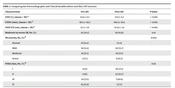
Functional echocardiographic characteristics showed significant improvements following CRT, as summarized in Table 2. Although the increase in the number of patients with moderate-to-severe TR following CRT was not significant, the exacerbation of TR degree after the procedure was significant (P = 0.002). Moreover, the NYHA class significantly improved after the CRT (P = 0.02).
| Characteristic | Pre CRT | Post CRT | P-Value |
|---|---|---|---|
| LVEFb | 23.0 ± 6.5 | 27.9 ± 8.2 | < 0.001 |
| LVESV (mm)b | 161.5 ± 69.3 | 140.1 ± 63.9 | < 0.001 |
| LVOT-VTI (cm)b | 13.7 ± 3.9 | 15.42 ± 3.9 | < 0.001 |
| Moderate to severe TR | 24 (34.2) | 30 (42.8) | 0.14 |
| TR severityc | 0.002 | ||
| Normal | 10 (14.3) | 1 (1.4) | |
| Mild | 36 (51.4) | 39 (55.7) | |
| Moderate | 19 (27.1) | 19 (27.1) | |
| Severe | 5 (7.1) | 11 (15.7) | |
| NYHA classc | 0.02 | ||
| I | 0 (0) | 26 (37.1) | |
| II | 0 (0) | 32 (45.7) | |
| III | 54 (77.1) | 7 (10.0) | |
| IV | 16 (22.9) | 5 (7.1) |
Comparing the Echocardiographic and Clinical Variables Before and After CRT Insertion a
The number of patients who had a clinical response to CRT based on the improvement in NYHA class was significantly higher among those who had no or mild TR at baseline (P = 0.003). Furthermore, the echocardiographic response to TR based on LVEF improvement was significantly lower in patients with moderate-to-severe TR (P = 0.001). However, the frequency of patients who had LVESV reduction following TR was not significantly different between the two groups (Table 3). Based on the TR observed in the post-CRT echocardiography, only the clinical response to CRT was significantly higher in patients who did not have TR following CRT. Tricuspid valve regurgitation was exacerbated in 23 (%33) patients following the procedure. Exacerbation of TR following CRT had no significant effect on the clinical and echocardiographic response to CRT (Table 3). The comparison of the clinical and echocardiographic response to CRT based on the degree of TR before or after CRT implantation is presented in Table 4.
| Characteristic | TR - | TR+ | P-Value |
|---|---|---|---|
| Pre-CRT TR | (n = 46) | (n = 24) | |
| NYHA class improvement | 43 (93.5) | 16 (66.7) | 0.003 |
| LVEF improvement | 32 (69.6) | 7 (29.2) | 0.001 |
| LVESV reduction | 22 (47.8) | 8 (33.3) | 0.24 |
| Post-CRT TR | (n = 40) | (n = 30) | |
| NYHA class improvement | 38 (95.0) | 21 (70.0) | 0.004 |
| LVEF improvement | 25 (62.5) | 14 (46.7) | 0.18 |
| LVESV reduction | 21 (52.5) | 9 (30.0) | 0.06 |
Clinical and Echocardiographic Response to CRT Based on the Presence of TR Before or After CRT Insertion
| Characteristic | Normal | Mild TR | Moderate TR | Severe TR | P-Value |
|---|---|---|---|---|---|
| Pre-CRT TR | (n = 10) | (n = 36) | (n = 19) | (n = 5) | |
| NYHA class improvement | 9 (90.0) | 34 (94.4) | 14 (73.7) | 2 (40.0) | 0.007 |
| LVEF improvement | 6 (60.0) | 26 (72.2) | 6 (31.6) | 1 (20.0) | 0.01 |
| LVESV reduction | 5 (50.0) | 17 (47.2) | 7 (36.8) | 1 (20.0) | 0.2 |
| Post-CRT TR | (n = 1) | (n = 39) | (n = 19) | (n = 11) | |
| NYHA class improvement | 1 (100) | 37 (94.9) | 16 (84.2) | 5 (45.5) | 0.001 |
| LVEF improvement | 0 (0) | 25 (64.1) | 11 (57.9) | 3 (27.3) | 0.11 |
| LVESV reduction | 0 (0) | 21 (53.8) | 7 (36.8) | 2 (18.2) | 0.12 |
Clinical and Echocardiographic Response to CRT Based on the Degree of TR Before or after CRT Insertion
5. Discussion
We observed that TR is common among CHF patients who are candidates for CRT, and it is sometimes exacerbated following CRT implantation. Moreover, clinical and echocardiographic response to CRT was higher in patients who did not have TR. Tricuspid regurgitation may develop from a primary tricuspid valve deformity, raised pulmonary artery pressure, and/or right ventricular (RV) dysfunction, with dilatation of the tricuspid valve annulus (15, 16). In the present study, mild or more TR was found in 60 (85.7%) patients. This high prevalence can be attributable to advanced prolonged CHF (17). Additionally, some subjects had RV and LV dysfunction simultaneously. We detected that moderate or severe TR was an independent anticipant of echocardiographic nonresponse to CRT (10).
Patients with moderate or severe TR had higher end-systolic pulmonary artery pressure, greater RV dysfunction, and lower LVEF. These patients failed to show improvement in LV structure and function (10). Of our cases, 24 (34%) had moderate-to-severe TR, which is comparable with the study by Stassen et al. (11), in which 22% of the patients had moderate-to-severe TR. Pacing leads passing the tricuspid valve can produce or aggravate TR. New onset or exacerbated TR after lead implantation may happen due to the involvement of the tricuspid valve apparatus, leaflet perforation, or fibrosis and further cohesion of the lead to the leaflets (18, 19). Tricuspid valve regurgitation can also be a consequence of RV pacing with no direct effect of the lead on the valve structure and function (20). New onset or magnified TR after pacing or defibrillator lead implantation has been described in up to 24.2% of the patients in one study (10).
Patients with implantable cardioverter-defibrillators (ICDs) have an excessive amount of worsened TR compared to those with pacemakers. Moreover, the position of the ventricular leads can influence the valve mechanics. It has also been shown that right ventricular outflow tract pacing is a safe site for implanting ventricular leads and could reduce the probability of valvular changes after pacing, compared with the right ventricular apex position (21). Therefore, several factors can influence the structure and function of heart valves during CRT implantation and thereby alter the response to CRT; still, as shown in our study, exacerbation of TR after device implantation does not reduce the response to CRT. The number of moderate and severe TR cases increased in our study after CRT implantation (from 24 to 30), unlike the decrease in the number of moderate and severe TR after CRT implantation in Stassen et al.’s study (11).
The present study showed improvement in NYHA class even with TR exacerbation, while the study by Stassen et al. showed an increase in all-cause mortality with TR exacerbation (11). Raed Abu Sham'a et al. (Europace 2013 Feb) (10) also evaluated the effects of tricuspid valve regurgitation on clinical and echocardiographic outcomes in patients with cardiac resynchronization therapy and concluded that the presence of baseline moderate-to-severe TR was associated with increased mortality but did not predict the clinical or echocardiographic response to CRT. Patients with worsened TR following CRT are less likely to clinically respond to CRT, but as shown in our study, the exacerbation of TR after device implantation does not diminish the response to CRT.
5.1. Study limitations
This study had some limitations. First, data on the symptoms and signs of right heart failure were not methodically collected and included. We did not collect clinical data on chronic obstructive lung disease or obstructive sleep apnea, which may develop pulmonary hypertension and right ventricular dysfunction. Moreover, we did not evaluate the right ventricle function by echocardiography. Finally, the sample size was relatively small, and thus, more prospective studies with larger samples are required to verify our findings.
5.2. Conclusions
This study demonstrated that TR severity was exacerbated after CRT implantation. Furthermore, patients with moderate-to-severe TR had significantly less clinical and echocardiographic response to CRT when compared with patients with no or mild TR. Nevertheless, aggravation of TR following CRT implantation had no significant negative effect on the clinical and echocardiographic response to CRT. Therefore, it should not be considered a criterion for decision-making about CRT implantation other than acceptable criteria.
