Low-Dose Aspirin and Uterine Natural Killer Cells in Mice at Day Seven of Pregnancy

authors:

avatar Mandana Beigi Boroujeni 1 , * , avatar Nasim Beigi Boroujeni 2 , avatar Mohammad Reza Gholami 1 , avatar Masoud Beigi Boroujeni 3 , avatar Afsaneh Shafiei 3 , avatar Maysam Rasouli 4

Department of Anatomy, Lorestan University of Medical Sciences, Khorramabad, IR Iran
Razi Herbal Medicines Research Center, Lorestan University of Medical Sciences, Khorramabad, IR Iran
Department of Biochemistry, Payame Noor University Branches of Tehran Shargh, Tehran, IR Iran
Department of Anatomy, Lorestan University of Medical Sciences, Khorramabad, IR Iran

How To Cite Beigi Boroujeni M, Beigi Boroujeni N, Gholami M R, Beigi Boroujeni M, Shafiei A, et al. Low-Dose Aspirin and Uterine Natural Killer Cells in Mice at Day Seven of Pregnancy. Jentashapir J Cell Mol Biol. 2015;6(6):e27858. https://doi.org/10.17795/jjhr-27858.

Abstract

Background:

Inhibiting the effect of aspirin on prostaglandin results in corpus luteum maintenance in pregnant mice. Uterine natural killer (uNK) cells activate between 5 - 7 days of pregnancy. uNK cell differentiation is indirectly regulated by ovarian hormones. Immune cells have important roles in the endometrium at early pregnancy stages. Although low-dose aspirin is effective on ovarian hormones, the effect of low-dose aspirin on the uNK cell population at early pregnancy stages is unknown.

Objectives:

The aim of this study was to assess the effect of low-dose aspirin on uNK cells at day seven of pregnancy.

Materials and Methods:

Adult female mice were coupled and divided into two groups (control and experimental). Thereafter, mice in the experimental group received 7.5 mg/kg aspirin twice a day for one week. Blood samples were collected for the measurement of the level of ovarian hormones at day seven of pregnancy and finally the animals were sacrificed by cervical dislocation and uterine horns were removed for histological study. Periodic acid-schiff (PAS) staining was performed for uNK cell counting. Data analysis was conducted by SPSS software.

Results:

The ovarian hormones level showed a significant difference between the experimental and control groups (P < 0.05). Furthermore, the uNK cell population showed a significant difference between the two groups (P < 0.05).

Conclusions:

Increased ovarian hormones due to low-dose aspirin administration resulted in decline in the uNK cells number, which may be effective in successful pregnancy.

1. Background

Prostaglandins are produced by many tissues in the body; but in the present study, we were concerned primarily about the type produced by the uterine lining or endometrium. Uterine prostaglandins enter the blood stream and are eventually transported to the ovaries where they cause destruction or lysis of the corpus luteum (CL) and as a result, they cause a decrease in blood progestrone levels. Aspirin is one of the major drugs that inhibits the activity of prostaglandins. Prostaglandin inhibitor drugs inhibit the luteolysis process that leads to the prolongation of the CL activity in pregnant mice (1-6). Natural killer (NK) cells are one of the most important cell populations of the innate immune system with diverse effector functions. These cells are the main population of lymphocytes in the endometrium and especially in decidua. Decidual NK cells produce a number of cytokines and other secretory factors that help with trophoblast proliferation and differentiation and promote pregnancy success (7-9).

Considering the cyclic changes in the population of NK cells of uterus, it seems that their distributions and functions are under the control of ovarian hormones; estrogen and progesterone (9-11). Steroidal hormones cause major alterations in the endometrium and directly affect the uterus receptivity for accepting the blastocyst (4, 12). The differentiation of uterine NK (uNK) cells is regulated indirectly through estrogen and progesterone (7, 13). uNK cells are activated during days 5 - 7 of pregnancy, showing several alterations during this period such as change in superficial glycosylation of these cells, proliferation and simulation of IFNγ production, perforines, serines, estrases and other factors that are involved in angiogenesis (14).

Lee et al. stated that the immunological cells played an important role in the stabilization and retention of pregnancy (15). Immune cells show an important regulatory role in the uterus during early pregnancy and the mother has immune tolerance for the allo-antigenic tissue of the embryo (16). The embryo is considered to be semi-exotic to the mother, but the effect of ovarian hormones on the endometrium results in the differentiation of this tissue during the immunological response compared to other tissues; conversely, the embryo continues its growth (17).

Greenwood et al. and Guimond et al. reported that during pregnancy, uNK cells are activated and it seems that the secreted factors of these cells play an important regulatory role in the expression of vascular tissue genes as well as for the endometrial decidua (8, 18). Guimond et al. also investigated the role of NK cells during pregnancy and on the endometrial decidua (18). The role of NK cells in regulating the diferentiation of stroma cells and the response of related vessels during the implantation process has been demonstrated clearly (19-21). An increase in the amount of these cells results in the decrease of implantation and increases the abortion rate (10, 11, 22).

Based on the histological reports, low dosed of aspirin improve the percentage of implantation in females with thin endometrium who receive oocyst (23). There has been conflicting reports regarding the effect of low-dose aspirin on the hemodynamic condition of uterus (24-27). There has not been any comprehensive studies regarding the immunomodulatory role of aspirin on the NK cells of body tissues (28). In addition, there are conflicting reports regarding the effect of aspirin on the NK cells of other tissues. The studies investigated the effect of 660 mg of aspirin on the NK cells activity in patients with melanoma; the results showed that aspirin could suppress 80 - 100% of the NK cells (29).

However, another study using culture media showed that aspirin cannot suppress the NK cells activity stimulated by INFβ or spontaneus stimulation (30). These variations indicate that more research is needed to understand the effect of aspirin on the NK cells in different tissues (28). Based on the studies, low dosage of aspirin results in an alteration in the levels of ovarian steroidal hormones; however, the aspirin regulatory role in the NK cells in different tissues has not been investigated yet.

2. Objectives

Since pregnancy is one of the major factors in changing the amount of immune cells throught alterations in hormonal condition and regarding the fact that using low dosages of aspirin results in an improve in the implantation condition, this study was undertaken to investigate the effect of low dosage of aspirin on the amount of NK cells in the endometrium at the seventh day of pregnancy.

3. Materials and Methods

Adult female (6 - 8 weeks old) mice were cared and housed for two weeks in the laboratory for better adaptation. The female mice were placed beside the male mice and then, the mice with positive vaginal plug were separated using vaginal plug investigation method. The selected mice were divided accidentally into two study groups of control and experimental (using aspirin).

In the Experimental group, a low dosage of aspirin (7.5 mg/kg) was injected twice a day to the adult mice.

To determinate the amount of estrogen and progesterone, blood samples were obtained from the hearts of five pregnant mice selected from both groups on day seven of pregnancy. The concentrations of hormones in the sera were measured with enzyme-linked immunosorbent assay (ELISA) method.

Five pregnant mice were sacrificed by cervical dislocation in each group in the seventh day of pregnancy. Their uterine horns were isolated from any fat and mesentery; then, the tissue samples were prepared from the uterine horns. Paraffin sections of 5 µm thickness were prepared (by serial section method). For NK cell counting, the sections were stained using periodic acid Schiff (PAS) method. From the prepared histological slides, one section was selected from each of the five sections and five microscopic fields in each section were analyzed. In the next step, the average of uterine stroma NK cells was calculated separately in each group. The results obtained from each group were compared using Mann-Whithny test. Statistical analysis was performed using statistical package for the social sciences (SPSS) and P values of less than 0.05 were considered statistically significant.

4. Results

The results of hormonal evaluation using Mann-Whithny test showed that there was a significant difference in the estrogen levels between the experimental and control groups (P < 0.05). Furthermore, the results of hormonal evaluation using the same test showed that there was a significant difference in the progesterone levels between the experimental and control groups (P < 0.05) (Table 1). The statistical evaluation of the NK cells showed a significant difference between the proportion of these cells in both groups (P < 0.05) (Table 2).

Table 1.

The Levels of Estrogen and Progestrone in the Control and Experimental Groupsa

Level of HormoneControl GroupExperimental Group b
Estrogen, pg/mL69.79 ± 3.27114.29 ± 6.74
Progestrone, ng/mL38.37 ± 2.35118.45 ± 6.2
Table 2.

Uterine Natural Killer Cells Number in the Control and Experimental Groupsa

uNK Cells Number bControl GroupExperimental Group
Number11.4 ± 0.543.84 ± 0.33 c

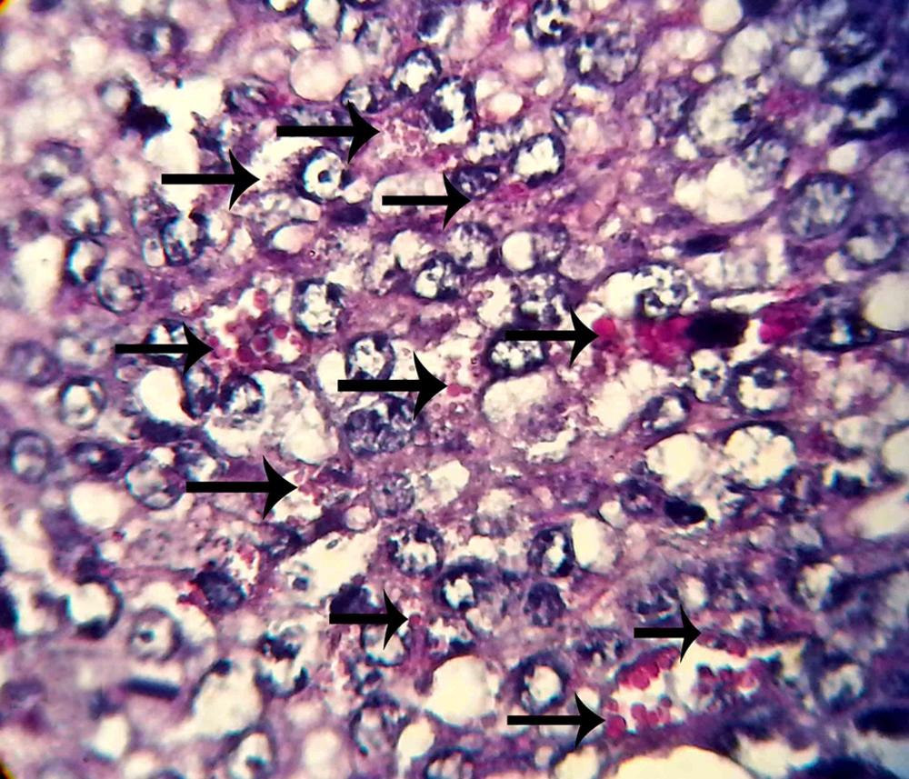
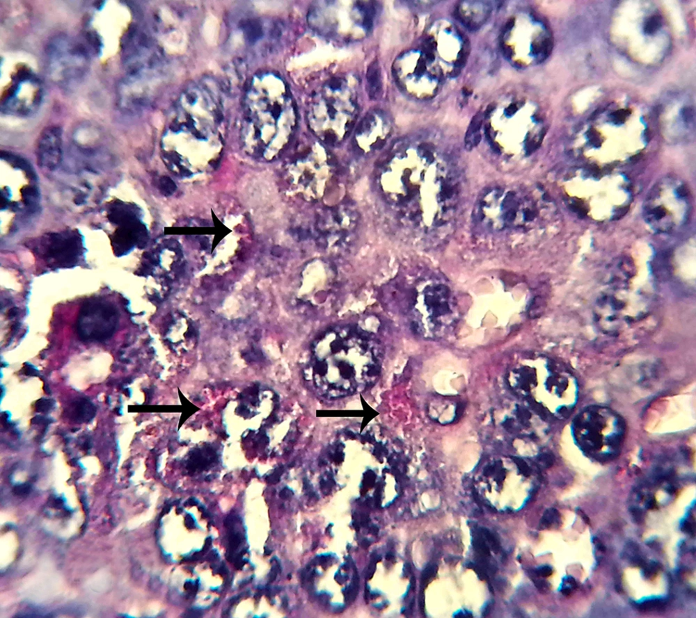
The micrographs indicated the presence of NK cells (positive PAS granules) in the uterine stroma of both groups (Figures 1 and 2).

uNK Cells in the Control Group
The black arrowhead shows NK cells and positive PAS granules (× 1000).
uNK Cells in the Experimental Group
The black arrowhead shows NK cells and positive PAS granules (× 1000).

5. Discussion

Aspirin is the inhibitor of prostaglandins. Prostaglandins affect the endometrium, resulting in the destruction of the CL and as a result, a decrease in blood progestrone levels (2, 3). Prostaglandin inhibitor drugs such as aspirin extend the duration of CL activity in pregnant mice due to the pause of luteolysis (4). The results of this study indicated that the administration of low-dose aspirin increased the level of estrogen compared to the control group, resulting in a significant statistical difference (P < 0.05); also, evaluating the results of progesterone determination showed a significant difference between the control and experimental groups (P < 0.05).

Al-Janabi showed that low dosage of aspirin resulted in the reinforcement of endometrium, size and quality of CL (31). Saki et al. demonstrated that using low-dose aspirin led to an increase in the number of CLs (32), as the results of the present study also confirmed the same findings.

Using low dosage of aspirin affects the ovaries, leads to an increase of estrogen and progesterone levels and alterations in the endometrium (33-35). Ovulation induction results in an increase in estrogen and progesterone levels and this phenomenon leads to a decrease in the number of NK cells (36-38).

The number of uNK cells in mice changes during sexual cycles and pregnancy. These cells can be found in the decidua during the implantation and are activated in day seven of pregnancy (39). Regarding the periodical changes of NK cells, it seems that they are under the control of the ovarian hormones (40, 41). NK cells play a fundamental role during the first stages of pregnancy, as the secretory factors of these cells are considered as the regulatory mediators in the expression of vascular tissue genes, decidualization reaction and survival of pregnancy; however, increase in the number of NK cells leads to an increase in abortion (8, 13, 15, 19, 42). The results of this study indicated that the treatment group using low-dose aspirin showed a significant statistical decrease in the number of uNK cells compared to the control group (P < 0.05).

In the present study, the administration of low dosage aspirin increased the level of estroidal hormones through affecting on the ovarian follicules. The results indicated that the injection of low-dose aspirin (7.5 mg/kg) caused significant differences in the levels of estrogen and progesterone. Seemingly, aspirin affected the ovaries and CL, leading to an increase in estrogen and progestrone levels. Finally, the endometrium underwent profound changes which were regulated by these hormones such as alterations in the number of NK cells.

It seems that the administration of low dosages of aspirin can lead to desirable alterations in endometrium and increase the pregnancy rate by affecting the proportion of NK cells within the uterus. Considering the important role of these cells in the establishment of a successful pregnancy, using a low dosage of aspirin (7.5 mg/kg) may affect the development of embryo and implantation.
References

  • 1.

    Schisterman EF, Gaskins AJ, Whitcomb BW. Effects of low-dose aspirin in in-vitro fertilization. Curr Opin Obstet Gynecol. 2009;21(3):275-8. [PubMed ID: 19300253]. https://doi.org/10.1097/GCO.0b013e32832a0673.

  • 2.

    Fuchs AR, Smitasiri Y, Chantharaksri U. The effect of indomethacin on uterine contractility and luteal regression in pregnant rats at term. J Reprod Fertil. 1976;48(2):331-40. [PubMed ID: 1033279].

  • 3.

    Strauss J3, Sokoloski J, Caploe P, Duffy P, Mintz G, Stambaugh RL. On the role of prostaglandins in parturition in the rat. Endocrinology. 1975;96(4):1040-3. [PubMed ID: 1168125]. https://doi.org/10.1210/endo-96-4-1040.

  • 4.

    Garris DR. Scanning electron microscopic and morphometric analysis of the guinea pig uterine luminal surface: Cyclic and ovarian steroid‐induced modifications. Anat Rec. 1998;252(2):205-14.

  • 5.

    Tavaniotou A, Smitz J, Bourgain C, Devroey P. Ovulation induction disrupts luteal phase function. Ann N Y Acad Sci. 2001;943:55-63. [PubMed ID: 11594558].

  • 6.

    Elnashar AM, Aboul-Enein GI. Endometrial receptivity. Middle East Fertil Soc J. 2004;9:10-24.

  • 7.

    Sharma R, Bulmer D, Peel S. Effects of exogenous progesterone following ovariectomy on the metrial glands of pregnant mice. J Anat. 1986;144:189-99. [PubMed ID: 3693044].

  • 8.

    Greenwood JD, Minhas K, di Santo JP, Makita M, Kiso Y, Croy BA. Ultrastructural studies of implantation sites from mice deficient in uterine natural killer cells. Placenta. 2000;21(7):693-702. [PubMed ID: 10985973]. https://doi.org/10.1053/plac.2000.0556.

  • 9.

    Dorfeshan P, Salehnia M, Moazzeni SM. Ovarian stimulation affects the population of mouse uterine NK cells at early pregnancy. Biomed Res Int. 2013;2013:182531. [PubMed ID: 24350248]. https://doi.org/10.1155/2013/182531.

  • 10.

    Lukassen HG, van der Meer A, van Lierop MJ, Lindeman EJ, Joosten I, Braat DD. The proportion of follicular fluid CD16+CD56DIM NK cells is increased in IVF patients with idiopathic infertility. J Reprod Immunol. 2003;60(1):71-84. [PubMed ID: 14568679].

  • 11.

    van den Heuvel MJ, Peralta CG, Hatta K, Han VK, Clark DA. Decline in number of elevated blood CD3(+) CD56(+) NKT cells in response to intravenous immunoglobulin treatment correlates with successful pregnancy. Am J Reprod Immunol. 2007;58(5):447-59. [PubMed ID: 17922698]. https://doi.org/10.1111/j.1600-0897.2007.00529.x.

  • 12.

    Dey S. K. H. H, Das SK, Reese J, Paria BC, Dai koka T. Molecular cuse to implantation. Endor. 2004;25:341-73.

  • 13.

    Peel S. Granulated metrial gland cells. Adv Anat Embryol Cell Biol. 1989;115:1-112. [PubMed ID: 2658485].

  • 14.

    Croy BA, Esadeg S, Chantakru S, van den Heuvel M, Paffaro VA, He H, et al. Update on pathways regulating the activation of uterine Natural Killer cells, their interactions with decidual spiral arteries and homing of their precursors to the uterus. J Reprod Immunol. 2003;59(2):175-91. [PubMed ID: 12896821].

  • 15.

    Lee JY, Lee M, Lee SK. Role of endometrial immune cells in implantation. Clin Exp Reprod Med. 2011;38(3):119-25. [PubMed ID: 22384430]. https://doi.org/10.5653/cerm.2011.38.3.119.

  • 16.

    Blois SM, Kammerer U, Alba Soto C, Tometten MC, Shaikly V, Barrientos G, et al. Dendritic cells: key to fetal tolerance? Biol Reprod. 2007;77(4):590-8. [PubMed ID: 17596562]. https://doi.org/10.1095/biolreprod.107.060632.

  • 17.

    Robertson SA. Control of the immunological environment of the uterus. Rev Reprod. 2000;5(3):164-74. [PubMed ID: 11006166].

  • 18.

    Guimond MJ, Wang B, Croy BA. Engraftment of bone marrow from severe combined immunodeficient (SCID) mice reverses the reproductive deficits in natural killer cell-deficient tg epsilon 26 mice. J Exp Med. 1998;187(2):217-23. [PubMed ID: 9432979].

  • 19.

    Croy BA, He H, Esadeg S, Wei Q, McCartney D, Zhang J, et al. Uterine natural killer cells: insights into their cellular and molecular biology from mouse modelling. Reproduction. 2003;126(2):149-60. [PubMed ID: 12887272].

  • 20.

    Hanna J, Goldman-Wohl D, Hamani Y, Avraham I, Greenfield C, Natanson-Yaron S, et al. Decidual NK cells regulate key developmental processes at the human fetal-maternal interface. Nat Med. 2006;12(9):1065-74. [PubMed ID: 16892062]. https://doi.org/10.1038/nm1452.

  • 21.

    Blois SM, Barrientos G, Garcia MG, Orsal AS, Tometten M, Cordo-Russo RI, et al. Interaction between dendritic cells and natural killer cells during pregnancy in mice. J Mol Med (Berl). 2008;86(7):837-52. [PubMed ID: 18506412]. https://doi.org/10.1007/s00109-008-0342-2.

  • 22.

    Krizan J, Cuchalova L, Sima P, Kralickova M, Madar J, Vetvicka V. Altered distribution of NK and NKT cells in follicular fluid is associated with IVF outcome. J Reprod Immunol. 2009;82(1):84-8. [PubMed ID: 19682753]. https://doi.org/10.1016/j.jri.2009.05.005.

  • 23.

    Haapsamo M, Martikainen H, Rasanen J. Low-dose aspirin and uterine haemodynamics on the day of embryo transfer in women undergoing IVF/ICSI: a randomized, placebo-controlled, double-blind study. Hum Reprod. 2009;24(4):861-6. [PubMed ID: 19153091]. https://doi.org/10.1093/humrep/den489.

  • 24.

    Khairy M, Banerjee K, El-Toukhy T, Coomarasamy A, Khalaf Y. Aspirin in women undergoing in vitro fertilization treatment: a systematic review and meta-analysis. Fertil Steril. 2007;88(4):822-31. [PubMed ID: 17509593]. https://doi.org/10.1016/j.fertnstert.2006.12.080.

  • 25.

    Lok IH, Yip SK, Cheung LP, Yin Leung PH, Haines CJ. Adjuvant low-dose aspirin therapy in poor responders undergoing in vitro fertilization: a prospective, randomized, double-blind, placebo-controlled trial. Fertil Steril. 2004;81(3):556-61. [PubMed ID: 15037402]. https://doi.org/10.1016/j.fertnstert.2003.07.033.

  • 26.

    Rubinstein M, Marazzi A, Polak de Fried E. Low-dose aspirin treatment improves ovarian responsiveness, uterine and ovarian blood flow velocity, implantation, and pregnancy rates in patients undergoing in vitro fertilization: a prospective, randomized, double-blind placebo-controlled assay. Fertil Steril. 1999;71(5):825-9. [PubMed ID: 10231040].

  • 27.

    Check JH, Dietterich C, Lurie D, Nazari A, Chuong J. A matched study to determine whether low-dose aspirin without heparin improves pregnancy rates following frozen embryo transfer and/or affects endometrial sonographic parameters. J Assist Reprod Genet. 1998;15(10):579-82. [PubMed ID: 9866064].

  • 28.

    Hussain M, Javeed A, Ashraf M, Zhao Y, Mukhtar MM, Rehman MU. Aspirin and immune system. Int Immunopharmacol. 2012;12(1):10-20. [PubMed ID: 22172645]. https://doi.org/10.1016/j.intimp.2011.11.021.

  • 29.

    Grohmann PH, Porzsolt F, Quirt I, Miller RG, Phillips RA. Stimulation of human NK cell activity by cultured cells. II Ingestion of aspirin by blood donors suppresses induced NK activity. Clin Exp Immunol. 1981;44(3):611-4. [PubMed ID: 7326871].

  • 30.

    Porzsolt F, Wolf W. Modulation of natural killer activity by aspirin: I. In vitro effect of aspirin. J Interferon Res. 1983;3(1):11-7. [PubMed ID: 6842038].

  • 31.

    Al-Janabi AS, Al-Zohyri AM, Al-Rubayai FK. Pharmacological effects of low-dose of aspirin on ovulation rate in mature cycling female mice. Middle East Fertil Soc J. 2007;12(1):97-106.

  • 32.

    Saki F, Beigi Boroujeni M, Sadoughi M, Adeli O. The effect of low dose (7.5 mg/kg) of Aspirin on ovary tissue during implantation period in mice (NMRI). Yafteh. 2012;14(3):85-90.

  • 33.

    Gunin A. Realization of estradiol effects in the uterus of ovariectomized rats under acute stress. Eur J Obstet Gynecol Reprod Biol. 1995;60(1):69-74. [PubMed ID: 7635235].

  • 34.

    Hosie MJ, Murphy CR. A scanning and light microscope study comparing the effects of clomiphene citrate, estradiol 17-beta and progesterone on the structure of uterine luminal epithelial cells. Eur J Morphol. 1995;33(1):39-50. [PubMed ID: 7710811].

  • 35.

    Deligdisch L. Hormonal pathology of the endometrium. Mod Pathol. 2000;13(3):285-94. [PubMed ID: 10757339]. https://doi.org/10.1038/modpathol.3880050.

  • 36.

    Oh MJ, Croy BA. A map of relationships between uterine natural killer cells and progesterone receptor expressing cells during mouse pregnancy. Placenta. 2008;29(4):317-23. [PubMed ID: 18289671]. https://doi.org/10.1016/j.placenta.2008.01.003.

  • 37.

    Lukassen HG, Joosten I, van Cranenbroek B, van Lierop MJ, Bulten J, Braat DD, et al. Hormonal stimulation for IVF treatment positively affects the CD56bright/CD56dim NK cell ratio of the endometrium during the window of implantation. Mol Hum Reprod. 2004;10(7):513-20. [PubMed ID: 15155817]. https://doi.org/10.1093/molehr/gah067.

  • 38.

    Junovich G, Mayer Y, Azpiroz A, Daher S, Iglesias A, Zylverstein C, et al. Ovarian stimulation affects the levels of regulatory endometrial NK cells and angiogenic cytokine VEGF. Am J Reprod Immunol. 2011;65(2):146-53. [PubMed ID: 20618181]. https://doi.org/10.1111/j.1600-0897.2010.00892.x.

  • 39.

    Moffett A, Loke C. Immunology of placentation in eutherian mammals. Nat Rev Immunol. 2006;6(8):584-94. [PubMed ID: 16868549]. https://doi.org/10.1038/nri1897.

  • 40.

    Croy BA, van den Heuvel MJ, Borzychowski AM, Tayade C. Uterine natural killer cells: a specialized differentiation regulated by ovarian hormones. Immunol Rev. 2006;214:161-85. [PubMed ID: 17100884]. https://doi.org/10.1111/j.1600-065X.2006.00447.x.

  • 41.

    DeLoia JA, Stewart-Akers AM, Brekosky J, Kubik CJ. Effects of exogenous estrogen on uterine leukocyte recruitment. Fertil Steril. 2002;77(3):548-54. [PubMed ID: 11872211].

  • 42.

    Walch KT, Huber JC. Progesterone for recurrent miscarriage: truth and deceptions. Best Pract Res Clin Obstet Gynaecol. 2008;22(2):375-89. [PubMed ID: 17964858]. https://doi.org/10.1016/j.bpobgyn.2007.08.009.