1. Background
Chronic sinusitis is diagnosed based on clinical symptoms. This diagnosis requires persistent complaints of the patient for more than 12 consecutive weeks. There are many causes for chronic rhinosinusitis and there is an overlap between most of them. Pathophysiological mechanisms include two groups of external (environmental) mechanisms and internal mechanisms that are associated with the host (1). Computed tomography (CT) scan is the standard imaging method for sinus examination and it is an excellent diagnostic tool for confirming the presence of inflammation and assessing their extent in sinuses. It provides information more than what can be achieved by endoscopic studies (2). It is very important to identify predisposing factors for chronic sinusitis. Paranasal anatomic variants have been investigated in several studies and have been proposed as a predisposing factor for sinusitis (3). The importance of an anatomic variation is dependent on its relationship with osteomeatal channels and nasal airways (1, 4). Some anatomical variations are considered pathogenic for chronic sinusitis and its recurrence (5).
Anatomic variations of middle meatus structures can complicate lateral nasal septum and anterior osteomeatal complex. Understanding these variations makes sinonasal surgery safer and more effective. Identifying these anatomical variations minimizes damages to critical structures such as the orbit and skull base (6, 7).
Anatomic variation in nasal septum is also common in children. It seems that local, systemic or environmental factors play a more important role in sinusitis of children compared to anatomic variation. Since no definite relationship exists between anatomical variation and sinus disease, the surgery in children should be avoided (8).
2. Objectives
This study aimed to examine the relationship between anatomic variations of anterior osteomeatal complex with evidence of chronic sinusitis in the radiological evaluation of patients. Based on the results of this study, the surgeon can predict the relationship between the mentioned anatomic abnormality and the disease. Therefore, the surgeon can add the required surgery method for removal of the disorder resulting in the pathology to the treatment plan, if needed.
3. Patients and Methods
In this cross-sectional and case-control study, all patients who were referred to the ear, nose and throat clinic of Imam Khomeini hospital, Ahvaz, Iran, in 2011 and diagnosed with chronic sinusitis based on clinical criteria and also criteria of Lanza and Kennedy (1) were examined. CT scans of paranasal sinuses in coronal, axial and sagittal sections were taken from the patients. If there were radiological changes confirming clinical diagnosis in maxillary ethmoid and frontal sinuses (sinuses around anterior osteomeatal complex), patients were entered into the study. In case of previous nose surgery or systemic diseases associated with current clinical symptoms, patients were excluded from the study. Clinical and radiological data of the patients were collected and recorded. Similarly, another group of patients who were referred to the ear, nose and throat clinic with other complaints were studied as the control group. Patients were undergone similar CT scans of paranasal sinuses and if there were similar radiological changes in chronic sinusitis in radiological images, they were excluded from the study. Afterwards, CT scans of all patients were reviewed by the radiologist for interpretation and examination of anatomical variations surrounding anterior osteomeatal complex. The examination was done in terms of anatomical variations including nasal deviation, concha bullosa, agger nasi, Haller’s cells, paradoxical middle turbinate, and maxillary hypoplasia. Data were analyzed using the chi-squared test and Fisher’s exact test with SPSS software version 18. P < 0.05 was considered statistically significant.
4. Results
From a total of 148 patients, 74 cases had chronic sinusitis (the case group) and 74 patients as the control group. In the chronic sinusitis group, 44 cases (56.4%) were males and 30 cases (43.3%) were females with the mean age of 34.2 years. In the control group, 40 cases (54.1%) were males and 34 cases (45.9%) were females with the mean age of 32.5 years.
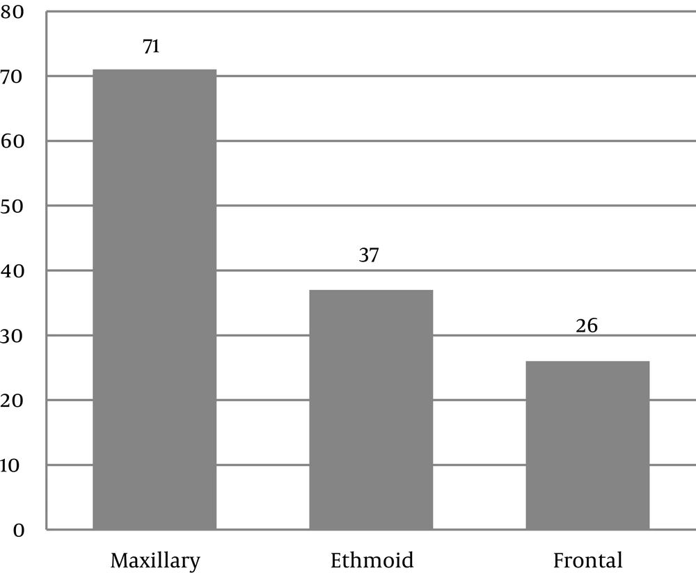
The CT scan results showed that the most common anatomic variations were nasal deviation, concha bullosa and agger nasi (Table 1). The most common involved sinus was maxillary sinus (Figure 1).
| Order | Anatomic Abnormality | Number | Percentage (of Total Cases) |
|---|---|---|---|
| 1 | Nasal deviation | 71 | 48 |
| 2 | Concha bullosa | 47 | 31.8 |
| 3 | Agger nasi | 19 | 23 |
| 4 | Lateralized uncinate | 34 | 12.8 |
| 5 | Haller’s cells | 12 | 8.1 |
| 6 | Paradoxical middle turbinate | 9 | 6.1 |
| 7 | Maxillary hypoplasia | 3 | 3.4 |
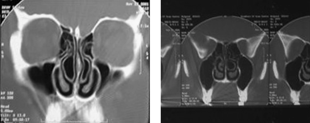
The following anatomical variations were reported: 1, nasal septum deviation: 71 patients had nasal deviation. In the patient group, 42 cases (59.2%) and in the control group, 29 cases (40.8%) had nasal deviation. Among 71 cases with nasal deviation, 42 patients had sinusitis while among 77 cases without nasal deviation, 32 patients had sinusitis. Nasal deviation was more common in patients with sinusitis and this difference was statistically significant (P = 0.032); 2, agger nasi, 34 patients had agger nasi (Figure 2). In the patient group, 20 cases (58.8%) and in the control group, 14 cases (41.2%) had agger nasi and this difference was not statistically significant (P = 0.241).
3, Concha bullosa: 47 patients had concha bullosa (Figure 3). In the patient group, 31 cases (66%) and in the control group, 16 cases (34%) had concha bullosa. Among 47 cases with concha bullosa, 31 patients had sinusitis while among 101 cases without oncha bullosa, 43 patients suffered from sinusitis. Concha bullosa was more common in patients with sinusitis and this difference was statistically significant (P = 0.008).
4, Others: chronic sinusitis was not associated with lateralized uncinate (P = 0.219), Haller’s cells (P = 0.547), paradoxical middle turbinate (P = 0.747), and maxillary hypoplasia (P = 0.245).
| Anatomic Variation | Without Chronic Sinusitis (Control) | With Chronic Sinusitis (Case) |
|---|---|---|
| Nasal deviation | ||
| Has | 29 | 42 |
| Has not | 45 | 32 |
| Concha bullosa | ||
| Has | 16 | 31 |
| Has not | 58 | 43 |
| Agger nasi | ||
| Has | 14 | 20 |
| Has not | 60 | 54 |
| Lateralized uncinate | ||
| Has | 7 | 12 |
| Has not | 67 | 62 |
| Haller's cells | ||
| Has | 5 | 7 |
| Has not | 69 | 67 |
| Paradoxical middle turbinate | ||
| Has | 5 | 4 |
| Has not | 69 | 70 |
| Maxillary hypoplasia | ||
| Has | 0 | 3 |
| Has not | 74 | 71 |
5. Discussion
Knowing the details of anatomical variations of paranasal sinuses is necessary for a surgeon who performs endoscopic sinus operations and also for a radiologist who conducts presurgery examination (9). Variations of anterior osteomeatal complex commonly occur. Variations may be a cause of chronic sinusitis. Therefore, proper planning is important for treatment of these variations in sinus surgery (10).
The most common reported variation was nasal septum deviation (48%), followed by concha bullosa (31.8%) and agger nasi cells (23%). Nasal septum deviation and concha bullosa were associated with chronic sinusitis (P < 0.01, and P = 0.032, respectively). Other variations were not associated with the occurrence of chronic sinusitis.
In the study of Danese (11) and Tonai (12), nasal deviation was associated with chronic sinusitis. This association was also shown in the current study (P = 0.032). Li believes that in terms of aerodynamics, in patients with nasal septum deviation, both sides of the nasal cavity are prone to chronic sinusitis (13). It seems that it is necessary to correct nasal deviation to address the predisposing factor in patients.
Lidov (14) and Clark (15) showed that there is an association between concha bullosa and chronic sinusitis. So, concha bullosa can be considered a predisposing factor for the occurrence of chronic sinusitis. Our study also confirmed it (P < 0.01), while in the study of Nadas (16) and Sivasli (17) no association was found between concha bullosa and chronic sinusitis. It is probably due to the definition of concha bullosa.
Agger nasi and chronic sinusitis were not associated in the study of Sivasli (17) and Tonai (12). The results of the current study did not show agger nasi as a predisposing factor for chronic sinusitis (P = 0.241). It seems that the modification of this variation cannot make any changes in the course of chronic sinusitis.
In the present study, chronic sinusitis was not associated with lateralized uncinate (P = 0.219), Haller’s cells (P = 0.547), paradoxical middle turbinate (P = 0.747) and maxillary hypoplasia (P = 0.245). Studies of Sivasli (17), Danese (11), Tonai (12) and Bolger (18) confirm the obtained results of the current study. However, in all studies, the small sample size can be considered as a limitation. Therefore, this group of variations cannot be definitively ineffective in the incidence of chronic sinusitis. Further studies with larger sample sizes are required to confirm the results and findings of our study.
In conclusion, we can state that chronic sinusitis treatment is based on eliminating predisposing factors. Among the mentioned variations which are more common in the general population (septal deviation, concha bullosa, and agger nasi), it seems that nasal septum deviation and concha bullosa are predisposing factors for chronic sinusitis. We recommend that patients with chronic sinusitis be treated by surgical procedures.


