


1. Background
Obstructive uropathy occurs when urine drainage from one or both kidneys is obstructed; this condition may be in renal, ureteral, bladder, or urethra. Supravesical obstruction is a blockage at a higher level than the bladder; for example, UVJ, or ureter or kidney. The major causes of supravesical obstructive uropathy include ureteral, pelvic, kidney and ureteral, pelvic and renal stones, as well as tumors (1, 2).
The presence of infectious conditions, increased creatinine, or prolonged blockage (2 to 4 weeks) indicates immediate intervention. Immediate renal drainage of the blocked kidney should be used to treatment of this condition. This not only helps reduce pain but also prevents renal function in the future (3, 4). Minimally invasive interventions and radiologic techniques lead to immediate kidney drainage until the final procedure is completed and in some cases treatment may be definitive. Percutaneous nephrostomy and internal stent are equally effective. The inner stent is less effective in the ureteral obstruction, with a high chance of failure, especially in malignant and metastatic disease, chemotherapy, radiotherapy, and kidney failure; therefore, nephrostomy is a more appropriate option (5, 6). In cases with coagulation disorder, ureteral stenosis is preferred. Nephrectomy is performed in the nonfunctional kidney, which has been drainage in 6 to 8 weeks, but has a less than 10% of function. However, if only one kidney functions, and with any degree of function, the patient does not require dialysis, it is not an indication for nephrectomy (7, 8).
The choice of surgical intervention method is based on the cause of obstruction, the kidneys condition and function, the patient’s age, and the overall medical condition of the patient. Due to the fact that there are obstructive electrolyte disorders in patients with obstructive uropathy and these can lead to an increased mortality rate; therefore, in cases with uremic symptoms it is recommended to preoperative hemodialysis for reduction of complications and mortality of surgery (9, 10).
2. Objectives
Based on the context, importance of obstructive uropathy condition was cleared, thus, the aim of this study was comparing the percutaneous nephrostomy and hemodialysis in patients with obstructive uropathy.
3. Methods
3.1. Study Setting
This was a hospital-based study who conducted on patients with obstructive uropathy in the Imam Khomeini Hospital.
3.2. Study Population
Study populations were all patients with obstructive uropathy diagnosis. The study group was based on 30 patients.
3.3. Measurements
The primary variables evaluated in this study were Bun/Cr, Blood pressure, Weight, Height, Body Mass Index, which was evaluated at the time of admission by a trained person. In following the patients were randomly divided into two groups of A and B.
3.3.1. Group A
Hemodialysis catheter was placed in these patients and the patient was under hemodialysis, according to the internal medicine protocol. BUN/Cr, Na, and K were measured every 6 hours and patients were under hemodialysis if necessary in order to be able to adequately control elective surgery in terms of water and electrolyte disturbances.
3.3.2. Group B
Patients who were undergoing percutaneous nephrostomy, unilateral, or bilateral depend on the case conditions. The first hour and then every 6 hours, the BUN/Cr, Na, and K levels were evaluated before surgery and every six hours in order to achieve an optimal electrolyte condition for elective surgery.
Complications of both groups include pain, bleeding, pneumothorax, failed hemodialysis, failure of nephrostomy percutaneous, and other unpredictable complications that are recorded in both groups; patients’ satisfaction is assessed based on VAS criteria.
At the time of T1, T2, and T3 intervals, the degree of pain, satisfaction of patients, and the complications of the operation will be compared in the two groups.
T1 was the duration from patient admission to the proper condition of water and electrolyte for operation, T2 was the duration from the patient’s surgery to discharge from hospital, and T3 was total admission time.
3.4. Ethical Considerations
Ethical issues were completely observed by authors. The study group adheres to the principles of medical ethics introduced by the Health Ministry and the Declaration of Helsinki and Legislation in the Medical Ethics Committee of Ahvaz University of Medical Sciences. In addition, the Ethical Committee of Ahvaz University of Medical Sciences approved the protocol of study. In addition, the IRCT code was IRCT-20150509022168 N5.
3.5. Statistical Analysis
The data were analyzed by SPSS program and P < 0.05 was considered as significant value. The Kolmogorov-Smirnovtest and Q-Q plot were used to evaluate normality of the data. In case of non-normality, the conversion of the variable was used. Nonparametric samples were used even if the change of variable did not allow normal distribution of the variable. Mann-Whitney, t-test, chi-Square, Fisher exact test, ANOVA, Pearson correlation coefficient, and Spearman test were used for single-variable analysis of data. Multiple linear regression analysis was used to analyze the data. In addition, we consider t-test for quantitative variables and χ2 test for qualitative variables.
4. Results
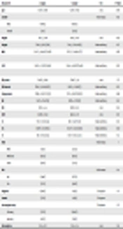
We evaluated 30 patients as our study groups, of these, mean ± SD of age were 54.9±16.8 years in group A and 54.6 ± 17.6 years in group B, therefore, there are no significant differences between the two groups (P = 0.94). In addition, we observed the gender of the patients in group A, 14 patients were male and 1 patient were female and in group B 10 patients were male and 5 patients were female; there are no significant differences for the gender of patients. In addition, other indicators have no significant difference in two groups, including BMI (P = 0.87), GFR (P = 0.74), systolic blood pressure (P=0.7), diastolic blood pressure (P = 0.5), temperature (P = 0.9), respiratory rate (P = 0.5), heart rate (P = 0.4), BUN (0.5), Cr (0.5), potassium (P = 0.6), pain (P = 1), and oliguria (P = 0.7). Sodium was the only variable that has a significant difference between the two groups (P = 0.02) (Table 1). In T1 evaluation we found that not one of evaluated indicators has a significant difference in the two groups (Table 2). However, we observed that T2 and T3 has a significant difference in two group of patients (Tables 3 and 4).
| Parameters | Group A | Group B | Test | P Value |
|---|---|---|---|---|
| Age | 54.93 ± 16.83 | 54.67 ± 17.66 | t test | 0.96 |
| Gender | Fischer-exact | 0.16 | ||
| Male | 14 (93.3) | 10 (66.7) | ||
| Female | 1 (6.7) | 5 (33.3) | ||
| Height | 170.2 ± 5.80 | 165.8 ± 14.9 | t test | 0.29 |
| Weight | 73.80 ± 12.8; 73 (20) | 70.6 ± 15.91; 69 (19) | Mann-withney | 0.71 |
| BMI | 25.47 ± 4.24; 25.7 (4.82) | 25.37 ± 3.8; 24.4 (2.7) | Mann-withney | 0.87 |
| GFR | 10.05 ± 5.2; 9.75 (6.94) | 10.26 ± 4.81; 7.87 (6.69) | Mann-withney | 0.74 |
| BP-systole | 126.67 ± 14.96 | 128.67 ± 14 | t test | 0.7 |
| BP-diastole | 78.67 ± 8.34; 80 (20) | 80.33 ± 5.8; 80 (5) | Mann-withney | 0.53 |
| Temperature | 37.09 ± 0.12; 37.1 (0.2) | 37.12 ± 0.19; 37.10 (0.3) | Mann-withney | 0.98 |
| RR | 19.47 ± 1.76; 17 (4) | 19.87 ± 1.7; 20 (2) | Mann-withney | 0.53 |
| HR | 83.13 ± 6.6 | 80.93 ± 8.5 | t test | 0.43 |
| BUN | 102.87 ± 25.18 | 96.93 ± 31.7 | t test | 0.57 |
| Cr | 9.71 ± 3.33; 9.1 (6) | 8.81 ± 3.47; 7 (5.7) | Mann-withney | 0.53 |
| Na | 138.87 ± 2.9; 138 (5) | 135.07 ± 5.05; 134 (9) | Mann-withney | 0.02 |
| K | 4.8 ± 0.9; 5.2 (1.5) | 5.07 ± 0.65; 5.1 (1.1) | Mann-withney | 0.6 |
| Pain | Fischer-exact | 1 | ||
| Mild | 3 (20) | 2 (13.3) | ||
| Moderate | 8 (53.3) | 8 (53.3) | ||
| Sever | 4 (26.7) | 5 (33.3) | ||
| DM | Fischer-exact | 0.6 | ||
| No | 13 (86.7) | 11 (73.3) | ||
| Yes | 2 (13.3) | 4 (26.7) | ||
| Oliguria | 10 (66.7) | 9 (60) | Chi-square | 0.7 |
| Anuria | 5 (33.3) | 6 (40) | Chi-square | |
| One way/two way | Chi-square | 0.7 | ||
| One way | 5 (33.3) | 10 (66.7) | ||
| two way | 6 (40) | 9 (60) | ||
| Parenchyma | 15.8 ± 2.11 | 15.9 ± 2.6 | t test | 0.8 |
| Parameters | R or Mean ± SD | Test | P Value |
|---|---|---|---|
| Group | t test | ||
| A | 4.33 ± 2.47; 4 (2) | 0.6 | |
| B | 3.87 ± 1.8; 3 (2) | 0.6 | |
| Age | 0.225 | Pearson | 0.3 |
| BMI | -0.21 | Pearson | 0.2 |
| GFR | -0.3 | Pearson | 0.2 |
| BP-systole | 0.26 | Pearson | 0.1 |
| BP-diastole | 0.049 | Pearson | 0.7 |
| T | 0.21 | Pearson | 0.2 |
| RR | 0.22 | spearman | 0.2 |
| HR | 0.1 | Pearson | 0.5 |
| BUN | 0.009 | Pearson | 0.9 |
| Cr | 0.346 | Pearson | 0.06 |
| Na | 0.006 | Pearson | 0.9 |
| K | 0.241 | Pearson | 0.1 |
| Pain | ANOVA | 0.4 | |
| Mild | 5.2 ± 2.1; 4 (4) | ||
| Moderate | 3.88 ± 1.6; 4 (2) | ||
| Sever | 3.89 ± 2.9; 3 (2.5) | ||
| DM | t test | 0.8 | |
| No | 4.1 ± 2.3; 4 (2.7) | ||
| Yes | 4 ± 1.1; 4 (1.5) | ||
| Oliguria | 4.21 ± 2.3; 4 (2) | t test | 0.6 |
| Anuria | 3.9 ± 2.1; 4 (2) | t test | 0.6 |
| One way/two way | t test | 0.1 | |
| One way | 3.3 ± 1.9; 3 (2) | ||
| Two way | 4.5 ± 2.2; 4 (3) | ||
| Parenchyma | -0.18 | Pearson | 0.3 |
| Gender | t test | 0.8 | |
| Male | 4.1 ± 2.3; 4 (2) | ||
| Female | 3.8 ± 1.3; 4 (1.7) |
| Parameters | R or Mean ± SD | Test | P Value |
|---|---|---|---|
| Group | t test | < 0.001 | |
| A | 4 ± 1.31; 4 (2) | ||
| B | 1.73 ± 0.88; 2 (1) | ||
| Age | 0.044 | Pearson | 0.819 |
| BMI | 0.284 | Pearson | 0.128 |
| GFR | 0.128 | Pearson | 0.499 |
| BP-systole | -0.027 | Pearson | 0.887 |
| BP-diastole | -0.183 | Pearson | 0.334 |
| T | 0.121 | Pearson | 0.525 |
| RR | 0.19 | Spearman | 0.30 |
| HR | 0.28 | Pearson | 0.12 |
| BUN | -0.045 | Pearson | 0.813 |
| Cr | 0.04 | Pearson | 0.82 |
| Na | 0.34 | Pearson | 0.06 |
| K | -0.07 | Pearson | 0.68 |
| Pain | ANOVA | 0.23 | |
| Mild | 3.40 ± 1.34; 4 (2.50) | ||
| Moderate | 3.13 ± 1.78; 3 (3.50) | ||
| Sever | 2.11 ± 1.17; 2 (2) | ||
| DM | t test | 0.63 | |
| No | 2.96 ± 1.68; 2.50 (2.75) | ||
| Yes | 2.50 ± 1.22; 2 (2.25) | ||
| Oliguria | 2.79 ± 1.47; 2 (2) | t test | 0.83 |
| Anuria | 3 ± 1.84; 2 (4) | t test | |
| One way/two way | t test | 0.282 | |
| One way | 2.55 ± 1.86; 2 (3) | ||
| Two way | 3.05 ± 1.43; 3 (2) | ||
| Parenchyma | -0.10 | Pearson | 0.57 |
| Gender | t test | 0.06 | |
| Male | 3.13 ± 1.60; 3 (2) | ||
| Female | 1.83 ± 1.17; 1.5 (1.5) | ||
| Satisfaction | 4.6 ± 1.18 | 7.13±1.64 | 0.05 |
| Parameters | R or Mean ± SD | Test | P Value |
|---|---|---|---|
| Group | t test | 0.003 | |
| A | 8.33 ± 2.50; 8 (4) | ||
| B | 5.60 ± 2.29; 5 (2) | ||
| Age | 0.221 | Pearson | 0.239 |
| BMI | 0.045 | Pearson | 0.81 |
| GFR | -0.128 | Pearson | 0.499 |
| BP-systole | 0.167 | Pearson | 0.377 |
| BP-diastole | -0.075 | Pearson | 0.69 |
| T | 0.196 | Pearson | 0.30 |
| RR | 0.204 | Spearman | 0.166 |
| HR | 0.254 | Pearson | 0.176 |
| BUN | -0.009 | Pearson | 0.96 |
| Cr | 0.282 | Pearson | 0.132 |
| Na | 0.212 | Pearson | 0.260 |
| K | 0.129 | Pearson | 0.49 |
| Pain | ANOVA | 0.22 | |
| Mild | 8.60 ± 2.61; 8 (4.50) | ||
| Moderate | 7.00 ± 2.68; 6.50 (3.75) | ||
| Sever | 6 ± 2.74; 5 (1.5) | ||
| DM | t test | 0.77 | |
| No | 7.08 ± 2.99; 6 (4.75) | ||
| Yes | 6.50 ± 1.38; 6.50 (3) | ||
| Oliguria | 7 ± 2.92; 6 (4) | t test | 0.99 |
| Anuria | 6.91 ± 2.51; 6 (4) | t test | |
| One way/two way | t test | 0.09 | |
| One way | 5.91 ± 2.34; 6 (4) | ||
| Two way | 7.58 ± 2.81; 7 (5) | ||
| Parenchyma | -0.238 | Pearson | 0.22 |
| Gender | t test | 0.22 | |
| Male | 7.29 ± 2.88; 6 (4.75) | ||
| Female | 5.67 ± 1.63; 5.50 (3.25) |
5. Discussion
We observed that there was no significant difference between T1 in both PCN and hemodialysis groups. However, there was a significant difference between T2 and T3 in both PCN and hemodialysis groups. In the PCN group, T2 and T3 decreased compared to hemodialysis. The satisfaction rate in patients with PCN was better than hemodialysis. In addition, concentration of potassium and creatinine in the PCN method decreased more than hemodialysis. There were no significant differences in the trend of reduction of other indicators such as sodium concentration and systolic and diastolic blood pressure in both methods. Therefore, in general we found that overall hospitalization time in the PCN method has a significant reduction compared to hemodialysis, this method may be more appropriate for the reduction of pre-operative electrolytes condition in patients with obstructive uropathy, so that PCN can be an alternative, appropriate, inexpensive, and less complicated method for people with obstructive uropathy. In following, other studies have been evaluated. In a study, Wilson et al., showed that in obstructive uropathy patients treated with PCN, the duration of hospitalization was not related to the age group, in addition, it was mentioned that 76% of patients were able to be discharged from the hospital (11). Our results showed that with increasing Cr and systolic blood pressure, T1 increased significantly. Therefore, increasing Cr and systolic blood pressure increases the time from the onset of the patient admission to the proper condition of water and electrolyte to surgery. In addition Clarkson et al. indicated that the increase in Cr was associated with a renal failure condition and an increase in the time of admission (12). According to our results T2 was significantly reduced in the PCN group compared to the hemodialysis group. In addition, the results of this study showed that T2 had a significant decrease in female patients compared to male patients, which indicates a decrease in the duration of the patient’s main surgery until discharge in women. In addition, Bhangu et al. mentioned that the average length of hospitalization in the post-surgical hospital by PCN method was 1.5 days, of which 60% of the patients were discharged after 24 hours. Kara et al. recorded a mean time of admission in the PCN and standard group as 1.5 and 3.2 days, respectively. In addition, Karami and Gholamrezaie recorded a mean time of admission in the PCN group as 1.5 days (1 - 3 days) and in the standard group as 4 days (3 - 7 days) (13). Also, Borges et al. reported longer admission and longer urinary drainage in the PCNL group (14). Istanbulluoglu et al. observed a significant difference between the duration of maintenance of patients in the PCN group (15). Also, Crook et al. reported the duration of hospitalization in PCN patients less frequently and without major complications (16). In addition, we observed that with increasing BMI and increased blood pressure, T2 time was significantly increased. Some studies have shown that increased BMI and hypertension can increase the risk of kidney disease and increase the formation of kidney stones (17). Based on this, PCN can be an alternative method for treatment of obstructive uropathy condition.
5.1. Conclusions
Considering that the overall hospitalization time in the PCN method has a significant reduction in comparison to hemodialysis, this method may be more appropriate for the reduction of pre-operative electrolytes condition in patients with obstructive uropathy, so that PCN can be an alternative, appropriate, inexpensive, and less complicated method for people with obstructive uropathy.
