


1. Introduction
Tinea favosa is an anthropophilic dermatophyte infection primarily caused by Trichophyton schoenleinii and transmitted by human contact. Tinea favosa is a relatively rare inflammatory infection that mainly affects children, and it is characterized by yellowish cup-shaped crusts (scutula) on the scalp (1). Direct examination, wood lamp examination, and fungal culture can be performed to confirm the diagnosis. Fluconazole, griseofulvin, itraconazole, and terbinafine are suggested regimens for treating Tinea favosa. Although tinea favosa was more common in the past, it is now limited to some endemic regions due to improved socio-economic conditions and hygiene. Tinea favosa has been observed in Southern and Northern Africa, Pakistan, the United Kingdom, Australia, South America, the Middle East, and Poland (2).
Herein, we report three severe cases of tinea favosa in Afghan immigrants, all immunocompetent residing in Shiraz city in Iran.
2. Case Presentation
2.1. Case 1
A 15-year-old boy from Afghanistan presented with patchy hair loss and yellowish cup-shaped crusts on his scalp since 2.5 years ago. He was a construction worker and had immigrated to Iran 4 years ago. He had never been referred to a dermatologist. His family history was negative. Mycological culture was positive for T. schoenleinii. The patient responded to a 6-week course of griseofulvin (20 mg/kg/day, a fungistatic agent that inhibits fungal cell mitosis and nuclear acid synthesis).
2.2. Case 2
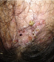
A 14-year-old girl from Afghanistan with a 3-year history of scalp lesions and hair loss was referred to our clinic for further evaluation. A dermatological examination revealed a wide patch of cicatricial alopecia on the crown, perifollicular erythema, and some yellow cup-shaped crusts on the frontal region of her scalp (Figure 1).

Close-up view of yellow cup-shaped crusts on the frontal region of the scalp
A skin biopsy had been performed on her lesions two years ago. Histopathological examination was in favor of folliculitis decalvans at that time. Then, The patient was treated for folliculitis decalvans using rifampicin, prednisolone, minoxidil solution, and topical clobetasol solution for 2 years before admission. On admission, mycological culture and tests were positive for T. schoenleinii (2).
Oral administration of griseofulvin (21 mg/kg/day) for 8 weeks and antifungal shampoo (2% ketoconazole, a fungistatic and fungicidal agent that alters cellular membranes, resulting in growth inhibition) every other day for 8 weeks resulted in disappearance of all active lesions together with little regrowth of the hairs in the clinically evident cicatricial areas.
2.3. Case 3
Since childhood, a 22-year-old man from Afghanistan was referred to our clinic with extensive yellowish cup-shaped crusts on his scalp. He had been a shepherd in Afghanistan and had immigrated to Iran 8 years ago. He had never been referred to a physician because of his low socioeconomic status. His family history was negative. Wood's light scalp examination showed a pale green fluorescence of the infected hairs. Mycological examination of the infected hairs clarified with lactophenol revealed fungal mycelia within the hair shaft and was positive for T. schoenleinii. The lesions completely disappeared after a 6-week of griseofulvin (15 mg/kg/day).
The patients signed informed consent to permit the publication of the case report. The institutional ethics committee approved the case report (ethics code: IR.SUMS.MED.REC.1402.113)
3. Discussion
Tinea favosa is one type of tinea capitis, characterized by yellow cup-shaped crusts known as scutula (3). It can be caused by T. schoenleinii. Trichophyton violaceum, T. verrucosum, T. mentagrophytes, and Microsporum canis. In addition, geophilic M. gypseum has also been recovered from favic lesions. Tinea favosa is typically a childhood disease, yet adult cases are not uncommon (4). Its transmission can occur through infected persons, hairs, animal vectors, and fomites (5).
Tinea favosa can mimic seborrheic dermatitis, psoriasis, lichen planus, or tinea amiantacea. Poppe et al. presented a case of favus closely mimicking lichen planus (6). Khaled et al. presented a case misdiagnosed as psoriasis (1), and one of our patients was treated for folliculitis decalvans. Because of a non-inflammatory presentation, it may remain undiagnosed for years, resulting in permanent scarring (6).
Tinea favosa is relatively common in Mediterranean countries, southern Asia (7), Greenland, and South Africa (8). It is now rare throughout Europe and most parts of India except the Kashmir valley (1, 9). In addition, the incidence of favus has decreased in Libya (10).
In a Tunisia study, only 1.6% of tinea capitis cases were due to T. schoenleinii (11). Of 372 patients with tinea capitis in Saudi Arabia, favus was found only in one patient (12).
Studies from Iran show that the frequency of reported favosa among all dermatophytoses is higher in the country's northern cities, for example, 16.8% in Rasht (13), versus southern cities, for example, 2.3% in Ahvaz (14). Although the reason is unclear, it might be due to a better reporting system in the North. Lifestyle, the level of hygiene, and socioeconomic status, as well as immigration, all influence the prevalence of dermatophyte infections. The heavy influx of immigrants from Afghanistan and the deterioration of the socioeconomic status of some families in some areas of the country could cause this infection's persistence in some cities of Iran. The scarcity of favus in southern Iran has led to the misdiagnosis or late diagnosis of this infection in those areas.
Considering the high potential of T. schoenleinii in causing inter-family epidemics, especially if left untreated, it can easily be transmitted to others. As demonstrated in all of the above cases, delay in diagnosis and misdiagnosis might cause permanent alopecia, and this will undoubtedly have a negative mental and physical impact on the patient's life.
The somewhat increasing trend of isolation of T. schoenleinii in the immigrant Afghan population in Iran should be considered a health problem that requires special attention, as it could be a potential source for re-emergence of this dermatophyte infection in Iran as well as in the region. The epidemiology of tinea favosa is likely to change with shifting patterns of migration, alterations in socioeconomic conditions, and the increase in international travel, as well as the overuse of drugs. A limitation of our study is that it only surveyed participants from one area of the country. This study emphasizes the importance of accurate and timely diagnosis of tinea favosa to prevent individual complications and highlights its regional spread. The distribution of dermatophyte species varies across different geographical regions. Therefore, the increasing trend of isolation of T. schoenleinii in a specific region and accurate identification of new cases can provide valuable insights into its epidemiological aspects and its impact on public health, so we should raise awareness about tinea favosa, its symptoms, and the importance of seeking timely medical attention. Targeting both immigrant communities and healthcare providers is crucial.
