1. Background
One of the aims of ergonomics is to create an environment consistent with the dimensions of the human body and physical and mental capacities. The physiology of work is a part of the knowledge of ergonomics that considers the coordination of man with work in energy consumption and physiological parameters during activity.
On the topic of work physiology, the maximum amount of oxygen consumption (VO2-max) is considered as a criterion to determine the aerobic capacity and the physical work endurance in people (1). There is a linear relationship between oxygen consumption with heart rate and the amount of activity (2). Also, studies show that the aerobic capacity index, the maximum volume of oxygen that a person can consume within a minute, can be a good criterion to determine the physical work capacity (PWC) and endurance; hence, the maximum oxygen consumption is accepted as a criterion to measure the cardiac and respiratory capacity (1, 3). In some studies, VO2-max is used to assess the health of the cardiovascular system and suitability of physical work (4-6). But, this index cannot be used directly, because man can work with maximum capacity for just a few minutes. Therefore, many researchers determined an acceptable limit for workload in an 8-hour shift and believed that the average level of effort during work should not exceed it. For example, Astrand, Michelle, and Bink proposed 40%, 35%, and 33% of the maximum aerobic capacity as PWC, respectively (7, 8). PWC is the amount of maximum energy that a person can consume during 8 hours work without damage to its health; it is defined kcal/minute (8). The Researches showed that PWC is approximately 33% of the maximum aerobic capacity (AC) (9). The maximum aerobic capacity is calculated by the maximum oxygen consumption (VO2-max) (9).
On the other hand, the results of some studies show a relationship between maximum oxygen consumption (VO2-max) with the forced expiratory volume during the first second (FEV1) and some obstructive respiratory diseases (10, 11). FEV1 is the maximum volume of the air exhaled from the lungs that may be removed during the first second (12). The decreased pattern of the FEV1 shows obstructive respiratory diseases (12).
One of the obstructive respiratory diseases is the chronic obstructive pulmonary disease (COPD), which is a broad classification of diseases associated with the chronic obstruction of air flow into or out of the lung. 15 The COPD is the most common cause of death and disability related to lung diseases. Such diseases are usually progressive and associated with an abnormal inflammatory response of the lung in exposure to harmful gases and particles. COPD include diseases such as chronic bronchitis, emphysema, and asthma (13). Hence, it is expected that VO2-max and PWC index also reduce in patients with COPD and reduced FEV1. To the authors’ best knowledge, no study evaluated the direct effect of different degrees of asthma on PWC. Therefore, according to the results of other studies, the mentioned items, and the prevalence of COPD such as asthma, the current study aimed at investigating the relationship between the PWC index and the different degrees of the asthma in workers.
2. Methods
The current cross sectional study was performed on male workers at a steel company in Isfahan, Iran. In the current study, 88 subjects were equally divided into 2 groups including healthy subjects and subjects with different degrees of asthma. The severity of asthma was determined by an occupational medicine specialist, based on the spirometry documentation. The subjects with asthma were divided into 3 groups of mild, moderate, and severe asthma. The current study employed the non-probabilistic sampling method. The inclusion criteria were no history of other respiratory diseases such as emphysema and bronchitis, neuromuscular, and musculoskeletal diseases, epilepsy, seizure, no consumption of high blood pressure controlling or heart rate affecting drugs, ceased consumption of coffee, caffeine, and alcohol 12 hours before the test. As the environmental conditions such as temperature and noise may also affect heart rate, all of the participants performed the test under the same conditions and location.
At first, the test procedure was explained to the workers and they signed the consent form developed by the ethics committee of Isfahan University of Medical Sciences. A demographic questionnaire including height, weight, body mass index (BMI), physical activity, smoking, age, and cardiovascular disease was completed by the researcher through interviewing the subjects. Then, each of the subjects rested for 15 minutes, and then, his heart rate was measured. In the next step, the weight without shoes, hat, and gloves was measured by a digital scale of Hamilton model with an accuracy of 0.1 kg; the height of the subject was measured by a stadiometer. In the current study, Tuxworth and Shahnavaz method was used to measure the VO2-max (7). There are different methods to measure VO2-max and numerous studies are conducted on these methods in different populations (14-16). One of the available simple methods is the step test; the Tuxworth and Shahnavaz method is an appropriate step test to measure VO2-max in Iranian population. Therefore, this method was used in the current study to measure the maximum aerobic capacity.
In this method, the subject goes up and down a 40-cm height step at a rate of 25 steps per minute for 5 minutes; then, the subject sits down and after 30 seconds his heart rate is measured at different intervals of 30 to 60, 90 to 120, and 150 to 180 seconds. In the current study, participants used this method while they take a rest and their heart rate was measured by a sports polar tester. Also, the exclusion criteria of the study were suffering from fatigue during the test and heart rate increase over 180 beats/minute.
After the test, b index was calculated by the following formula:
Index b = [(the heart rate in seconds (180 - 150) + (120 - 90) + (60 - 30)) × 2] / body weight in kilograms]
Then, the VO2-max was calculated using the following equation (7) (X is the b index):
Y = - 0.378 X + 4.67
When the VO2-max was estimated, the following formula was used to calculate the maximum PWC:
VO2-max × 5
PWC (kcal/minute) = maximum PWC (kcal/min) × 0.33
In this formula, the number 5 shows that burning a liter of oxygen in the body produces 5 kcal of energy.
Finally, data was analyzed with SPSS version 16, using independent t test and ANCOVA. The significance level was 0.05 for all the tests.
| Group | Parameter | Range | Mean (SD) |
|---|---|---|---|
| Without asthma | Age | 26 - 52 | 37.30 (6.86) |
| Height (cm) | 170 - 183 | 176 (4.59) | |
| Weight (kg) | 66 - 95 | 82 (8.68) | |
| BMI (kg/m2) | 20.15 - 30.67 | 26.25 (2.75) | |
| Rate of smoking (cigarette/d) | 0 | 0 | |
| Physical activity (h/wk) | 0 - 10 | 3.73 (2.68) | |
| With asthma | Age | 24 - 53 | 40.38 (7.50) |
| Height (cm) | 167 - 192 | 174 (5.74) | |
| Weight (kg) | 59 - 102 | 79.38 (9.08) | |
| BMI (kg/m2) | 19.94 - 30.79 | 26.18 (2.77) | |
| Rates of smoking (cigarettes/d) | 0 - 8 | 0.57 (1.81) | |
| Physical activity (h/wk) | 0 - 5 | 2.73 (1.86) |
Demographic Characteristics in the Study Groups
| Group | Parameter | Range | Mean (SD) |
|---|---|---|---|
| Without asthma | Resting heart rate (beat/ min) | 60 - 80 | 70.17 (5.89) |
| Maximum PWC (kcal/ min) | 13.90 - 18.77 | 15.60 (1.94) | |
| PWC (kcal/min) | 4.59 - 6.20 | 15.5 (0.36) | |
| With asthma | Resting heart rate (beat/ min) | 60 - 90 | 78.96 (7.71) |
| Maximum PWC (kcal/ min) | 11.43 - 17.10 | 14.51 (1.65) | |
| PWC (kcal/min) | 3.77 - 5.64 | 4.79 (0.54) |
Comparison Between the Study Groups
3. Results
Some demographic characteristics and conditions of the workers in the 2 groups of with/without asthma are shown in Table 1. There was no significant difference between the study groups in the mean of age, weight, height, physical activity, BMI, and smoking rates (P > 0.05). None of the study participants had cardiovascular disease.
The means, ranges, and SD values of the rest heart rate, as well as maximum PWC and PWC in the 2 study groups are illustrated in Table 2. ANCOVA showed a significant difference between the study groups in the resting heart rate, maximum aerobic capacity, and PWC (P <0.05).
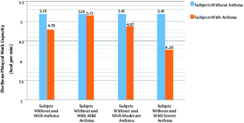
The mean (SD) PWC in the subjects with mild, moderate, and severe asthma was 5.15 (0.29), 4.87 (0.60), and 4.28 (0.34), respectively. The statistical analysis showed no significant difference between the groups of without asthma and with mild asthma in the mean PWC (P > 0.05). Mean PWC in the subjects without asthma was significantly higher than that of the ones with moderate asthma (P < 0.05); but, there was a significant difference between the 2 groups of without asthma and with severe asthma in the mean PWC (P < 0.05). Also, there was a significant difference in the mean PWC between the subjects with mild/moderate asthma and the ones with severe asthma (P < 0.05). The mean PWC in the subjects with mild asthma was significantly higher than that of the ones with moderate asthma (P < 0.05). The comparison of the mean PWC between the subjects with mild, moderate, and severe asthma and the ones without asthma are shown in Figure 1.
4. Discussion
In the current study, PWC of the subjects with asthma was significantly lower than that of the ones without asthma. All of the participants performed the test under the same conditions and location, but the environmental factors such as temperature and noise may affect their heart rate. However, there was no significant difference between the study groups regarding the age, weight, height, physical activity level, BMI, and smoking parameters that can affect heart rate and PWC index. None of the participants had heart disease in the current study. Results of the study showed a significant correlation between VO2-max and height, weight, BMI, doing exercise, and smoking variables; since these factors could affect the aerobic capacity of the subjects (15, 17). Also, some other studies showed that the aerobic capacity decreased with age increase. Similarly, cardiovascular diseases and disorders can affect the aerobic capacity of subjects (18). Therefore, it can be concluded that the PWC index are affected by asthma.
The results of the current study showed that the resting heart rate of the subjects with asthma was significantly higher than that of the subjects without asthma. This can be due to irritants in the air of the workplace. The irritants cause reduction in the diameter of the airways and airflow obstruction in the subjects with asthma. This reduction in ventilation stimulates the chemical receptors of the medulla oblongata, since such receptors are sensitive to the increase of carbonic anhydride. Then, the stimulation of these receptors increases the heart rate through exciting the sympathetic nervous system (19). Also, results of the current study showed a correlation between PWC and asthma due to the decreased FEV1 in subjects with asthma; there was also a relationship between VO2-max and FEV1, based on the results of some studies. It was also expected that VO2-max and the PWC index reduce in subjects with asthma. The results of Ganju et al., showed a significant correlation between %FEV1 and %VO2-max in 3 groups of the subjects with mild, moderate, and severe COPD (10). In addition, a study by Carter showed a significant correlation between FEV1 and VO2-max in males and females with COPD (11). Also, some studies showed that the level of VO2-max in the subjects with COPD was significantly lower than the ones without COPD. For example, results of a study by Clark et al., on 44 subjects with mild to moderate asthma and 64 healthy subjects showed that VO2-max in males and females with asthma was significantly lower than the healthy subjects (20). Also, a study by Wakayama et al., showed a significant correlation between VO2-max and the score of emphysema, a COPD (21). These findings were in agreement to those of the current study. Results of the present study also showed that PWC more reduced when the severity of asthma increased. It can be due to the more constriction of air ducts followed by heart rate increase in the subjects with severe asthma. A study by Baurerla et al., showed that variability of the ventilatory responses during exercise is one of the main determinants of variability of the exercise capacity in subjects with COPD (22). In addition, results of the current study showed a significant difference in PWC between the subjects with moderate and severe asthma, and the healthy subjects, but the difference was insignificant between the subjects with mild asthma and healthy subjects. It can be due to the low constriction of air ducts in subjects with mild asthma, as the chemical receptors of the medulla oblongata were not highly stimulated.
4.1. Conclusion
Based on the results of the current study, PWC in subjects with asthma was lower than the ones without asthma; hence, asthma is an important factor impressing PWC. Hence, it is suggested to identify patients before recruitment, screen workers in periodic examinations, and do not assign such patients to the tasks require high PWC. Also, the employed step test method can be used to trace asthma in industrial environments, although the environmental conditions should also be considered. Further study is suggested to prepare a database for PWC and confounding variables.

