Risk Factors for Mucormycosis in COVID-19 Patients

authors:

avatar Zahra Taghinejad ORCID 1 , avatar Mohammad Asgharzadeh ORCID 2 , avatar Vahid Asgharzadeh 3 , avatar Abdolhassan Kazemi ORCID 4 , *

Hematology and Oncology Research Center, Tabriz University of Medical Sciences, Tabriz, Iran
Biotechnology Research center and Faculty of Paramedicine, Tabriz University of Medical Sciences, Tabriz, Iran
Student Research Committee, Tabriz University of Medical Sciences, Tabriz, Iran
Medical Philosophy and History Research Center, Tabriz University of Medical Sciences, Tabriz, Iran

How To Cite Taghinejad Z , Asgharzadeh M, Asgharzadeh V, Kazemi A. Risk Factors for Mucormycosis in COVID-19 Patients. Jundishapur J Microbiol. 2021;14(8):e117435. https://doi.org/10.5812/jjm.117435.

Abstract

Opportunistic infections, such as mucormycosis, in coronavirus disease 2019 (COVID-19) patients has become a new health challenge. Since opportunistic infections can exacerbate COVID-19 patients' status, it is vital to identify the risk factors to prevent, diagnose, and treat them as soon as possible. Viral, fungal, environmental, and host factors may be responsible for this situation. Long hospital stays, impaired host immune system function due to viral infection, and excessive consumption of glucocorticoids in managing COVID-19 patients are the main risk factors for the increased risk of mucormycosis in COVID-19 patients. Educating health care workers and considering the association between mucormycosis of the paranasal sinuses and different strains of severe acute respiratory syndrome coronavirus 2 (SARS-CoV-2) as the cause of COVID-19 can help prevent invasive fungal sinusitis in COVID-19 patients.

1. Context

Coronavirus disease 2019 (COVID-19) pandemic caused by a novel coronavirus, severe acute respiratory syndrome coronavirus 2 (SARS-CoV-2), emerged in late 2019 in Wuhan, China, and soon spread throughout the world (1). As different factors like age, gender, co-morbidities, education status, and viral load play a role in COVID-19 severity, SARS-CoV-2-infected patients reveal a wide range of clinical features from asymptomatic infection or mild upper respiratory tract symptoms to multiple organ failure (MOF) and even death (2). In COVID-19 patients, especially those who are critically ill, bacterial or fungal opportunistic co-infections with worse outcomes and higher mortality rates have been reported and evolved into a new concern (3).

Mucormycosis is an invasive fungal infection commonly caused by Rhizopus and Rhizomucor genus belonging to the Mucorales order. Immunocompromised people have higher risks of contracting Mucorales infection, and in several reports (4, 5), these infections have been diagnosed concurrently with COVID-19 infection (6-8). During the last two to three months, several reports have shown concurrent mucormycosis (Black fungus’ disease) with COVID-19 (9), and recently it has been reported in Iran among COVID-19 patients (10, 11).

The occurrence of this mycose in COVID-19 patients in Iran and its widespread coverage in the social media have caused social concerns (12, 13). The inhalation of spores or inoculation of wounds may result in Mucorales infection (14, 15). Because of vascular invasion and tissue infarction, this aggressive infection leads to death in 50% of infected people and leaves survivors with severe long-term consequences such as ocular traumas (14). Severe and irreversible consequences of opportunistic infections and the frequent reports of mucormycosis in COVID-19 emphasize the need for identifying the risk factors for mucormycosis in COVID-19 patients and determining its prevention, diagnosis, and therapeutic strategies. Thus, in this study, we reviewed the host, environmental, and microbial risk factors to make preventative decisions about opportunistic co-infections in COVID-19 patients and reduce their complications.

2. Evidence Acquisition

2.1. SARS-CoV-2 Transmission

SARS-CoV-2 is a highly transmissible zoonotic novel coronavirus with the reproduction rate of 3.28 (16). Humans were initially exposed to SARS-CoV-2 at Wuhan's Huanan seafood market, and while the intermediate host of SARS-CoV-2 is unknown, bats are considered as the natural reservoirs of SARS-CoV-2 due to the high sequence homology of their coronavirus strains with SARS-CoV-2 (17). As a result of the rapid spread of SARS-CoV-2 to over a hundred nations around the world, on March 11, 2020, COVID-19 was declared a pandemic by the World Health Organization (18).

SARS-CoV-2 may transmit among humans and even from humans to animals in different ways. Notably, close contact with respiratory droplets and airborne transmission routes are the major ones (2, 19). As these transmission routes accelerate transmission from a small number of infected cases to many other people in indoor crowded spaces with poor ventilation, keeping social distancing and following health protocols are recommended (20). Other forms of transmission like fomite transmission and fecal-oral ways have been also reported in several studies, which emphasizes the importance of adhering to health rules (21, 22). Although 80% of COVID-19 patients are asymptomatic or only have mild upper respiratory symptoms, in 20% of cases pneumonia is followed by fever, cough, shortness of breath, nausea, acute respiratory distress syndrome (ARDS), and MOF (23). The rising incidence of fatal coinfections like mucormycosis in patients with COVID-19 is now becoming another challenge for health systems (3).

2.2. Mucormycosis and Its Clinical Features

Human zygomycosis is caused by two Zygomycetes orders, namely Mucorales and Entomophthorales. Mucorales are divided into six families, each of which can cause cutaneous or deep infections known as mucormycosis in humans, particularly those who are immunocompromised (24). Oral, nasal, and cutaneous routes are the possible forms of mucormycosis transmission and as it cannot spread between people or between people and animals it is not contagious (25). Inhaling fungal spores that are commonly found in soil, plants, and decaying fruits in the environment causes individuals to contract lung/sinus mucormycosis. A skin infection also can arise after the fungus entry to the skin through a burn blister or other types of skin injury (14, 15, 26). As the fungus can spread through the bloodstream and invade other tissues, mucormycosis is majorly characterized by vascular invasion and is categorized into the six groups of rhinocerebral, pulmonary, cutaneous, gastrointestinal, disseminated, and rare presentations as a result of thrombosis and tissue infarction/necrosis of particular anatomic sites (27).

The most common form of mucormycosis is rhinocerebral involvement, which affects the sinuses, nose, eyes, and brain (28). The symptoms could vary based on the infected site, but facial pain, headache, jaw pain, blurring of vision, double vision, cough, dyspnea, fever, and blackened skin lesions on the nasal bridge or upper inside of the mouth are the common signs and symptoms of mucormycosis, but a conclusive diagnosis requires histology examination and microbiological investigations (29). The major therapeutic techniques for slowing fungal invasion are antifungal medicines and rigorous surgical debridement of the infected region (30). Despite the use of various therapeutic approaches, the death rate for mucormycosis varies depending on the patient's underlying illnesses, stage of infection, fungus species, and affected anatomical location, but the overall mortality rate is reported to be 50% (31). Because of the high mortality rate, long-term consequences of aggressive therapeutic surgeries, and rising prevalence of mucormycosis in COVID-19 patients, identifying mucormycosis risk factors can help in developing preventative, diagnostic, and therapeutic strategies for this time-sensitive opportunistic infection.

2.3. Host Risk Factors

The following are the key host risk factors that increase mucormycosis susceptibility in COVID-19 patients.

2.3.1. Corticosteroid Usage

Glucocorticoids are often used to manage inflammatory and autoimmune diseases. They have been also demonstrated to enhance the survival rate in hypoxemic COVID-19 patients in the absence of specific and effective anti-COVID-19 medications. Due to the beneficial survival effects, inexpensiveness, and wide accessibility of glucocorticoids, they have been used with other interventions such as remdesivir, tocilizumab, and mechanical ventilatory support for managing COVID-19 patients (32). However, glucocorticoids suppress both the innate and adaptive immune systems and increase the risk of infections, especially opportunistic ones, by suppressing pro-inflammatory mediators, impairing immune cell migration and phagocytic utility, and up-regulating anti-inflammatory factors (33). They also induce hyperglycemia by prompting insulin resistance that facilitates the germination of Mucorales spores and worsens the infection. Overall, 76.3% of COVID-19 individuals diagnosed with mucormycosis had a history of corticosteroid usage (34). Thus, there is a need to limit the excessive administration and usage of glucocorticoids in COVID-19 patients.

2.3.2. Immunocompromising States

Owing to strong immune system functionality, healthy individuals appear to have a low chance of improving mucormycosis. Phagocytes of the innate immunity are the first line of defense when the spores enter the body and then the adaptive immune system limits this invasive fungal infection (35). As in COVID-19 patients, there is a reduction in the cluster of differentiation (CD)4-positive and CD8-positive T cell levels and impaired innate immune responses, secondary opportunistic fungal infections are common to be diagnosed (34). Pre-existing hereditary or acquired immunodeficiencies in COVID-19 patients can also elevate the risk of mucormycosis in them (36).

2.3.3. Diabetes

Diabetes mellitus is a group of metabolic syndromes marked by persistent hyperglycemia caused by impaired insulin production, activity, or both (37, 38). Apart from the conventional consequences of diabetes like neuropathy or nephropathy, its effects on T cell response, inflammatory cytokines, neutrophil function, and humoral immune activity are associated with immunosuppression (39). Diabetes is one of the risk factors for COVID-19 severity, which prolongs the hospitalization and recovery period and increases the probability of immunosuppressive medication for patients with moderate to severe forms of COVID-19 (40, 41). Due to these reasons, secondary infections have a significantly higher probability of developing in diabetic COVID-19 patients with or without diabetic ketoacidosis. Mucormycosis is observed to be more common in people with uncontrolled diabetes and hyperglycemic conditions (38). In a study reported by Corzo-Leon et al. in 2017, 68% of patients with mucormycosis were diabetic (42).

2.3.4. Malignancy

Cancer is a genetic disorder that is eventually the outcome of environmental circumstances. Mutations in DNA, especially in proto-oncogenes or tumor suppressor genes’ regions, can lead to uncontrolled proliferation of cells (43). Cancer cells can modulate the immunological responses through several strategies, like spreading into the bone marrow and impairing white blood cell proliferation. Even cancer therapies, such as chemotherapy, radiotherapy, and high-dose corticosteroids, can potentially eliminate the immune response (44). Because neutrophils are vital in the host defense against Mucorales, cancer or chemotherapy-induced neutropenic patients are more likely to develop mucormycosis (45). Furthermore, since the onset of severe forms of COVID-19, which are linked to long-term hospital stays and immuno-suppressive treatments, is more common in cancer patients, focused susceptibility to clinical status is essential (2, 46).

2.3.5. Malnutrition

Malnutrition is defined as an imbalance between the required and received nutrients. It can be classified as protein-energy malnutrition or micronutrient deficiency that increases susceptibility to a variety of infections and typically delays recovery (47). It has been reported that vitamin B12, folic acid deficiency (micronutrient deficiency), or severe protein-calorie malnutrition (protein-energy malnutrition) can negatively impact immune responses and can be associated with neutropenia (48). According to the study, mucormycosis, particularly the gastrointestinal variant, has been linked to severe malnutrition (49).

2.3.6. High Body Mass Index

Body mass index (BMI) is the most widely used metric for determining anthropometric height/weight features in humans. It is calculated by dividing weight in kilograms by height in meters squared. By BMI index people are divided into underweight (BMI under18.5 kg/m2), normal weight (BMI 18.5 to 25), overweight (BMI 25 to 30), and obese (BMI over 30) (50). Obesity is a low-grade inflammatory disease that has been associated with poor immunological function and reduction in lung capacity. Thus, obese patients are at an increased risk of aggravating bacterial, viral, and fungal respiratory infections due to the chronic systematic inflammation and the higher risk of immune exhaustion (51). Also, obesity is a key risk factor for comorbidities linked to COVID-19 severity, such as diabetes, hypertension, and cardiovascular diseases (52). According to research, compared with non-obese COVID-19 patients, obese ones were at 2.26-fold risk of developing severe forms of infection (P = 0.006) and 1.51-fold risk of increased mortality (P = 0.006) (53).

2.3.7. Other Host Factors

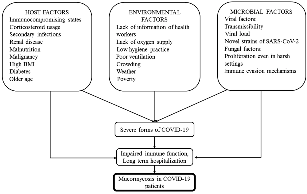
Renal diseases, older age, and secondary infections are other risk factors for mucormycosis because of impaired immune system function. There have been reports of fatal occurrences of systemic mucormycosis associated with acute or chronic renal failures (54) (Figure 1).

Risk factors for mucormycosis in COVID-19 patients

2.4. Environmental Risk Factors

2.4.1. Crowding

Although mucormycosis is not contagious as crowding is a major route of transmission of respiratory infections and COVID-19, it can indirectly increase the risk of mucormycosis by increasing the proportion of infected people and individuals with severe respiratory infections and decreased lung capacity (2).

2.4.2. Weather

Climatic conditions play an important role in increasing the risk of various pathogenic infections. In mucormycosis, the relationship between temperature and humidity has been investigated, and it has been found more common in summer and fall than in winter or spring. According to studies, the most prevalent season for the incidence of mucormycosis was winter. This discrepancy can be attributed to the fact that winter in their studied regions was like autumn in Middle Eastern nations (55).

2.4.3. Lack of Information of Health Workers

Proper and sufficient training is a critical aspect in prevention and timely diagnosis and treatment of mucormycosis. Poor education about caring for people with skin wounds, such as burns and skin lesions, as well as poor information about the potency of dust, soil, and even water for infecting people with Mucorales can lead to unconscious transmission and worsening disease status (2).

2.4.4. Shortage of Anti-fungal Drugs

Amphotericin B is an antifungal medication that is used to treat serious, life-threatening mucormycosis. The shortage of antifungal drugs in India has led to an exacerbation of mucormycosis progression in COVID-19 patients. Also, a shortage of anti-fungal medications can lead to increased commuting that is one of the risk factors of COVID-19 and secondary infections (56-58).

2.4.5. Lack of Oxygen Supply

Countries are struggling to provide medical oxygen due to the increased transmissibility of novel SARS-CoV-2 variants, the increased number of newly infected patients, and the requirement for oxygenation and ventilation for managing hypoxic COVID-19 patients. Hypoxia in COVID-19 patients can worsen their condition and cause immune system impairment. Hypoxia, hyperglycemia, and immunosuppression give mucormycosis the chance to thrive, and in vitro analyses have shown the suppression of fungal growth in hyperbaric oxygen conditions (59). One of the key points about hyperbaric oxygen therapy is humidification because hyperbaric oxygenation without humidification can cause damage to the inner lining of the lungs. This humidification should be done with frequently changing sterilized water because non-sterilized water is potentially a source of mucormycosis (60).

2.4.6. Other Environmental Factors

As shown in Figure 1, poor ventilation, poverty, and low hygiene practice, especially in hospitals, can lead to an increased risk of developing mucormycosis and COVID-19.

2.5. Microbial Risk Factors

Microbial factors associated with developing mucormycosis in COVID-19 patients can be divided into viral and fungal factors.

2.5.1. Viral Factors

There is some evidence for the association between SARS-CoV-2 strains and mucormycosis development (61, 62). COVID-19 prevalence and severity are linked to parameters such as transmissibility, viral load, and the development of novel strains of SARS-CoV-2, such as delta and delta plus, particularly from India (2, 63). Also, it has been reported that the use of steroids, such as prednisolone and antibiotics, and some traditional medicines could play a role in increased opportunistic mycoses during the COVID-19 epidemic (61). Besides the above factors, complex immune dysregulation in COVID-19 patients with severe respiratory failure would create a suitable condition for invasive mycoses, like mucormycosis and aspergillosis (64). These factors can eventually be associated with impaired immune system function, long-term hospitalization, and raised risk of secondary infections, such as mucormycosis (2).

2.5.2. Fungal Factors

Mucorales have various characteristics that allow them to avoid recognition by the host immune system. Different species of opportunistic fungi like the Mucorales, Aspergillus (Mucor, Rhizomucor, Rhizopus, Saksenaea, Cunninghamella, Apophysomyces, Absidia, Aspergillus fumigates, A. flavous, and A. tereus) are microflora of the atmosphere (65); therefore, at-risk individuals such as COVID-19 patients could be contaminated with these species during inhalation of etiologic agents of invasive mycoses, including spores of Aspergillus and Mucorales (66). The rapid growth of all these species in human tissues and phospholipase B secretion are also considered as important factors for the pathogenesis of these species in COVID-19 patients (67, 68). According to research, they develop resistance to host innate immune system activity by compromising phagocytic activity (69, 70). They can also proliferate in harsh settings, such as hypoxia, and this increased fungal load can lead to more infracted tissues and organs (71), as shown in Figure 1.

3. Conclusions

It can be concluded that as mucormycosis has a close linkage to diabetes, using cortone and conditions that modulate the immune system, identifying COVID-19 patients with risk factors for mucormycosis, and reducing corticosteroids administration in non-vital conditions can help in the prevention, fast diagnosis, and treatment of this opportunistic infection. Providing anti-fungal and oxygen supply and re-informing medical staff and individuals about the transmission routes of Mucorales and caring for skin wounds, especially burns, can limit the prevalence of this infection. Eventually, finding the role of viral factors in SARS-CoV-2 and Mucorales infection immune pathogenesis can help reduce the mortality rate of mucormycosis in COVID-19 patients.

References

  • 1.

    Rabaan AA, Al-Ahmed SH, Haque S, Sah R, Tiwari R, Malik YS, et al. SARS-CoV-2, SARS-CoV, and MERS-COV: A comparative overview. Infez Med. 2020;28(2):174-84. [PubMed ID: 32275259].

  • 2.

    Rashedi J, Mahdavi Poor B, Asgharzadeh V, Pourostadi M, Samadi Kafil H, Vegari A, et al. Risk factors for COVID-19. Infez Med. 2020;28(4):469-74. [PubMed ID: 33257620].

  • 3.

    Yang S, Hua M, Liu X, Du C, Pu L, Xiang P, et al. Bacterial and fungal co-infections among COVID-19 patients in intensive care unit. Microbes Infect. 2021;23(4-5):104806. [PubMed ID: 33684520]. [PubMed Central ID: PMC7933791]. https://doi.org/10.1016/j.micinf.2021.104806.

  • 4.

    Nashibi R, Afzalzadeh S, Mohammadi MJ, Yari AR, Yousefi F. Epidemiology and treatment outcome of mucormycosis in Khuzestan, Southwest of Iran. Arch Clin Infect Dis. 2016;12(1). https://doi.org/10.5812/archcid.37221.

  • 5.

    Dioverti MV, Cawcutt KA, Abidi M, Sohail MR, Walker RC, Osmon DR. Gastrointestinal mucormycosis in immunocompromised hosts. Mycoses. 2015;58(12):714-8. [PubMed ID: 26456920]. https://doi.org/10.1111/myc.12419.

  • 6.

    Mehta S, Pandey A. Rhino-orbital mucormycosis associated with COVID-19. Cureus. 2020;12(9). e10726. [PubMed ID: 33145132]. [PubMed Central ID: PMC7599039]. https://doi.org/10.7759/cureus.10726.

  • 7.

    Werthman-Ehrenreich A. Mucormycosis with orbital compartment syndrome in a patient with COVID-19. Am J Emerg Med. 2021;42:264 e5-8. [PubMed ID: 32972795]. [PubMed Central ID: PMC7493738]. https://doi.org/10.1016/j.ajem.2020.09.032.

  • 8.

    Mekonnen ZK, Ashraf DC, Jankowski T, Grob SR, Vagefi MR, Kersten RC, et al. Acute invasive rhino-orbital mucormycosis in a patient with COVID-19-associated acute respiratory distress syndrome. Ophthalmic Plast Reconstr Surg. 2021;37(2):e40-80. [PubMed ID: 33229953]. [PubMed Central ID: PMC7927902]. https://doi.org/10.1097/IOP.0000000000001889.

  • 9.

    Kuchay B. ‘Black fungus’ new scare in India as second COVID wave ebbs. 2021. Available from: https://www.aljazeera.com/news/2021/6/8/black-fungus-new-scare-in-india-as-second-covid-wave-ebbs.

  • 10.

    Tabarsi P, Khalili N, Pourabdollah M, Sharifynia S, Naeini AS, Ghorbani J, et al. Case report: Coronavirus disease 2019-associated Rhinosinusitis Mucormycosis caused by Rhizopus arrhizus: A rare but potentially fatal infection occurring after treatment with corticosteroids. Am J Trop Med Hyg. 2021;105(2):449-53. [PubMed ID: 34237015]. [PubMed Central ID: PMC8437195]. https://doi.org/10.4269/ajtmh.21-0359.

  • 11.

    IRNA. [Detected cases of suspected black fungal infection in Zanjan are being treated]. 2021. Persian. Available from: www.irna.ir/news/84448862/.

  • 12.

    ISNA. [What is the story of corona patients getting "black fungus"?]. 2021. Persian. Available from: https://www.isna.ir/news/1400060302079/.

  • 13.

    IRIBnews. [Black fungus caused by corona]. 2021. Persian. Available from: https://www.iribnews.ir/00D6Gg.

  • 14.

    Sharma S, Grover M, Bhargava S, Samdani S, Kataria T. Post coronavirus disease mucormycosis: A deadly addition to the pandemic spectrum. J Laryngol Otol. 2021;135(5):442-7. [PubMed ID: 33827722]. [PubMed Central ID: PMC8060545]. https://doi.org/10.1017/S0022215121000992.

  • 15.

    Alekseyev K, Didenko L, Chaudhry B. Rhinocerebral mucormycosis and COVID-19 pneumonia. J Med Cases. 2021;12(3):85-9. [PubMed ID: 33984095]. [PubMed Central ID: PMC8040444]. https://doi.org/10.14740/jmc3637.

  • 16.

    Liu Y, Gayle AA, Wilder-Smith A, Rocklov J. The reproductive number of COVID-19 is higher compared to SARS coronavirus. J Travel Med. 2020;27(2). [PubMed ID: 32052846]. [PubMed Central ID: PMC7074654]. https://doi.org/10.1093/jtm/taaa021.

  • 17.

    Zhou P, Yang XL, Wang XG, Hu B, Zhang L, Zhang W, et al. A pneumonia outbreak associated with a new coronavirus of probable bat origin. Nature. 2020;579(7798):270-3. [PubMed ID: 32015507]. [PubMed Central ID: PMC7095418]. https://doi.org/10.1038/s41586-020-2012-7.

  • 18.

    WHO. WHO Director-General's opening remarks at the media briefing on COVID-19-11 March 2020. 2020. Available from: https://www.who.int/director-general/speeches/detail/who-director-general-s-opening-remarks-at-the-media-briefing-on-covid-19---11-march-2020.

  • 19.

    WHO. Transmission of SARS-CoV-2: Implications for infection prevention precautions. World Health Organization; 2020.

  • 20.

    Chen B, Jia P, Han J. Role of indoor aerosols for COVID-19 viral transmission: A review. Environ Chem Lett. 2021:1-18. [PubMed ID: 33462543]. [PubMed Central ID: PMC7805572]. https://doi.org/10.1007/s10311-020-01174-8.

  • 21.

    Xiao F, Sun J, Xu Y, Li F, Huang X, Li H, et al. Infectious SARS-CoV-2 in feces of patient with severe COVID-19. Emerg Infect Dis. 2020;26(8):1920-2. [PubMed ID: 32421494]. [PubMed Central ID: PMC7392466]. https://doi.org/10.3201/eid2608.200681.

  • 22.

    van Doremalen N, Bushmaker T, Morris DH, Holbrook MG, Gamble A, Williamson BN, et al. Aerosol and surface stability of SARS-CoV-2 as compared with SARS-CoV-1. N Engl J Med. 2020;382(16):1564-7. [PubMed ID: 32182409]. [PubMed Central ID: PMC7121658]. https://doi.org/10.1056/NEJMc2004973.

  • 23.

    Huang C, Wang Y, Li X, Ren L, Zhao J, Hu Y, et al. Clinical features of patients infected with 2019 novel coronavirus in Wuhan, China. Lancet. 2020;395(10223):497-506. [PubMed ID: 31986264]. [PubMed Central ID: PMC7159299]. https://doi.org/10.1016/S0140-6736(20)30183-5.

  • 24.

    Mastroianni A. Paranasal sinus mucormycosis in an immunocompetent host: Efficacy and safety of combination therapy with Liposomal Amphotericin B and adjuvant rHuGM-CSF. Infez Med. 2004;12(4):278-83. [PubMed ID: 15729020].

  • 25.

    Camara-Lemarroy CR, Gonzalez-Moreno EI, Rodriguez-Gutierrez R, Rendon-Ramirez EJ, Ayala-Cortes AS, Fraga-Hernandez ML, et al. Clinical features and outcome of mucormycosis. Interdiscip Perspect Infect Dis. 2014;2014:562610. [PubMed ID: 25210515]. [PubMed Central ID: PMC4158140]. https://doi.org/10.1155/2014/562610.

  • 26.

    Reid G, Lynch JP, Fishbein MC, Clark NM. Mucormycosis. Semin Respir Crit Care Med. 2020;41(1):99-114. [PubMed ID: 32000287]. https://doi.org/10.1055/s-0039-3401992.

  • 27.

    Petrikkos G, Skiada A, Lortholary O, Roilides E, Walsh TJ, Kontoyiannis DP. Epidemiology and clinical manifestations of mucormycosis. Clin Infect Dis. 2012;54 Suppl 1:S23-34. [PubMed ID: 22247442]. https://doi.org/10.1093/cid/cir866.

  • 28.

    Dolatabadi S, Ahmadi B, Rezaei-Matehkolaei A, Zarrinfar H, Skiada A, Mirhendi H, et al. Mucormycosis in Iran: A six-year retrospective experience. J Mycol Med. 2018;28(2):269-73. [PubMed ID: 29545123]. https://doi.org/10.1016/j.mycmed.2018.02.014.

  • 29.

    Brown J. Zygomycosis: an emerging fungal infection. Am J Health Syst Pharm. 2005;62(24):2593-6. [PubMed ID: 16333056]. https://doi.org/10.2146/ajhp050188.

  • 30.

    Vaezi A, Moazeni M, Rahimi MT, de Hoog S, Badali H. Mucormycosis in Iran: A systematic review. Mycoses. 2016;59(7):402-15. [PubMed ID: 26906121]. https://doi.org/10.1111/myc.12474.

  • 31.

    Roden MM, Zaoutis TE, Buchanan WL, Knudsen TA, Sarkisova TA, Schaufele RL, et al. Epidemiology and outcome of zygomycosis: A review of 929 reported cases. Clin Infect Dis. 2005;41(5):634-53. [PubMed ID: 16080086]. https://doi.org/10.1086/432579.

  • 32.

    Garg D, Muthu V, Sehgal IS, Ramachandran R, Kaur H, Bhalla A, et al. Coronavirus disease (Covid-19) associated mucormycosis (CAM): Case report and systematic review of literature. Mycopathologia. 2021;186(2):289-98. [PubMed ID: 33544266]. [PubMed Central ID: PMC7862973]. https://doi.org/10.1007/s11046-021-00528-2.

  • 33.

    Gensler LS. Glucocorticoids: Complications to anticipate and prevent. Neurohospitalist. 2013;3(2):92-7. [PubMed ID: 23983891]. [PubMed Central ID: PMC3726115]. https://doi.org/10.1177/1941874412458678.

  • 34.

    Singh AK, Singh R, Joshi SR, Misra A. Mucormycosis in COVID-19: A systematic review of cases reported worldwide and in India. Diabetes Metab Syndr. 2021;15(4):102146. [PubMed ID: 34192610]. [PubMed Central ID: PMC8137376]. https://doi.org/10.1016/j.dsx.2021.05.019.

  • 35.

    Ghuman H, Voelz K. Innate and adaptive immunity to mucorales. J Fungi (Basel). 2017;3(3). [PubMed ID: 29371565]. [PubMed Central ID: PMC5715954]. https://doi.org/10.3390/jof3030048.

  • 36.

    Moreira J, Varon A, Galhardo MC, Santos F, Lyra M, Castro R, et al. The burden of mucormycosis in HIV-infected patients: A systematic review. J Infect. 2016;73(3):181-8. [PubMed ID: 27394402]. https://doi.org/10.1016/j.jinf.2016.06.013.

  • 37.

    Kharroubi AT, Darwish HM. Diabetes mellitus: The epidemic of the century. World J Diabetes. 2015;6(6):850-67. [PubMed ID: 26131326]. [PubMed Central ID: PMC4478580]. https://doi.org/10.4239/wjd.v6.i6.850.

  • 38.

    Nezafati S, Kazemi A, Asgari K, Bahrami A, Naghili B, Yazdani J. Rhinocerebral mucormycosis, risk factors and the type of oral manifestations in patients referred to a University Hospital in Tabriz, Iran 2007-2017. Mycoses. 2018;61(10):764-9. [PubMed ID: 29896908]. https://doi.org/10.1111/myc.12802.

  • 39.

    Casqueiro J, Casqueiro J, Alves C. Infections in patients with diabetes mellitus: A review of pathogenesis. Indian J Endocrinol Metab. 2012;16 Suppl 1:S27-36. [PubMed ID: 22701840]. [PubMed Central ID: PMC3354930]. https://doi.org/10.4103/2230-8210.94253.

  • 40.

    Wu S, Xue L, Legido-Quigley H, Khan M, Wu H, Peng X, et al. Understanding factors influencing the length of hospital stay among non-severe COVID-19 patients: A retrospective cohort study in a Fangcang shelter hospital. PLoS One. 2020;15(10). e0240959. [PubMed ID: 33085709]. [PubMed Central ID: PMC7577449]. https://doi.org/10.1371/journal.pone.0240959.

  • 41.

    Sen M, Lahane S, Lahane TP, Parekh R, Honavar SG. Mucor in a viral land: A tale of two pathogens. Indian J Ophthalmol. 2021;69(2):244-52. [PubMed ID: 33463566]. [PubMed Central ID: PMC7933891]. https://doi.org/10.4103/ijo.IJO_3774_20.

  • 42.

    Corzo-Leon DE, Chora-Hernandez LD, Rodriguez-Zulueta AP, Walsh TJ. Diabetes mellitus as the major risk factor for mucormycosis in Mexico: Epidemiology, diagnosis, and outcomes of reported cases. Med Mycol. 2018;56(1):29-43. [PubMed ID: 28431008]. https://doi.org/10.1093/mmy/myx017.

  • 43.

    Lodish H, Berk A, Zipursky SL, Matsudaira P, Baltimore D, Darnell J. Proto-oncogenes and tumor-suppressor genes. Molecular cell biology. 4th ed. New York: W. H. Freeman; 2000.

  • 44.

    Wargo JA, Reuben A, Cooper ZA, Oh KS, Sullivan RJ. Immune effects of chemotherapy, radiation, and targeted therapy and opportunities for combination with immunotherapy. Semin Oncol. 2015;42(4):601-16. [PubMed ID: 26320064]. [PubMed Central ID: PMC4955940]. https://doi.org/10.1053/j.seminoncol.2015.05.007.

  • 45.

    Pagano L, Ricci P, Tonso A, Nosari A, Cudillo L, Montillo M, et al. Mucormycosis in patients with haematological malignancies: a retrospective clinical study of 37 cases. GIMEMA Infection Program (Gruppo Italiano Malattie Ematologiche Maligne dell'Adulto). Br J Haematol. 1997;99(2):331-6. [PubMed ID: 9375750]. https://doi.org/10.1046/j.1365-2141.1997.3983214.x.

  • 46.

    Liang W, Guan W, Chen R, Wang W, Li J, Xu K, et al. Cancer patients in SARS-CoV-2 infection: A nationwide analysis in China. Lancet Oncol. 2020;21(3):335-7. [PubMed ID: 32066541]. [PubMed Central ID: PMC7159000]. https://doi.org/10.1016/S1470-2045(20)30096-6.

  • 47.

    Ghosh S. Factors responsible for childhood malnutrition: A review of the literature. Curr Res Nutr Food Sci. 2020:360-70. https://doi.org/10.12944/crnfsj.8.2.01.

  • 48.

    Newburger PE, Dale DC. Evaluation and management of patients with isolated neutropenia. Semin Hematol. 2013;50(3):198-206. [PubMed ID: 23953336]. [PubMed Central ID: PMC3748385]. https://doi.org/10.1053/j.seminhematol.2013.06.010.

  • 49.

    Irawan R. Protein calorie malnutrition and immune response in children. Folia Medica Indonesiana. 2008;44(2):119-26.

  • 50.

    Nuttall FQ. Body mass index: Obesity, BMI, and health: A critical review. Nutr Today. 2015;50(3):117-28. [PubMed ID: 27340299]. [PubMed Central ID: PMC4890841]. https://doi.org/10.1097/NT.0000000000000092.

  • 51.

    Frasca D, McElhaney J. Influence of obesity on pneumococcus infection risk in the elderly. Front Endocrinol (Lausanne). 2019;10:71. [PubMed ID: 30814978]. [PubMed Central ID: PMC6381016]. https://doi.org/10.3389/fendo.2019.00071.

  • 52.

    Stefan N, Birkenfeld AL, Schulze MB, Ludwig DS. Obesity and impaired metabolic health in patients with COVID-19. Nat Rev Endocrinol. 2020;16(7):341-2. [PubMed ID: 32327737]. [PubMed Central ID: PMC7187148]. https://doi.org/10.1038/s41574-020-0364-6.

  • 53.

    Hoong CWS, Hussain I, Aravamudan VM, Phyu EE, Lin JHX, Koh H. Obesity is associated with poor covid-19 outcomes: A systematic review and meta-analysis. Horm Metab Res. 2021;53(2):85-93. [PubMed ID: 33395706]. https://doi.org/10.1055/a-1326-2125.

  • 54.

    Gupta KL, Radotra BD, Sakhuja V, Banerjee AK, Chugh KS. Mucormycosis in patients with renal failure. Ren Fail. 1989;11(4):195-9. [PubMed ID: 2485482]. https://doi.org/10.3109/08860228909054931.

  • 55.

    Bartzokas CA. Relationship between the metereological conditions and the air-borne fungal flora of the Athens metropolitan area. Mycopathologia. 1975;57(1):35-8. [PubMed ID: 1207718]. https://doi.org/10.1007/BF00431176.

  • 56.

    Mathew BP, Nath M. Recent approaches to antifungal therapy for invasive mycoses. ChemMedChem. 2009;4(3):310-23. [PubMed ID: 19170067]. https://doi.org/10.1002/cmdc.200800353.

  • 57.

    IRIBnews. [The drug has become rare in patients with black fungal infections]. 2021. Persian. Available from: https://www.iribnews.ir/fa/news/3200704/.

  • 58.

    ISNA. [Black market for black fungus medicine]. 2021. Persian. Available from: https://www.isna.ir/news/1400060403005/.

  • 59.

    Ferguson BJ, Mitchell TG, Moon R, Camporesi EM, Farmer J. Adjunctive hyperbaric oxygen for treatment of rhinocerebral mucormycosis. Rev Infect Dis. 1988;10(3):551-9. [PubMed ID: 3393782]. https://doi.org/10.1093/clinids/10.3.551.

  • 60.

    Barbet JP, Chauveau M, Labbe S, Lockhart A. Breathing dry air causes acute epithelial damage and inflammation of the guinea pig trachea. J Appl Physiol (1985). 1988;64(5):1851-7. [PubMed ID: 3391888]. https://doi.org/10.1152/jappl.1988.64.5.1851.

  • 61.

    Kumar M, Sarma DK, Shubham S, Kumawat M, Verma V, Singh B, et al. Mucormycosis in COVID-19 pandemic: Risk factors and linkages. Curr Res Microb Sci. 2021;2:100057. [PubMed ID: 34396355]. [PubMed Central ID: PMC8349419]. https://doi.org/10.1016/j.crmicr.2021.100057.

  • 62.

    Ashour MM, Abdelaziz TT, Ashour DM, Askoura A, Saleh MI, Mahmoud MS. Imaging spectrum of acute invasive fungal rhino-orbital-cerebral sinusitis in COVID-19 patients: A case series and a review of literature. J Neuroradiol. 2021;48(5):319-24. [PubMed ID: 34087330]. [PubMed Central ID: PMC8168306]. https://doi.org/10.1016/j.neurad.2021.05.007.

  • 63.

    Alexandar S, Ravisankar M, Kumar RS, Jakkan K. A comprehensive review on covid-19 delta variant. Int J Pharmacol Clin Res. 2021;5:83-5.

  • 64.

    Lahmer T, Kriescher S, Herner A, Rothe K, Spinner CD, Schneider J, et al. Invasive pulmonary aspergillosis in critically ill patients with severe COVID-19 pneumonia: Results from the prospective AspCOVID-19 study. PLoS One. 2021;16(3). e0238825. [PubMed ID: 33730058]. [PubMed Central ID: PMC7968651]. https://doi.org/10.1371/journal.pone.0238825.

  • 65.

    Vaezi A, Walther G, Kurzai O, Mahdi D, Dadashzadeh M, Nasri E, et al. Frequency of occurrence, seasonal variation and antifungal susceptibility of opportunistic Mucorales isolated from hospital soils in Iran. Mycoses. 2021;64(7):780-7. [PubMed ID: 33835599]. https://doi.org/10.1111/myc.13283.

  • 66.

    Kazemi A, Ahmadpour E, Naghili B, Mahmoudabadi AZ, Jafari A, Ayatollahi AM. Airborne fungi in Tabriz, comparing airborne and clinical samples of A. fumigatus (2011), survey and literature review. Jundishapur J Microbiol. 2013;6(4). e4997. https://doi.org/10.5812/jjm.4997.

  • 67.

    Nicolas FE, Murcia L, Navarro E, Navarro-Mendoza MI, Perez-Arques C, Garre V. Mucorales species and macrophages. J Fungi (Basel). 2020;6(2). [PubMed ID: 32604972]. [PubMed Central ID: PMC7344864]. https://doi.org/10.3390/jof6020094.

  • 68.

    Kazemi A. Phospholipases of Aspergillus fumigatus. United Kingdom: The University of Manchester; 2002.

  • 69.

    Chamilos G, Lewis RE, Lamaris G, Walsh TJ, Kontoyiannis DP. Zygomycetes hyphae trigger an early, robust proinflammatory response in human polymorphonuclear neutrophils through toll-like receptor 2 induction but display relative resistance to oxidative damage. Antimicrob Agents Chemother. 2008;52(2):722-4. [PubMed ID: 18025115]. [PubMed Central ID: PMC2224765]. https://doi.org/10.1128/AAC.01136-07.

  • 70.

    Gil-Lamaignere C, Simitsopoulou M, Roilides E, Maloukou A, Winn RM, Walsh TJ. Interferon- gamma and granulocyte-macrophage colony-stimulating factor augment the activity of polymorphonuclear leukocytes against medically important zygomycetes. J Infect Dis. 2005;191(7):1180-7. [PubMed ID: 15747255]. https://doi.org/10.1086/428503.

  • 71.

    Katragkou A, Walsh TJ, Roilides E. Why is mucormycosis more difficult to cure than more common mycoses? Clin Microbiol Infect. 2014;20 Suppl 6:74-81. [PubMed ID: 24279587]. https://doi.org/10.1111/1469-0691.12466.