1. Background
Many protozoan parasites can transmit to humans via ingestion of contaminated meat with various types of parasite cysts. In point of view, Toxoplasma, Sarcocystis, and Trichenella are some examples of these agents. Sarcocystis species are obligatory apicomplexan intracellular parasites in a wide range of vertebrate including mammals, birds, and reptiles. The parasite has 2 hosts, which spend their sexual life cycle in definitive host (predator) and asexual life cycle in intermediate host (prey). Sexual life takes place in an intestinal epithelial cell of final host, which finally produce oocysts and passes from feces and contaminate environment. In the intermediate host, parasite proliferates in various types of cells by merogony division and finally produces macroscopic or microscopic cyst in muscles as well as brain, according to species (1, 2). Approximately, 150 species of Sarcocystis with variety of clinical signs were introduced in the world (3). There are 4 predominant Sarcocystis species in sheep with worldwide distribution including, Sarcocystis tenella, S. gigantea (S. ovifelis), S. arieticanis, and S. medusiformis (4).
There are 3 species of Sarcocystis in cattle including S. bovifelis (S. hirsuta), S. cruzi (S. bovicanis), and S. hominis (S. bovihominis) (5). The severity of clinical signs in domesticated animals depends on the species of parasite and number of digested oocysts. Clinical signs included fever, anorexia, tachypnoe, tachycardia, anemia, encephalitis, encephalomyelitis, and hemorrhage, which can cause death in infected animals (6). The other feature of infection is abortion and fetal death in pregnant sheep (7). Sarcocystis tenella is a pathogen species in sheep; However, S. ovifelis is not. Additional economic losses are a condemnation of whole or partly carcasses in a slaughterhouse, reduction of animal products such as milk, meat, wool, and decreasing fertility (8). Economic losses in Spain have been estimated 20 million Euros annually due to condemnation of carcasses (9).
Humans can be infected as intermediate and final hosts by S. hominis, S. porcihuminis, and S. lindemanni (5). Biopsy or necropsy finding revealed that human serves as an intermediate host for at least 7 species of Sarcocystis (3). Many infections in humans are asymptomatic, however, some clinical signs in infected individuals were reported, which include vomiting, nausea, acute or chronic enteritis according to the species, and a number of ingested cyst (3). The other aspect of human sarcocystosis is muscular sarcocystosis. The most muscular sarcocystosis patients are asymptomatic and only 10 cases were reported with acute inflammation of muscles in the world (10, 11). The disease is endemic in some part of tropical country, especially in Malaysia (12).
Elevation of hepatic enzymes ALT (alanine aminotransferase), AST (aspartate aminotransferase), CRP (C reactive protein), ESR (erythrocyte sedimentation rate), LDH (lactic dehydrogenase), CK (creatinine phosphokinase), and Eosinophilia has been reported in eosinophilic myositis caused by sarcocystosis (13, 14). The conventional diagnosis of different species of Sarcocystis is based on the structure study of the cyst wall by using light or transmission electron microscopic (15). This method is not very precise because of existence changes of cysts during tissue processing and age of cyst (3).
2. Objectives
The goal of this study was conducted to identify the species of Sarcocystis by RFLP-PCR and nucleotide sequencing method in sheep and cattle to identify public health problem in human community.
3. Methods
3.1. Sampling
Fifty samples were collected from the diaphragm, heart, skeletal muscles of sheep and cattle carcasses (each animal 25 samples) in industrial abattoir of Ahvaz, and 50 mg of each sample were considered for DNA extraction using commercial DNA extract kit (QIAamp DNA Mini Kit, Qiagen, Germany). DNA was extracted according to manufacture instructions and stored in -20 centigrade until used.
3.2. PCR-RFLP
In this study 2 pair primers were used. A variable region of the 18S ribosomal RNA gene was used as a good marker for determining sheep Sarcocystis species (16, 17), which included 18S forward (5/ GGA TAA CCG TGG TAA TTC TAT G3/) and 18S reverse (5/ TCC TAT GTC TGG ACC TGG TGA G3/). The primer amplify a part of 18s ribosomal gene with 1100 bp length (18). The PCR protocol was performed 5 minutes primary denaturation at 94°C follow by 35 cycles of 94°C for 1 minute, 50°C for 1 minute, and 72°C for 90 seconds, as well as a final extension of 72°C for 10 minutes (19). The final volume of PCR reaction was 25 μL including 5 μL of the sample DNA, 20 pmol of each primer,12.5 μL of PCR Master Mix (Ampliqon, Denmark) , and 5.5 μL distillated water.
The second pair primer was designed for detecting cattle Sarcocystis species, which almost amplify a 600 bp segment. The primer was Sar F 5/ GCA CTT GAT GAA TTC TGG CA 3/ and Sar R 5/ CAC CAC CCA TAG AAT CAA G 3/ (20). The PCR steps included 94°C for 5 minutes followed by 40 cycles of 94°C for 2 minutes, annealing at 55°C for 1 minute, and extension step 72°C for 90 seconds, followed by a final extension step at 72°C for 5 minutes. The final volume of PCR reaction was 25 μL including 3 μL of the sample DNA, 20 pmol of each primer, and 12.5 μL of PCR Master Mix and 7.5 μL distillated water. The PCR bands appear by using gel electrophoresis in 1% agarose, ethidium bromide, and gel documentation apparatus.
The PCR products were digested using the restriction fragment length polymorphism (RFLP) method by 3 nucleotide enzymes including Haemophilus influenza Rf endonuclease enzyme (Hinf), Moraxella bovis endonuclease enzyme (Mbo1), and Escherichia coli RY13 endonuclease enzyme (EcoR1). The RFLP reaction was carried out by 5 μL PCR product, 10 unit of each enzyme, and buffer. The reaction are incubated 24 hours at 37°C. Eight samples of PCR yields of sheep and cattle (each one 4 samples) was sent to Bioneer company South Korea for nucleotide sequencing. The nucleotide sequences were compared with other registered nucleotide sequencing in the National Center for Biotechnology Information (NCBI).
4. Results
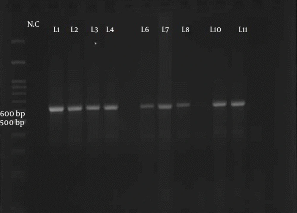
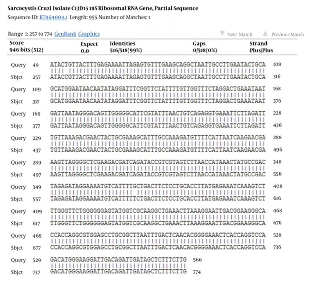
Fifty samples of sheep and cattle (each 25 samples) were selected for PCR-RFLP molecular technique. These samples were previously examined for Sarcocystis species contamination by using the digestive method with HCL and pepsin; the result showed that all samples were infected by Sarcocystis (21). Two pair primers were selected to identify Sarcocystis species in sheep and cattle. PCR finding presented that a nucleotide segment with 600 base per was amplified for cattle samples (Figure 1). The comparison of nucleotide sequencing with other registered nucleotides sequencing in NCBI confirmed that they belong to S. cruzi with 99% homology (Figure 2). There is no difference between isolates when aligned together with Mega 6 software (Figure 2). The use of 3 restricted enzymes for the RFLP study presented that Eco1enzyms cannot break down the PCR yield. The Mbo restricted enzyme can divide the PCR product to 2 bands approximately 300 and 330 base pair. The Hinf enzymes produced 2 bands including 550 and 50 bp (Figure 3).
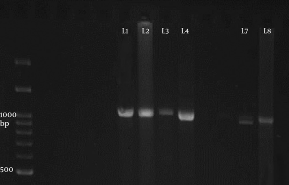
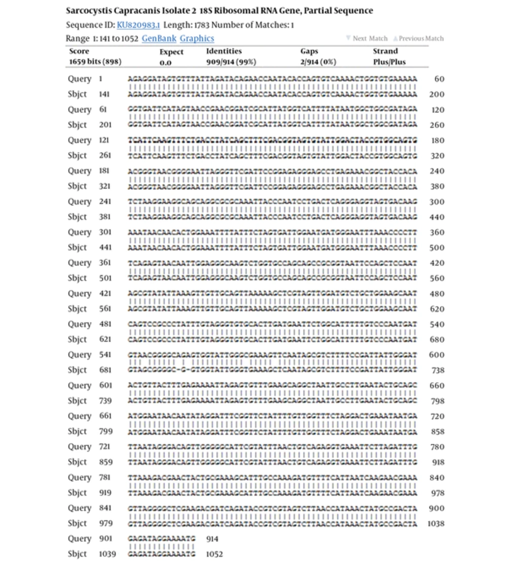
The PCR of 18S ribosomal RNA gene in sheep samples amplified a 1100 base pair and 900 bp nucleotide (Figure 4), which pertained to S. tenella and S. capriovis, respectively. The comparison of nucleotide sequence with other nucleotide in NCBI revealed that they have 100% homology with S. tenella and S. capricanis. The use of 3 restricted enzymes for the RFLP study on S. tenella PCR presented that Eco1enzyms break down the PCR yield to 2 fractions, including 600 and 400 BP (Figure 5). The Mbo restricted enzyme can divide the PCR yield to 3 bands approximately 750, 250, and 150 base pair. The Hinf enzymes produced 2 bands including 750 and 400 (Figure 5). The comparison of nucleotide sequences of PCR product of Sarcocystis in sheep samples with NCBI data revealed that 100% homology for S. tenella and 99% for S. capricanis (Figure 6).
5. Discussion
Some Sarcocystis species can cause clinical signs in the human population. Men serve as intermediated hosts with myositis symptoms and a definitive host with gastrointestinal symptoms. Livestock and carnivorous have an important role in transporting sarcocystosis in humans. In a previously study, we detected that 100% of 100 sheep and cattle samples had been infected by Sarcocystis species using digestion and microscopic method (21). For identifying species of Sarcocystis, 50 samples of sheep and cattle samples (each one 25 samples) were selected to determinate species of the parasite using the PCR-RFLP method. The reason of performing this study was to distinguish the risk of human sarcocystosis due to consumption of contaminated meat as a public health problem.
Currently, the identification of Sarcocystis species in animals and humans is carried out by using transmission electron microscopy to study structure of cyst wall (22). However, the use of this method has some limitations in the extended epidemiology study and detection of little morphology variation in species (23). Therefore, many investigators use the molecular approach for identification of Sarcocystis species variation. At this point, 2 genes were presented, which included 18S ribosomal RNA and small subunit ribosomal RNA gene. In this study we used 18S r RNA gene for distinguishing Sarcocystis species in studied animals. We find that all Sarcocystis isolates in cattle samples belonged to S. cruzi. The results of the PCR of cattle samples presented a 600-nucleotide bp fragment. Comparison of studied nucleotide sequencing with other sequence of Sarcocystis species in the gene bank revealed 99% homology with S. cruzi with only 2 nucleotide different (Figure 2).
The nucleotides sequence of S. cruzi was booked to the NCBI gene bank as LC214880, LC214881, LC214882, and LC214883 accession numbers. This finding should be considered by the veterinary organization due to the fact that S. cruzi has severe pathogenicity in livestock and can cause severe clinical sign, abortion, and loss of animal products, however, without any pathologic effects on the human population. Controversy, Agholi et al. described that S. cruzi was detected in fecal samples of one women immunocompromised patient (HIV positive) using 18S r DNA gene amplifying and phylogenic analysis (24). The S. cruzi has worldwide distribution and is frequently reported. Sarcocystiscruzi, S. hirsute, and S. hominis cysts were detected on imported cattle meat from Argentina to Norway in 2009 (25). Pritt et al. showed that 31 samples of 48 (64.5%) beef meat samples are infected by S. cruzi using the molecular and histology method and indicated high prevalence S. cruzi in USA. More et al. in Germany, indicated that 52% of 275 beef samples were infected by S. cruzi, following 37% by S. sinensis (26). Additionally, water buffalo can also serve as an intermediated host for S. cruzi (27, 28). In Iran, there are many articles confirmed that the predominant species of Sarcocystis in cattle is S. cruzi (29-31).
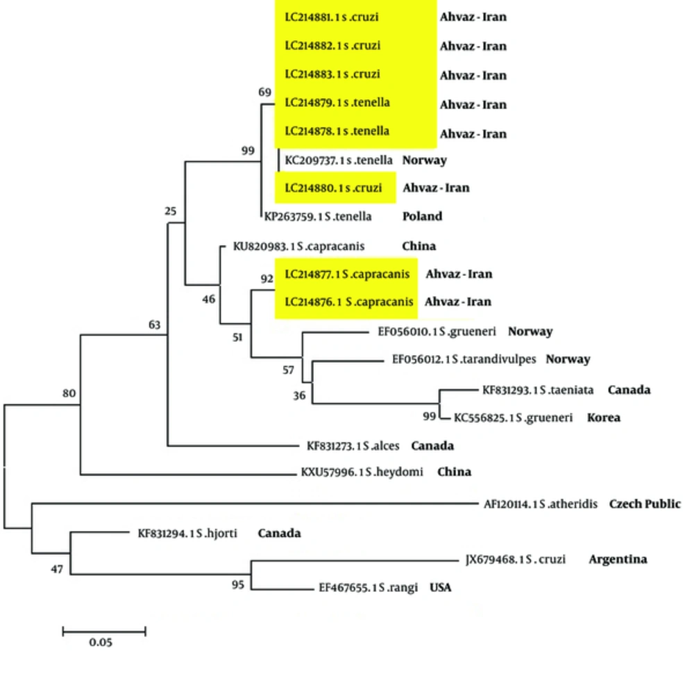
The present study indicated that all sheep samples were predominantly infected by S. tenella (80%) and followed by S. capracanis (20%). Amplifying of the18S r RNA gene by PCR showed a 1100 and 900 bp nucleotide segments for S. tenella and S. capracanis, respectively. The comparison of nucleotide sequence with other booked nucleotide in the gene bank revealed that there are > 99% homology with 2 mentioned Sarcocystis species with 5 different nucleotide for S. capracanis (Figure 7). Sarcocystis tenella is one of the pathogen Sarcocystis species in sheep. The abortion in sheep currently happened in this area and it seems that S. tenella should be considered as important agents for abortion and loses of animal production. Three restricted enzymes included EcoR1, Mbo, and Hinf can break down the PCR yield to 2 or 3 fragments. The PCR and RFLP analyzing of sheep samples showed that 2 species of Sarcocystis, S. tenella and S. capracanis, exists in this area.
In this study, we have not seen any macroschizont on sheep carcasses caused by S. gigantea. Controversy, Aghaeipour et al. found no microschizont cyst of S. tenella or S. capracanis in goat, however, they presented macroschizont from S. moulei in Tehran and Ghazvin province of Iran (32). Bittencourt et al. found that 95.8% of sheep and 91.6% of goats in Brazil are infected by S. tenella, S. arieticanis, and S. capracanis, respectively. They reported that the macroschizont, due to S. gigantea or S. medusiformis, was very rare in Brazil (33). Dubey et al. presented that S. arieticanis and S. capracanis are main Sarcocystis species in sheep, in USA. Farhang-Pajuh et al. reported that 29.3% and 7.52% of sheep were infected by S. gigantea and S. medusiformis, respectively, using the RFLP-PCR method.
The relationship phylogeny between some species of Sarcocystis in the gene bank was compared with isolated Sarcocystis in the current study by drawing a phylogeny tree (Figure 7). The phylogeny finding presented that there is a 99% relationship between isolated S. tenella and 92% between S. capricanis. The relationship between S. cruzi and S. tenella was 69%. Other Sarcocystis species showed various relationships (Figure 7). This study showed that Sarcocystis species in infected sheep and cows in this area are not pertained to human Sarcocystis species and cannot induce sarcocystosis in human population. Further studies on patients with gastrointestinal symptoms such as persistence diarrhea, especially in immunocompromised patients are needed.
5.1. Conclusions
In this study we presented that all isolated Sarcocystis species in cattle and sheep belonged to S. cruzi, S. tenella, and S. capracanis in this area and none of them have an important role for transmission human sarcocystosis. However, S. cruzi and S. tenella, as pathogen species, should be considered for causing economic loss in livestock animals by veterinary office.
