Dear Editor,
Candida species have emerged as important opportunistic pathogens in recent years (1, 2). Candida albicans may activate monocytes to release proinflammatory cytokines, triggering mucosal candidiasis or sepsis in the host (3, 4); however, whether C. parapsilosis also has a similar effect is still unknown. The release of TNFα as a response to infection by C. albicans (5) has been studied in THP-1 cells. The present study examined the levels of TNFα and IL-1β released from THP-1 cells infected with several different ratios of C. parapsilosis or C. albicans to obtain a better understanding of the pathogenesis of candidemia caused by C. parapsilosis.
Candida parapsilosis (ATCC 90018) or Candida albicans (ATCC 14053) were co-cultured at several different ratios [multiplicity of infection (MOI)] with THP-1 cells (ATCC TIB-202) in RPMI-1640 (Sigma-Aldrich, St. Louis, MO, USA) containing 5% fetal bovine serum (FBS; Gibco, USA) and 1µg/ml endotoxin. Escherichia coli O111 (B4 LPS, Sigma-Aldrich, USA) was used as the positive control (6). After 24 hours, the reaction was stopped in an ice bath and the cells were centrifuged at 13000 × g for 2 minutes at 4°C. Aliquots of the endotoxin-free supernatant (the Limulus Amebocyte Lysate assay, BioWhittaker, Walkersville, MD, USA) were stored at -70 OC. TNFα and IL-1β were quantitated (Quantikine, R&D Systems, Minneapolis, MN, USA) using an enzyme-linked immunosorbent assay (ELISA microplate reader, Dynatech MRX, USA). Each assay was run in duplicate and three independent experiments were conducted. A value of P < 0.05, determined by the general linear model (GLM) repeated measure test (SPSS Version 14.0), was considered statistically significant.
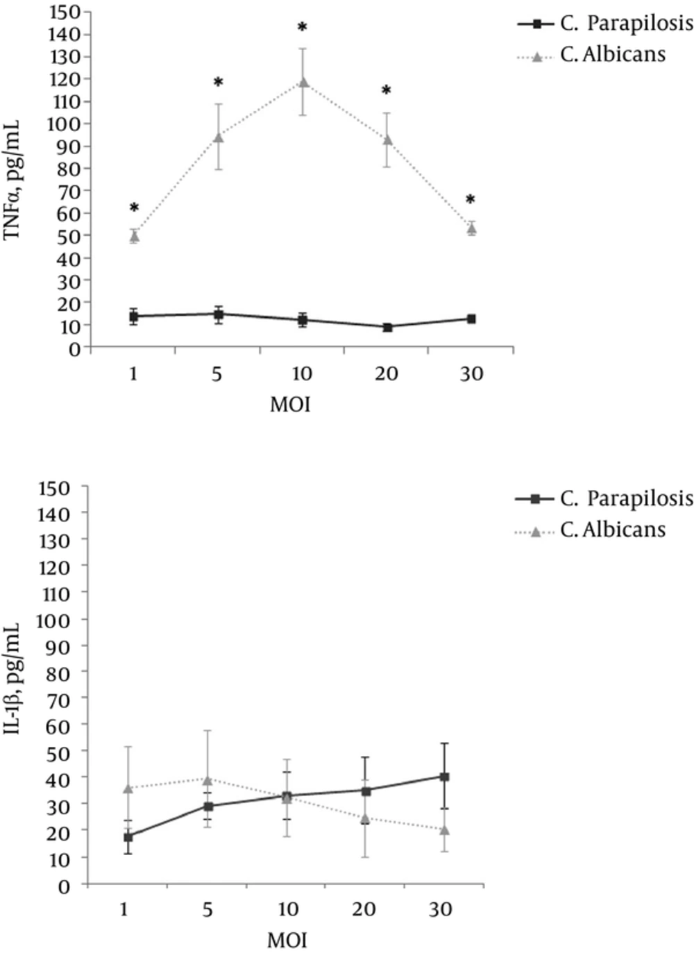
In this study, C. parapsilosis stimulated THP-1 cells to release a significantly lower level of TNFα than was observed for C. albicans at the same MOIs (P ≤ 0.001, P = 0.001, P ≤ 0.001, P ≤ 0.001, and P ≤ 0.001 at MOI = 1, 5, 10, 20, and 30, respectively). The level of TNFα released from C. albicans-infected cells gradually increased as the MOI was increased (MOI = 1, 5, and 10) and reached the highest level of TNFα (mean ± SD = 118.93 ± 15.13 pg/ml) at MOI = 10. Subsequently, the level of TNFα gradually decreased as the MOI was increased (MOI = 20 and 30) (Figure 1A). However, the level of TNFα released from C. parapsilosis-infected cells was always lower (mean < 15 pg/ml at all MOIs) and reached the highest level (mean ± SD = 14.55 ± 3.77 pg/ml) at MOI = 5.
The levels of IL-1β released from the THP-1 cells infected with C. parapsilosis and C. albicans showed no significant differences at the same MOIs (P ≥ 0.005 at MOI = 1, 5, 10, 20, 30). Infection with C. albicans at the lowest MOI (MOI = 1) stimulated THP-1 cells to release a higher level of IL-1β (mean ± SD = 36.07 ± 15.38 pg/ml). The highest release of IL-1 β (mean ± SD = 39.17 ± 18.35 pg/ml) was reached at MOI = 5. Subsequently, the level of IL-1 β gradually decreased as the MOI increased (Figure 1B). However, in C. parapsilosis-infected cells, the level of IL-1 β released from the cells gradually increased in a dose-dependent manner as the MOI was increased. Although C. parapsilosis at a lower MOI (MOI = 1 and 5) stimulated the cells to release a lower level of IL-1β than was observed with C. albicans, it stimulated the cells to release the highest level of IL-1β (mean ± SD = 40.27 ± 12.45 pg/ml) at the highest MOI (MOI = 30). This level was even higher than the highest level released by the cells infected with C. albicans.
Similar to previous studies (7, 8), our study demonstrated that the TNFα release was significantly lower from C. parapsilosis-infected than from C. albicans-infected THP-1 cells at all MOIs. Some studies have reported upregulation of IL-1 gene expression in C. parapsilosis-infected monocytes (9). In our study, the IL-1 β release from C. parapsilosis-infected cells at a higher MOI was similar to that observed with C. albicans-infected cells. Furthermore, the mean IL-1 β release was higher from C. parapsilosis-infected THP-1 cells at the highest MOI than from C. albicans-infected cells at any MOI. This revealed that infection with a sufficient amount of C. parapsilosis could result in a similar course to that seen with C. albicans. These findings also indicated that a biofilm was an important virulence factor and was responsible for the high mortality observed with C. parapsilosis infection (10). These results provide more information regarding the pathogenesis of C. parapsilosis infection and should be useful for its treatment or prevention.
