Abstract
Background:
Acinetobacter baumannii is an important cause of nosocomial pneumonia in patients requiring long-term mechanical ventilation. Besides, extensively drug-resistant (XDR) strains cause infection in intensive care unit (ICU) patients. Chronic infections of A. baumannii and antimicrobial resistance are associated with biofilm formation. Several virulence genes, such as blaPER-1, pgaA, and bap, are involved in biofilm formation.Objectives:
The current study examines the expression levels of biofilm formation-related genes in pneumonia patients.Methods:
The sputum samples were collected from patients hospitalized in the ICU, and A. baumannii ATCC 19606, the reference strain, was isolated and cultured on blood agar, eosin methylene blue agar, and chocolate agar medium. The media were then incubated at 37°C for 18 - 24 hours. Next, Gram-Thirty XDR A. baumannii isolates were collected from the sputum samples of ICU patients at Besat Hospital in Tehran, Iran. Bacterial isolates were characterized for antibiotic resistance patterns and biofilm-forming ability. Subsequently, RNA was extracted from the biofilm-forming isolates. A real-time polymerase chain reaction (PCR) assay was performed to evaluate the expression levels of the blaPER-1, pgaA, and bap genes. Transcripts were normalized to 16S rRNA as an internal control, and gene expression fold changes were calculated. Statistical analysis was performed using an unpaired two-tailed t-test (P < 0.05) with SPSS (V. 16).Results:
The disk diffusion susceptibility test revealed that all 30 (100%) isolates were resistant to piperacillin-tazobactam, cefepime, ceftriaxone, ceftazidime, gentamicin, imipenem, meropenem, levofloxacin, and ciprofloxacin. All 30 isolates from ICU-admitted patients (100%) were classified as XDR, and 27 (90%) isolates demonstrated the ability to form biofilms. The obtained results indicated a significant difference in gene expression levels. The fold change in expression for blaPER-1, bap, and pgaA was 7.473, 11.964, and 5.277, respectively.Conclusions:
In our study, XDR A. baumannii primarily caused ventilator-associated pneumonia, and an observed increase in the expression of biofilm-related genes was noted in these strains. Healthcare centers should implement appropriate infection control programs to manage nosocomial infections, particularly in the ICU.Keywords
1. Background
Acinetobacter baumannii, an aerobic gram-negative coccobacillus with extensively drug-resistant (XDR), belongs to the Moraxellaceae family. It is a major cause of nosocomial infections and thrives under aerobic conditions. (1, 2). This bacterium exhibits high resistance to ultraviolet rays and chemical disinfectants and can survive on the surface of dry objects for more than 25 days (3). It is responsible for various infections, including hospital-acquired pneumonia, ventilator-associated pneumonia, urinary tract infections, meningitis, bacteremia, gastritis, and skin wound infections (1, 4, 5). Numerous virulence factors contribute to the pathogenicity of this species and contribute to its high prevalence of infections, particularly in immunocompromised patients (6, 7).
Biofilm formation is one of the most significant properties involved in bacterial colonization and the spread of infections (8). Biofilms are complex communities of surface-attached microorganisms composed of extracellular polymeric substances (EPS), including polysaccharides, secreted proteins, and extracellular DNA (9). Acinetobacter baumannii strains readily form biofilms on body tissues, such as the skin. Biofilm formation requires a significant investment of cellular resources and energy (10). Extracellular polymeric substances facilitates intercellular interactions and horizontal gene transfer, enhances bacterial adhesion to surfaces, and provides protection against external factors like water and nutrient deprivation (8, 11).
Biofilm formation is a multi-step process that begins with reversible attachment to surfaces through intermolecular and hydrophobic bonds, ultimately leading to the production of EPS, enabling cells to adhere permanently (12). The formation of biofilms has undesirable consequences in the food industry, as pathogenic bacteria can form biofilms inside processing equipment, leading to food spoilage and posing risks to consumer health (13, 14). Moreover, in hospital environments, biofilms can persist on the surfaces of medical devices, catheters, and patient tissues, causing persistent infections (15).
There are several genes associated with A. baumannii’s ability to form biofilms. Numerous studies have demonstrated the correlation between the expression of these genes and the virulence of XDR strains (16). The biofilm-associated protein (Bap), encoded by the bap gene, is a high molecular weight surface protein that contains tandem repeats of domains involved in intercellular adhesion on bacterial cell surfaces (17). It is also associated with biofilm thickness and antibiotic resistance (18-20). Mutation in the bap gene in A. baumannii reduces biofilm growth and diminishes adhesion to human bronchial epithelial cells and neonatal keratinocyte cells (21). Bap plays a role in increasing the hydrophobicity and adhesion properties of bacterial cell surfaces (22). Another gene involved in biofilm formation in A. baumannii is β-lactamase PER-1 (blaPER-1) (23).
Adhesion to both bronchial epithelial cells and plastic surfaces is enhanced by the presence and expression of the blaPER-1 gene, although its precise mechanism of action remains unclear (24). The presence and expression of antibiotic resistance traits in A. baumannii for biofilm formation are heavily influenced by the blaPER-1 gene. The expression of the Poly-β-1,6-N-acetyl-d-glucosamine A (pgaA) gene is another factor in biofilm formation (25). This gene is involved in adhesion to abiotic surfaces, intercellular adhesion, protection against innate host defenses such as phagocytosis and antimicrobial peptides, and virulence (26). Different forms of pgaA exhibit variations in molecular weight, the degree of N-deacetylation of GlcNAc residues, and the presence of O-succinate substituents (27). Currently, the most pressing task is to disseminate knowledge about the risk factors associated with multidrug-resistant bacteria among medical professionals and prevent their spread in hospitals (28). Research on the clinical epidemiology and drug resistance mechanisms of A. baumannii, particularly its resistance to meropenem, has revealed the challenging situation healthcare providers face in treating this bacterium (10).
2. Objectives
This study aimed to examine the expression of these genes, evaluate virulence factors, and assess their impact on biofilm formation.
3. Methods
3.1. Case Definition
Thirty isolates of A. baumannii were collected from sputum samples of ICU-admitted patients at Besat Hospital in Tehran, Iran, between April and September 2020. Informed consent was obtained from each patient, and a questionnaire was completed. The antibiotic susceptibility pattern was determined using the disk diffusion method following the guidelines of the Clinical and Laboratory Standards Institute (CLSI 2019). The exclusion criteria for the study were non-Acinetobacter pneumonia and cases where pneumonia was caused by A. baumannii and other organisms simultaneously.
3.2. Bacterial Isolation and Antibiotic Sensitivity Test
The sputum samples were collected from patients hospitalized in the ICU, and the reference strain A. baumannii ATCC 19606 was isolated and cultured on blood agar, eosin methylene blue agar, and chocolate agar medium. The media were incubated at 37°C for 18 - 24 hours. Subsequently, gram-negative Bacillus colonies were characterized using standard bacteriological tests, including oxidase, catalase, oxidative/fermentation (OF) glucose test, triple sugar iron (TSI), urease, arginine dihydrolase, malonate, and motility and growth tests at 42°C. The antibiotic susceptibility test was conducted using imipenem, ciprofloxacin, gentamicin, levofloxacin, ceftazidime, cefepime, meropenem, ampicillin-sulbactam, piperacillin-tazobactam, amikacin, and ceftriaxone (Rosco, Padtan Teb Company, Tehran, Iran), following the CLSI 2019 guidelines. The minimum inhibitory concentration (MIC) of colistin (Rosco, Padtan Teb Company, Tehran, Iran) was also determined (29). The reference strain Escherichia coli ATCC 25922 was used as the standard strain in this test.
3.3. Phenotypic Evaluation of Biofilm Production by Microtiter Plate Test
For this purpose, a bacterial suspension equivalent to 0.5 McFarland was prepared in nutrient broth. Then, 200 μL of the suspension was added to each well of a 96-well polystyrene microtiter plate. The strains were incubated at 37°C for 24 hours. Subsequently, the content of each well was aspirated, and the wells were washed five times with PBS to remove all planktonic cells. After drying the wells, biofilm formation was assessed using the crystal violet method. To do this, 200 μL of 2% crystal violet was added to each well, and after 5 minutes, the wells were aspirated. The wells were then washed with water, and 160 μL of acetic acid was added to the dried wells. Finally, the optical density (OD) was measured at 650 nm using an ELISA plate reader. The results were interpreted with a positive control and reported as follows: High (4 × OD(c) < OD), medium (2 × OD(c) < OD ≤ 4 × OD(c)), low (OD(c) < OD ≤ 2 × OD(c)), and absence of biofilm formation (OD ≤ OD(c)). In this study, A. baumannii 19606 was used as a positive control. The test was repeated three times for accuracy.
3.4. RNA Extraction and Evaluation of Biofilm formation-related gene expression by Real-Time PCR
Several colonies were collected from the culture plates of each sample and grown overnight in tryptic soy broth (TSB) at 37°C to extract RNA from bacterial colonies. The cells were then centrifuged at 10,000 rpm and washed in a sterile saline solution (0.9% NaCl). The concentrations of the harvested colonies were determined by spectrophotometry (Thermo Fisher Scientific, Konica) at 640 nm to achieve a final concentration of 1×10⁵ CFU/mL. The total RNA of A. baumannii isolates and the A. baumannii ATCC 19606 reference strain was purified using the RNeasy Mini Kit (Qiagen, Valencia, CA, USA), following the manufacturer’s instructions.
The quality and quantity of purified RNAs were assessed using agarose gel electrophoresis and a NanoDrop 1000TM spectrophotometer (Thermo Scientific, Waltham, MA, USA). The absorbance ratios (A260/A280 and A260/A230) were measured. Reverse transcription (RT)-PCR was performed using a Takara cDNA synthesis kit in a final volume of 20 µL with random hexamers and oligo (dT) primers. The expression of blaPER-1, bap, and pgaA in the isolated and standard samples were evaluated using WizPure PCR 2X Master (Malaysia) in a Rotor-Gene 6000 real-time PCR system (CFX 96 Bio-Rad, USA). The primer efficiency of the primer sets was also examined using the standard curve method for specific genes, with the 16s RNA gene serving as the internal control. The specific primer sequences are provided in Table 1.
The List of Primers Used for the Evaluation of blaPER-1, bap, and pgaA Gene Expression by Real-time PCR Assay
Primers | Sequences (5’ - 3’) | Reference |
---|---|---|
Bap | (26) | |
Forward | ATGCCTGAGATACAAATTAT | |
Reverse | GTCAATCGTAAAGGTAACG | |
blaPER-1 | (30) | |
Forward | ATGAATGTCATTATAAAAGC | |
Reverse | AATTTGGGCTTAGGGCAGAA | |
pgaA | (24) | |
Forward | ACCGATAATAAAATACGCCCATCAACTGAC | |
Reverse | GGCCTTTATAAACAGGCCGATTACTCTGCT | |
16S rRNA | (31) | |
Forward | ACTCCTACGGGAGGCAGCAGT | |
Reverse | TATTACCGCGGCTGCTGGC |
3.5. Statistical Analysis
The relative expression levels of the chosen genes were measured in this study. The 2-∆∆Ct method was used to assess fold changes in the expression of virulence genes compared to the standard A. baumannii ATCC 19606 strain. Additionally, an unpaired t-test was conducted to identify significant differences between the patients’ samples and the standard strain. The P-value and 95% confidence interval (CI) were calculated accordingly. A P-value below 0.05 was considered statistically significant (P < 0.05).
4. Results
4.1. Bacterial Isolation, Characterization, and Antibiotic Sensitivity Test
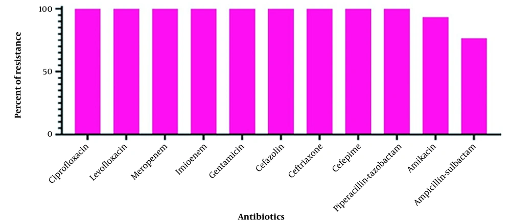
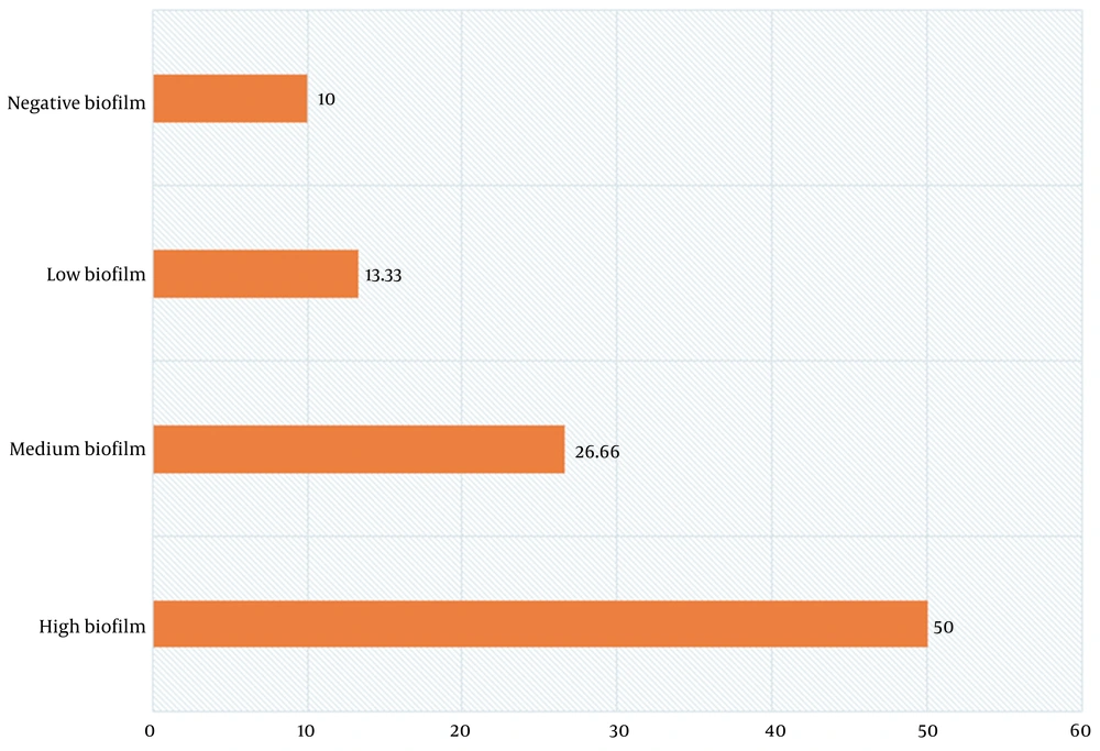
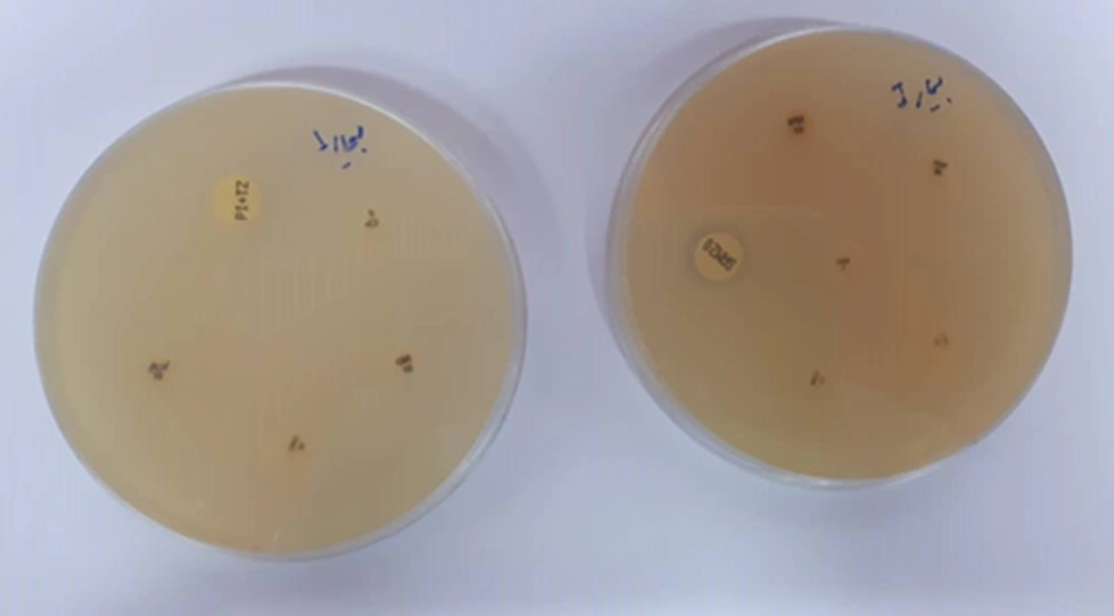
The oxidase-negative, catalase-positive, urease-negative, arginine dihydrolase-positive, malonate-positive, non-motile, and non-fermenting bacilli with inert TSI were identified as A. baumannii. The disk diffusion susceptibility test revealed that all 30 (100%) isolates were resistant to piperacillin-tazobactam, cefepime, ceftriaxone, ceftazidime, gentamicin, imipenem, meropenem, levofloxacin, and ciprofloxacin. Among them, 28 (93.3%) isolates were resistant to amikacin, and 23 (76.6%) isolates were resistant to ampicillin-sulbactam. All 30 isolates from ICU-admitted patients (100%) were identified as XDR (Figure 1). The susceptibility results are shown in Figure 2. The biofilm-forming ability of the isolates was evaluated, and the results are illustrated in Figure 3. In general, 27 out of 30 isolates (90%) formed biofilm, confirming the significant role of biofilm in bacterial pathogenicity. Among the isolates, 15 (50%) exhibited high biofilm-forming ability, eight (26.66%) showed medium biofilm-forming ability, and four (13.33%) displayed low biofilm-forming ability, while three isolates (10%) had no biofilm-forming properties.
All 30 isolates from ICU-admitted patients (100%) defined as MDR
Antibiotic resistance of Acinetobacter baumannii isolates
Frequency of biofilm formation among Acinetobacter baumannii isolates (Percentages)
4.2. RNA Extraction and Evaluation of Biofilm Formation-related Gene Expression by Real-time PCR
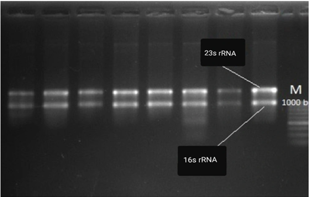
The RNA concentrations of A. baumannii isolates ranged from 1000 to 1500 ng/μL in a bacterial concentration of 1×10⁵ CFU/mL. The RNA concentration of each isolate was normalized for cDNA synthesis. The integrity of the extracted RNA was confirmed based on the 16S/23S RNA band pattern. Furthermore, the expression levels of bap, blaPER-1, and pgaA genes in XDR A. baumannii isolates were evaluated using SYBR green Real-time PCR and the 2-∆∆Ct method. Relative quantification via Real-time PCR was performed to assess the invasive potency of isolates compared to the standard strain.
As indicated in Table 2, the expression of biofilm formation-related genes was significantly higher in the patients’ isolates compared to the standard strain. This finding suggests the increased invasive potency of A. baumannii isolates during colonization in pneumonia patients. Despite the patients’ isolates and the standard strain being XDR, the higher expression levels of biofilm formation-related genes in the colonizing bacteria indicate significant pathogenicity. Additionally, an unpaired t-test confirmed the significant upregulation of biofilm formation-related genes in the patients’ samples relative to the standard strain. The 16S rRNA housekeeping gene was used as the internal control for the relative quantifications (Figure 4). The expression levels of blaPER-1, pgaA, and bap were 7.47, 5.27, and 11.96 times higher in the patients’ samples compared to the standard ATCC 19606 strain, respectively (P < 0.05).
Evaluation of Biofilm Formation-related Gene Expression (blaPER-1, bap, and pgaA genes)
Genes | Type | Reaction Efficiency | Expression | Std. Error | 95% CI | Results |
---|---|---|---|---|---|---|
16S rRNA | REF | 0.91 | 1.000 | - | - | - |
blaPER | TRG | 0.95 | 7.473 | 2.197 - 26.566 | 0.707 - 75.040 | Overexpression |
pgaA | TRG | 0.93 | 5.277 | 1.051 - 36.239 | 0.010 - 159.195 | Overexpression |
bap | TRG | 0.96 | 11.964 | 1.993 - 72.428 | 0.430 - 425.961 | Overexpression |
The RNA integrity was confirmed based on 16S/23S RNA band pattern
5. Discussion
Acinetobacter baumannii infection is commonly observed in immunosuppressed patients with underlying diseases requiring intensive care or those undergoing invasive procedures. This bacterium has become increasingly prevalent as a causative agent of ventilator-associated pneumonia, bacteremia, meningitis, urinary tract infections, as well as infections in the skin, soft tissues, central nervous system, and bones (30). It has been reported in previous studies that XDR A. baumannii has the ability to form biofilms on medical devices and biological surfaces, leading to decreased susceptibility to antibiotics (25, 31-33).
Biofilms exhibit significant resistance to antibiotics, host immune responses, desiccation, and UV light, enabling them to persist in challenging environments (34). The formation of biofilms and the development of antibiotic resistance create favorable conditions for A. baumannii to cause respiratory tract infections among hospitalized patients and military personnel (35). Deslandes et al. conducted a study showing that injured soldiers contracted infections from A. baumannii in military field hospitals (36), which is consistent with previous similar studies (37-39).
In this study, the isolated strains from patients hospitalized in the ICU demonstrated resistance to a broad spectrum of antibiotics. This observation aligns with previous studies that have reported the high resistance of A. baumannii isolates to various antimicrobial agents in neurosurgical and neonatal ICUs (40-42). Efflux pumps, such as adeA and adeS, are recognized as significant mechanisms of resistance in A. baumannii, as they actively expel toxic substances, including antibiotics, from the bacterial cell (43). The extensive antibiotic resistance observed in A. baumannii is attributed to the presence and proliferation of multiple antibiotic-resistance genes. Numerous studies have documented the resistance of A. baumannii to most beta-lactam antibiotics (44, 45) and quinolones (46, 47), which is consistent with our findings. Additionally, there is a growing trend of resistance to aminoglycosides, as highlighted by Mortazavi et al. (48). The ability of A. baumannii to acquire or enhance antimicrobial resistance has contributed to the global prevalence of XDR strains (49-51).
The microtiter plate method employed in our study revealed that 90% of A. baumannii isolates were capable of forming biofilms. Moreover, the expression of key genes such as blaPER-1, bap, and pgaA was found to be crucial for adherence to the epithelial cell surface during respiratory tract infections. We observed significantly higher expression levels of blaPER-1, pgaA, and bap in the patients’ samples compared to the standard ATCC 19606 strain, with fold changes of 7.47, 5.27, and 11.96, respectively. This suggests a notable correlation between the upregulation of these genes involved in biofilm formation and the strains isolated from the ICU department of the hospital.
These findings align with earlier investigations that reported a high prevalence of biofilm-forming A. baumannii strains among ICU isolates (40, 52-54). The upregulation of biofilm formation-related gene expression contributes to the development of biofilms with adequate thickness. Consequently, this association between gene expression and extensively drug-resistant (XDR) strains impedes the diffusion of antimicrobial agents through the biofilm matrix and hinders their interaction with the biofilm itself. Additionally, enzyme-mediated resistance and the levels of metabolic activity within the biofilm are other factors that contribute to the emergence of XDR strains, as reported by Rahmati et al. (13). While biofilm-forming genes play a significant role in nosocomial infections, it should be noted that other virulence genes may also contribute to the overall pathogenicity of A. baumannii, despite 90% of the isolated strains exhibiting biofilm-forming ability.
5.1. Conclusions
Our study has confirmed that biofilm formation-related genes were upregulated in bacterial isolates obtained from sputum samples of pneumonia patients. A. baumannii has emerged as a significant cause of nosocomial pneumonia worldwide and exhibits extensive drug resistance to commonly used antimicrobial agents, resulting in a high mortality rate. Ventilator-associated pneumonia is predominantly caused by XDR A. baumannii. It is crucial for healthcare facilities, particularly in ICUs, to implement effective infection control programs to manage nosocomial infections. For instance, further research is warranted to determine whether droplet precautions should be implemented for patients with A. baumannii in their sputum or if more attention should be given to ventilator maintenance and management protocols.
Acknowledgements
References
-
1.
Kyriakidis I, Vasileiou E, Pana ZD, Tragiannidis A. Acinetobacter baumannii antibiotic resistance mechanisms. Pathogens. 2021;10(3). [PubMed ID: 33808905]. [PubMed Central ID: PMC8003822]. https://doi.org/10.3390/pathogens10030373.
-
2.
Chang PY, Hsueh PR, Wu PS, Chan PC, Yang TT, Lu CY, et al. Multidrug-resistant acinetobacter baumannii isolates in pediatric patients of a university hospital in Taiwan. J Microbiol Immunol Infect. 2007;40(5):406-10. [PubMed ID: 17932600].
-
3.
Wang X, Qin L. A review on Acinetobacter baumannii. J Acute Disease. 2019;8(1):16.
-
4.
Basatian-Tashkan B, Niakan M, Khaledi M, Afkhami H, Sameni F, Bakhti S, et al. Antibiotic resistance assessment of Acinetobacter baumannii isolates from Tehran hospitals due to the presence of efflux pumps encoding genes (adeA and adeS genes) by molecular method. BMC Res Notes. 2020;13(1):543. [PubMed ID: 33213526]. [PubMed Central ID: PMC7678095]. https://doi.org/10.1186/s13104-020-05387-6.
-
5.
Rit K, Saha R. Multidrug-resistant acinetobacter infection and their susceptibility patterns in a tertiary care hospital. Niger Med J. 2012;53(3):126-8. [PubMed ID: 23293410]. [PubMed Central ID: PMC3531029]. https://doi.org/10.4103/0300-1652.104379.
-
6.
Malekzadegan Y, Abdi A, Heidari H, Moradi M, Rastegar E, Sedigh Ebrahim-Saraie H. In vitro activities of colistin, imipenem and ceftazidime against drug-resistant Pseudomonas aeruginosa and Acinetobacter baumannii isolates in the south of Iran. BMC Res Notes. 2019;12(1):301. [PubMed ID: 31138309]. [PubMed Central ID: PMC6540545]. https://doi.org/10.1186/s13104-019-4344-7.
-
7.
Patel SJ, Oliveira AP, Zhou JJ, Alba L, Furuya EY, Weisenberg SA, et al. Risk factors and outcomes of infections caused by extremely drug-resistant gram-negative bacilli in patients hospitalized in intensive care units. Am J Infect Control. 2014;42(6):626-31. [PubMed ID: 24725516]. [PubMed Central ID: PMC4083852]. https://doi.org/10.1016/j.ajic.2014.01.027.
-
8.
Rahmati F. Microencapsulation of Lactobacillus acidophilus and Lactobacillus plantarum in Eudragit S100 and alginate chitosan under gastrointestinal and normal conditions. Applied Nanoscience. 2020;10(2):391-9.
-
9.
Mahto KU, Priyadarshanee M, Samantaray DP, Das S. Bacterial biofilm and extracellular polymeric substances in the treatment of environmental pollutants: Beyond the protective role in survivability. J Clean Prod. 2022:134759.
-
10.
Abdumohsin BMA, Al-Daraghi WA. Biochemical and Molecular Identification of Acinetobacter baumannii Isolated from Patients in Baghdad Hospitals. Kerbala J Pharm Sci. 2020;(18).
-
11.
Michaelis C, Grohmann E. Horizontal gene transfer of antibiotic resistance genes in biofilms. Antibiotics (Basel). 2023;12(2). [PubMed ID: 36830238]. [PubMed Central ID: PMC9952180]. https://doi.org/10.3390/antibiotics12020328.
-
12.
Treccani L. Interactions between surface material and bacteria: From biofilm formation to suppression. Surface‐functionalized ceramics: For biotechnological and environmental applications. 2023:283-335.
-
13.
Rahmati F, Hosseini SS, Mahuti Safai S, Asgari Lajayer B, Hatami M. New insights into the role of nanotechnology in microbial food safety. 3 Biotech. 2020;10(10):425. [PubMed ID: 32968610]. [PubMed Central ID: PMC7483685]. https://doi.org/10.1007/s13205-020-02409-9.
-
14.
Rahmati F. Impact of microencapsulation on two probiotic strains in alginate chitosan and Eudragit S100 under gastrointestinal and normal conditions. Open Biotech J. 2019;13(1).
-
15.
Gedefie A, Demsis W, Ashagrie M, Kassa Y, Tesfaye M, Tilahun M, et al. Acinetobacter baumannii biofilm formation and its role in disease pathogenesis: A review. Infect Drug Resist. 2021;14:3711-9. [PubMed ID: 34531666]. [PubMed Central ID: PMC8439624]. https://doi.org/10.2147/IDR.S332051.
-
16.
Abdi-Ali A, Hendiani S, Mohammadi P, Gharavi S. Assessment of biofilm formation and resistance to imipenem and ciprofloxacin among clinical isolates of acinetobacter baumannii in Tehran. Jundishapur J Microbiol. 2014;7(1). e8606. [PubMed ID: 25147652]. [PubMed Central ID: PMC4138664]. https://doi.org/10.5812/jjm.8606.
-
17.
Schiffer CJ, Schaudinn C, Ehrmann MA, Vogel RF. SxsA, a novel surface protein mediating cell aggregation and adhesive biofilm formation of Staphylococcus xylosus. Mol Microbiol. 2022;117(5):986-1001. [PubMed ID: 35072960]. https://doi.org/10.1111/mmi.14884.
-
18.
Brossard KA, Campagnari AA. The Acinetobacter baumannii biofilm-associated protein plays a role in adherence to human epithelial cells. Infect Immun. 2012;80(1):228-33. [PubMed ID: 22083703]. [PubMed Central ID: PMC3255684]. https://doi.org/10.1128/IAI.05913-11.
-
19.
Mima T, Joshi S, Gomez-Escalada M, Schweizer HP. Identification and characterization of TriABC-OpmH, a triclosan efflux pump of Pseudomonas aeruginosa requiring two membrane fusion proteins. J Bacteriol. 2007;189(21):7600-9. [PubMed ID: 17720796]. [PubMed Central ID: PMC2168734]. https://doi.org/10.1128/JB.00850-07.
-
20.
De Gregorio E, Del Franco M, Martinucci M, Roscetto E, Zarrilli R, Di Nocera PP. Biofilm-associated proteins: News from Acinetobacter. BMC Genomics. 2015;16:933. [PubMed ID: 26572057]. [PubMed Central ID: PMC4647330]. https://doi.org/10.1186/s12864-015-2136-6.
-
21.
Schiffer C, Hilgarth M, Ehrmann M, Vogel RF. Bap and cell surface hydrophobicity are important factors in staphylococcus xylosus biofilm formation. Front Microbiol. 2019;10:1387. [PubMed ID: 31293539]. [PubMed Central ID: PMC6603148]. https://doi.org/10.3389/fmicb.2019.01387.
-
22.
Shukla SK. Molecular Studies on Biofilm-associated-protein (Bap) in Staphylococcus aureus. 2015.
-
23.
Upmanyu K, Haq QMR, Singh R. Factors mediating Acinetobacter baumannii biofilm formation: Opportunities for developing therapeutics. Curr Res Microb Sci. 2022;3:100131. [PubMed ID: 35909621]. [PubMed Central ID: PMC9325880]. https://doi.org/10.1016/j.crmicr.2022.100131.
-
24.
Colquhoun JM, Rather PN. Insights Into Mechanisms of Biofilm Formation in Acinetobacter baumannii and Implications for Uropathogenesis. Front Cell Infect Microbiol. 2020;10:253. [PubMed ID: 32547965]. [PubMed Central ID: PMC7273844]. https://doi.org/10.3389/fcimb.2020.00253.
-
25.
Amin M, Navidifar T, Shooshtari FS, Rashno M, Savari M, Jahangirmehr F, et al. Association Between Biofilm Formation, Structure, and the Expression Levels of Genes Related to biofilm formation and Biofilm-Specific Resistance of Acinetobacter baumannii Strains Isolated from Burn Infection in Ahvaz, Iran. Infect Drug Resist. 2019;12:3867-81. [PubMed ID: 31853190]. [PubMed Central ID: PMC6914661]. https://doi.org/10.2147/IDR.S228981.
-
26.
Kırmusaoğlu S. Staphylococcal biofilms: Pathogenicity, mechanism and regulation of biofilm formation by quorum sensing system and antibiotic resistance mechanisms of biofilm embedded microorganisms. IntechOpen; 2016.
-
27.
Nguyen HTT, Nguyen TH, Otto M. The staphylococcal exopolysaccharide PIA - Biosynthesis and role in biofilm formation, colonization, and infection. Comput Struct Biotechnol J. 2020;18:3324-34. [PubMed ID: 33240473]. [PubMed Central ID: PMC7674160]. https://doi.org/10.1016/j.csbj.2020.10.027.
-
28.
Rahmati F. Characterization of Lactobacillus, Bacillus and Saccharomyces isolated from Iranian traditional dairy products for potential sources of starter cultures. AIMS Microbiol. 2017;3(4):815-25. [PubMed ID: 31294191]. [PubMed Central ID: PMC6604970]. https://doi.org/10.3934/microbiol.2017.4.815.
-
29.
Shahbazi S, Shivaee A, Nasiri M, Mirshekar M, Sabzi S, Sariani OK. Zinc oxide nanoparticles impact the expression of the genes involved in toxin-antitoxin systems in multidrug-resistant Acinetobacter baumannii. J Basic Microbiol. 2022. [PubMed ID: 36086811]. https://doi.org/10.1002/jobm.202200382.
-
30.
Beigverdi R, Sattari-Maraji A, Emaneini M, Jabalameli F. Status of carbapenem-resistant Acinetobacter baumannii harboring carbapenemase: First systematic review and meta-analysis from Iran. Infect Genet Evol. 2019;73:433-43. [PubMed ID: 31176030]. https://doi.org/10.1016/j.meegid.2019.06.008.
-
31.
Nasiri MJ, Zamani S, Fardsanei F, Arshadi M, Bigverdi R, Hajikhani B, et al. Prevalence and mechanisms of carbapenem resistance in acinetobacter baumannii: A comprehensive systematic review of cross-sectional studies from Iran. Microb Drug Resist. 2020;26(3):270-83. [PubMed ID: 30822197]. https://doi.org/10.1089/mdr.2018.0435.
-
32.
Mirzaei B, Bazgir ZN, Goli HR, Iranpour F, Mohammadi F, Babaei R. Prevalence of multi-drug resistant (MDR) and extensively drug-resistant (XDR) phenotypes of Pseudomonas aeruginosa and Acinetobacter baumannii isolated in clinical samples from Northeast of Iran. BMC Res Notes. 2020;13(1):380. [PubMed ID: 32778154]. [PubMed Central ID: PMC7418330]. https://doi.org/10.1186/s13104-020-05224-w.
-
33.
Askari N, Momtaz H, Tajbakhsh E. Prevalence and phenotypic pattern of antibiotic resistance of Acinetobacter baumannii isolated from different types of raw meat samples in Isfahan, Iran. Vet Med Sci. 2020;6(1):147-53. [PubMed ID: 31576672]. [PubMed Central ID: PMC7036315]. https://doi.org/10.1002/vms3.199.
-
34.
Rahmati F. Identification and characterization of Lactococcus starter strains in milk-based traditional fermented products in the region of Iran. AIMS Agriculture and Food. 2018;3(1):12-25.
-
35.
Azimi L, Fallah F, Karimi A, Shirdoust M, Azimi T, Sedighi I, et al. Survey of various carbapenem-resistant mechanisms of Acinetobacter baumannii and Pseudomonas aeruginosa isolated from clinical samples in Iran. Iran J Basic Med Sci. 2020;23(11):1396-400. [PubMed ID: 33235696]. [PubMed Central ID: PMC7671419]. https://doi.org/10.22038/IJBMS.2020.44853.10463.
-
36.
Deslandes A, Meyer A, Soing-Altrach S, Giard M, Locher G, Jouzeau N, et al. Highly drug-resistant organisms in hospitalized civilians and soldiers from Ukraine in France, March-December 2022. J Hosp Infect. 2023. [PubMed ID: 36924899]. https://doi.org/10.1016/j.jhin.2023.03.006.
-
37.
Higgins PG, Kniel M, Rojak S, Balczun C, Rohde H, Frickmann H, et al. Molecular epidemiology of carbapenem-resistant acinetobacter baumannii strains isolated at the German Military Field Laboratory in Mazar-e Sharif, Afghanistan. Microorganisms. 2021;9(11). [PubMed ID: 34835355]. [PubMed Central ID: PMC8622437]. https://doi.org/10.3390/microorganisms9112229.
-
38.
Granzer H, Hagen RM, Warnke P, Bock W, Baumann T, Schwarz NG, et al. Molecular epidemiology of carbapenem-resistant acinetobacter baumannii complex isolates from patients that were injured during the Eastern Ukrainian Conflict. Eur J Microbiol Immunol (Bp). 2016;6(2):109-17. [PubMed ID: 27429793]. [PubMed Central ID: PMC4936333]. https://doi.org/10.1556/1886.2016.00014.
-
39.
Glover MP, Beaman LA, Berry-Caban CS. Acinetobacter baumannii hallux infection secondary to partial amputation in a US soldier a case report. J Am Podiatr Med Assoc. 2015;105(6):560-3. [PubMed ID: 26667510]. https://doi.org/10.7547/14-048.1.
-
40.
Zeighami H, Valadkhani F, Shapouri R, Samadi E, Haghi F. Virulence characteristics of multidrug resistant biofilm forming Acinetobacter baumannii isolated from intensive care unit patients. BMC Infect Dis. 2019;19(1):629. [PubMed ID: 31315572]. [PubMed Central ID: PMC6637494]. https://doi.org/10.1186/s12879-019-4272-0.
-
41.
Moosavian M, Ahmadi K, Shoja S, Mardaneh J, Shahi F, Afzali M. Antimicrobial resistance patterns and their encoding genes among clinical isolates of Acinetobacter baumannii in Ahvaz, Southwest Iran. MethodsX. 2020;7:101031. [PubMed ID: 32983919]. [PubMed Central ID: PMC7492985]. https://doi.org/10.1016/j.mex.2020.101031.
-
42.
Arjmand R, Porrostami K, Esteghamat SS, Chaghamirzayi P, Sharifian P, Zahmatkesh E, et al. Frequency and Antibiotic Susceptibility of Pseudomonas aeruginosa and Acinetobacter baumannii Infections in Pediatrics Intensive Care Unit of Imam Ali Hospital, Karaj, Iran During 2017- 2018. Int J Enteric Pathog. 2020;8(1):15-8. https://doi.org/10.34172/ijep.2020.04.
-
43.
Verma P, Tiwari M, Tiwari V. Efflux pumps in multidrug-resistant Acinetobacter baumannii: Current status and challenges in the discovery of efflux pumps inhibitors. Microb Pathog. 2021;152:104766. [PubMed ID: 33545327]. https://doi.org/10.1016/j.micpath.2021.104766.
-
44.
Ingti B, Upadhyay S, Hazarika M, Khyriem AB, Paul D, Bhattacharya P, et al. Distribution of carbapenem resistant Acinetobacter baumannii with bla(ADC-30) and induction of ADC-30 in response to beta-lactam antibiotics. Res Microbiol. 2020;171(3-4):128-33. [PubMed ID: 31988011]. https://doi.org/10.1016/j.resmic.2020.01.002.
-
45.
Lazaretti WY, Dos Santos EL, da-Conceicao Silva JL, Kadowaki MK, Gandra RF, Maller A, et al. Upregulation of the clpB gene in response to heat shock and beta-lactam antibiotics in Acinetobacter baumannii. Mol Biol Rep. 2020;47(2):1499-505. [PubMed ID: 31786767]. https://doi.org/10.1007/s11033-019-05209-4.
-
46.
Glover JS, Ticer TD, Engevik MA. Profiling antibiotic resistance in acinetobacter calcoaceticus. Antibiotics (Basel). 2022;11(7). [PubMed ID: 35884232]. [PubMed Central ID: PMC9312123]. https://doi.org/10.3390/antibiotics11070978.
-
47.
Ibitoye OB, Ajiboye TO. Ferulic acid potentiates the antibacterial activity of quinolone-based antibiotics against Acinetobacter baumannii. Microb Pathog. 2019;126:393-8. [PubMed ID: 30476577]. https://doi.org/10.1016/j.micpath.2018.11.033.
-
48.
Mortazavi SM, Farshadzadeh Z, Janabadi S, Musavi M, Shahi F, Moradi M, et al. Evaluating the frequency of carbapenem and aminoglycoside resistance genes among clinical isolates of Acinetobacter baumannii from Ahvaz, south-west Iran. New Microbes New Infect. 2020;38:100779. [PubMed ID: 33194209]. [PubMed Central ID: PMC7644744]. https://doi.org/10.1016/j.nmni.2020.100779.
-
49.
Weidensdorfer M, Ishikawa M, Hori K, Linke D, Djahanschiri B, Iruegas R, et al. The Acinetobacter trimeric autotransporter adhesin Ata controls key virulence traits of Acinetobacter baumannii. Virulence. 2019;10(1):68-81. [PubMed ID: 31874074]. [PubMed Central ID: PMC6363060]. https://doi.org/10.1080/21505594.2018.1558693.
-
50.
Rolain JM, Diene SM, Kempf M, Gimenez G, Robert C, Raoult D. Real-time sequencing to decipher the molecular mechanism of resistance of a clinical pan-drug-resistant Acinetobacter baumannii isolate from Marseille, France. Antimicrob Agents Chemother. 2013;57(1):592-6. [PubMed ID: 23070160]. [PubMed Central ID: PMC3535948]. https://doi.org/10.1128/AAC.01314-12.
-
51.
Giammanco A, Cala C, Fasciana T, Dowzicky MJ. Global assessment of the activity of tigecycline against multidrug-resistant gram-negative pathogens between 2004 and 2014 as Part of the tigecycline evaluation and surveillance trial. mSphere. 2017;2(1). [PubMed ID: 28124025]. [PubMed Central ID: PMC5244261]. https://doi.org/10.1128/mSphere.00310-16.
-
52.
Khoshnood S, Savari M, Abbasi Montazeri E, Farajzadeh Sheikh A. Survey on Genetic Diversity, Biofilm Formation, and Detection of Colistin Resistance Genes in Clinical Isolates of Acinetobacter baumannii. Infect Drug Resist. 2020;13:1547-58. [PubMed ID: 32547124]. [PubMed Central ID: PMC7266307]. https://doi.org/10.2147/IDR.S253440.
-
53.
Zhang D, Xia J, Xu Y, Gong M, Zhou Y, Xie L, et al. Biological features of biofilm-forming ability of Acinetobacter baumannii strains derived from 121 elderly patients with hospital-acquired pneumonia. Clin Exp Med. 2016;16(1):73-80. [PubMed ID: 25543268]. https://doi.org/10.1007/s10238-014-0333-2.
-
54.
Castilho SRA, Godoy CSM, Guilarde AO, Cardoso JL, Andre MCP, Junqueira-Kipnis AP, et al. Acinetobacter baumannii strains isolated from patients in intensive care units in Goiania, Brazil: Molecular and drug susceptibility profiles. PLoS One. 2017;12(5). e0176790. [PubMed ID: 28475585]. [PubMed Central ID: PMC5419545]. https://doi.org/10.1371/journal.pone.0176790.