High Prevalence of Antiseptic Resistance Encoding Genes and Reduced Phenotypic Antiseptic Susceptibility Among Antibiotic-Resistant Pseudomonas aeruginosa Isolates

authors:

avatar Mohadeseh Radmehr 1 , avatar Majid Moghbeli 1 , avatar Hamed Ghasemzadeh-Moghaddam ORCID 2 , * , avatar Amir Azimian ORCID 2 , avatar Alex van Belkum ORCID 3

Department of Microbiology, Damghan Branch, Islamic Azad University, Damghan, Iran
Vector-Borne Diseases Research Center, North Khorasan University of Medical Sciences, Bojnurd, Iran
Open Innovation & Partnerships, BaseClear, Leiden, Netherlands

how to cite: Radmehr M, Moghbeli M, Ghasemzadeh-Moghaddam H, Azimian A, van Belkum A. High Prevalence of Antiseptic Resistance Encoding Genes and Reduced Phenotypic Antiseptic Susceptibility Among Antibiotic-Resistant Pseudomonas aeruginosa Isolates. Jundishapur J Microbiol. 2023;16(3):e135911. https://doi.org/10.5812/jjm-135911.

Abstract

Background:

Multi-drug resistant (MDR) and extensively drug-resistant (XDR) Pseudomonas aeruginosa isolates are of clinical concern.

Objectives:

To determine the distribution of antiseptic resistance genes and the associated level of phenotypic antiseptic resistance against quaternary ammonium compounds and biguanide compounds, we studied MDR and XDR P. aeruginosa isolates collected from different infections among patients from a single hospital.

Methods:

Pseudomonas aeruginosa isolates were investigated in 2020 for in vitro susceptibility to benzethonium chloride (BTC), benzalkonium chloride (BKC), and chlorhexidine digluconate (CHG). The minimum inhibitory concentrations (MICs) against these antiseptic agents were determined using broth microdilution. Also, polymerase chain reaction (PCR)-mediated detection of qacE, qacEΔ1, and blaOXA-23 genes was used.

Results:

Isolates were largely non-clonal according to their phenotypical and genotypical non-similarity (35 overall data-combination types detected). Most P. aeruginosa infections occurred in intensive care unit (ICU) patients (n = 43, 61.4%). Extensively drug-resistant and MDR phenotypes were detected in 20% and 12.6%, respectively. Among the 70 isolates retained, 53 (75.7%) harbored at least one resistance gene, comprising 11 (20.7%) isolates with solely the qacEΔ1 gene; seven (13.2%) isolates harbored the qacE gene. Both the qacE and qacEΔ1 genes were detected simultaneously in 35 (66%) isolates. The mean MICs for BTC (24.0 versus 10.56 µg/mL), BKC (46.1 versus 17.22 µg/mL), and CHG (107.7 versus 29.4 µg/mL) were statistically significantly higher among antiseptic resistance gene harboring isolates than in other isolates without such genes.

Conclusions:

The significantly increased MICs against antiseptic agents among antibiotic-resistant P. aeruginosa isolates highlight the importance of monitoring such increases and implementing effective infection control.

1. Background

Pseudomonas aeruginosa is a saprophytic gram-negative bacterial species that can be isolated from soil, plants, and hospital environments (1). Pseudomonas aeruginosa-related infections cause death in patients who suffer from bacteremia (2), intensive care unit (ICU)-related septicemia (3), ventilator-related pneumonia, and even urinary tract infections (4, 5). Multiple drug-resistant (MDR) and even extensively drug-resistant (XDR) phenotypes among P. aeruginosa have been reported frequently and at elevated prevalence (4 - 60%) (6, 7). Reduced cytoplasmic membrane permeability, expression of efflux pump-related genes, the release of antibiotic-destroying enzymes such as beta-lactamases, and alginate production are the most common antibiotic resistance mechanisms among P. aeruginosa strains (8-10).

Efflux pumps are a group of proteins transferring energy packages, antibiotics, and other small molecules out of the bacterial cytoplasm or periplasmic space. Continuous transfer of antibiotics out of bacteria will increase the minimum inhibitory concentrations (MIC) for such antibiotics and antiseptic agents. It helps the bacteria continue growing while the antibiotic or antiseptic target is still present in the bacterial cytoplasm (8-10). Also, blaOXA-23, blaOXA-24, and blaOXA-40 are genes responsible for resistance against different carbapenem antibiotics (11).

Utilizing disinfectants with quaternary ammonium compounds (QAC) and biguanide compounds included is one of the most applied prevention strategies against nosocomial infection (12). Reduced susceptibility against the active agents of the mentioned antiseptics has been reported before (13, 14). Antiseptic resistance may result from the expression of chromosomal genes and plasmid-located genes, including biocide resistance genes (BRGs) and qac (15). The qacE gene has been reported as one of the genes encoding resistance against QACs and chlorhexidine digluconate (CHG) among Enterobacterales and different Pseudomonas spp. (16).

2. Objectives

The current study investigated the prevalence of antiseptic resistance genes and the resulting increase in MIC against QACs and CHG among P. aeruginosa isolates from different infections among patients in the Imam Hassan Hospital (IHH). The IHH is the biggest referral teaching and care hospital in North Khorasan province of Iran.

3. Methods

3.1. Study Samples

All P. aeruginosa isolates causing infections at different anatomical sites were collected by tracheal aspirate culture, blood culture, urine culture, and wound culture. Clinical specimens originated from hospitalized patients in various hospital wards (ICU, Cardiology, Emergency Department, Infectious Diseases Department, and Neurology) in the IHH during 2020. Bacteria were identified at the species level in the hospital laboratory, and this was confirmed in the microbiology laboratory at the Faculty of Medicine by Gram staining, oxidase testing, motility testing, and defining the ability of growth at 42°C according to Clinical and Laboratory Standard Institute (CLSI) guidelines (17). All isolates were stored at -30°C in trypticase soy broth (TSB) supplemented with 20% glycerol.

3.2. Antiseptic Susceptibility Testing

Bacterial susceptibilities to benzethonium chloride (BTC), benzalkonium chloride (BKC), and biguanide compounds such as CHG (Sigma-Aldrich, Steinheim, Germany) were determined using the Mueller-Hinton broth microdilution method (BMD) (18).

3.3. Antibiotic Susceptibility Testing

Antimicrobial susceptibility testing (AST) was performed on Mueller-Hinton agar (Merck, Germany) using the disk diffusion (Kirby-Bauer) technique, with zone size interpretation based on CLSI (2021) guidelines (17). The 11 antimicrobials agents used in characterizing the isolates of P. aeruginosa were: Carbapenems (doripenem, meropenem, imipenem), a tetracycline (tigecycline), aminoglycosides (amikacin, tobramycin), beta-lactamase/beta-lactamase inhibitor combinations (ampicillin + sulbactam, piperacillin + tazobactam), cephalosporins (cefepime, ceftazidime), and a fluoroquinolone (ciprofloxacin). Isolates were categorized as XDR when they revealed resistance to one or more antimicrobial agents in at least six categories or showed resistance to all except one or two antibiotics. Resistance to one or more agents in three or more categories was used for grouping the bacteria as MDR (19).

3.4. Detection of Genes

Chromosomal DNA was extracted using a commercial DNA extraction kit (Poyagene Azma, Iran) according to the manufacturer's instructions and kept at -30°C for further molecular investigations. All isolates were confirmed at the gene level as P. aeruginosa by detecting the gyrB gene using polymerase chain reaction (PCR) (20). All isolates were screened for antiseptic and antibiotic resistance genes such as qacE, qacEΔ1, and blaOXA-23 genes as described before (21, 22). The Supplementary File shows the primer sequences and related PCR protocols.

3.5. Typing of Bacterial Isolates

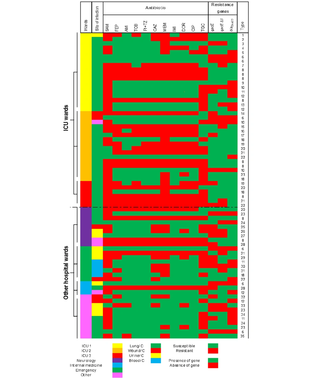
In order to verify that isolates were sufficiently heterogeneous and that past or ongoing outbreaks did not bias our study, we accumulated all data gathered above in a single figure. We defined the uniqueness of the strains by comparing phenotypic and PCR data (Figure 1). Typing the strains was performed using the combined similarities of isolates in their antibiotic resistance patterns and their resistance gene content. Similar P. aeruginosa isolates were placed under the same type and numbered accordingly.

Accumulation of all strain-specific variables determined in the current study

3.6. Statistical Analysis

A one-way ANOVA test was performed to evaluate possibly significant differences using SPSS statistics 21. P-values < 0.05 were considered significant.

4. Results

From 78 documented P. aeruginosa infections detected in 2020, 70 P. aeruginosa isolates were still available and subjected to the current study. These P. aeruginosa isolates were obtained from different clinical samples such as tracheal aspirates (n = 46; 65.7%), wounds (n = 6; 8.5%), blood (n = 4; 5.7 %), and urine (n = 10; 14.2 %) (Appendix 2 in the Supplementary File). Among the infected patients, 58.5% were male (n = 41/70). The mean age of the infected patients was 63.3 (2 - 92) years, with 61.8 (2 - 92) years for males and 64.9 (42 - 87) years for females (Table 1).

The vast majority of P. aeruginosa infections occurred in the ICU (n = 43, 61.4%), comprising ICU I (n = 19, 27.1%), ICU II (n = 18, 25.7%), and ICU III (n = 6, 8.5%). An extensive diversity among ICU isolates (23 out of 43 strains, 53.4%) was spotted when an accumulation of all the strain-specific variables determined was used (Figure 1). Of note, when all isolates were considered, we documented 35 different types among all 70 isolates included. The main isolation site of P. aeruginosa isolates was lung infection (65.7%), followed by urine infection (14.2%), wound infections (8.5%), blood infection (5.7%), and other (5.7%) (Supplementary File).

4.1. Antibiotic Susceptibility Test

Among the 11 tested antibiotics, the highest resistance rate was detected against ampicillin + sulbactam (77.1%), followed by tigecycline (64.3%). The lowest resistance rate was for amikacin (32.9%) (Table 1). Carbapenem-resistant P. aeruginosa (CRPA) was detected in 48.5% of all cases.

Table 1.

Antibiotic Resistance Patterns, Antiseptic, and Antibiotic Resistance Genes Distribution Among Pseudomonas aeruginosa Isolates a, b

Resistance PhenotypePatternValuesResistant AntibioticNo.SensitiveNo.Having at Least A GeneqacEqacEΔ1qacE + qacEΔ1blaOXA-23
XDRA14 (20)SAM, FEP, AMI, TOB, PI + TZ, CAZ, MEM, IMI, DOR, CIP, TGC11--9 (64.2)2 (14.28)2 (14.28)9 (64.2)14 (100)
MDRB3 (4.2)SAM, FEP, AMI, TOB, PI + TZ, CAZ, MEM, IMI, DOR, CIP10TGC11 (33.3)01 (33.3)2 (66.6)3 (100)
C2 (2.8)SAM, CAZ, MEM, TGC4FEP, AMI, TOB, PI + TZ, IMI, DOR, CIP71 (50)01 (50)01 (50)
D2 (2.8)SAM, MEM, TGC3FEP, AMI, TOB, PI + TZ, IMI, DOR, CIP, CAZ82 (100)002 (100)2 (100)
E2 (2.8)SAM, DOR, MEM, TGC4FEP, AMI, TOB, PI + TZ, IMI, CAZ, CIP72 (100)002 (100)2 (100)
Non-MDRF13(18.5)SAM, TGC2FEP, AMI, TOB, PI + TZ, CAZ, MEM, IMI, DOR, CIP94 (30.7)1 (7.6)3 (23)6 (46.1)0
G3 (4.2)SAM1FEP, AMI, TOB, PI + TZ, CAZ, MEM, IMI, DOR, CIP, TGC101 (33.3)001 (33.3)0
H9 (12.8)00SAM, FEP, AMI, TOB, PI + TZ, CAZ, MEM, IMI, DOR, CIP, TGC112 (22.2)1 (11.1)01 (11.1)1

Eight antibiotic resistance patterns were detected (A-H). Five contained XDR and MDR phenotypes (n = 23 isolates, 32.8%) (Table 1). Non-MDR patterns (F-H) were the dominant resistance phenotypes detected among 25 (35.7%) isolates. Also, P. aeruginosa showing pattern A (XDR) expressed resistance against 11 antibiotics (Table 1). The MDR isolates were spotted among pattern B isolates (10 antibiotics) (N = 3, 4.3%), pattern C (four antibiotics) (N = 2, 2.8%), pattern D (three antibiotics) (N = 2, 2.8%), and pattern E (four antibiotics) (N = 2, 2.8%) (Table 1). Twenty-two isolates showed a unique resistance pattern comprising 15 ICU-collected isolates and seven isolates of other wards. The majority of P. aeruginosa infections were observed among ICU patients (n = 46/70, 65.7%), comprising 29 (63%) male and 17 (36.9%) female patients. Moreover, MDR and XDR phenotypes were detected in 29/46 (63%).

4.2. Antiseptic Resistance Gene Distribution

Among 70 isolates, 53 isolates harbored at least one antiseptic resistance gene (75.7%), comprising 11 (20.7%) isolates with the qacE Δ1 gene alone and seven (13.2%) isolates with the qacE gene alone. Simultaneous occurrence of qacE and qacEΔ1 genes were spotted in 35 (66%) isolates. As stated above, grouping the isolates according to their antibiotic resistance phenotypes and resistance genes pattern similarity defined 35 overall combination types among the 70 (50%) strains we studied (Figure 1). The highest prevalence of antiseptic resistance genes (qacE and qacEΔ1) was detected in antibiotic resistance patterns B, D, and E (100%), followed by pattern A (64.2%). The most frequent single occurrence of qacE and qacEΔ1 (50%) genes was in MDR pattern C, followed by XDR pattern A (14.28%). The highest co-occurrence of resistance genes was observed in pattern D (50%) (Table 1).

4.3. Antibiotic Resistance Gene Distribution

The blaOXA-23 gene was spotted in 59 (84.3%) isolates. Most blaOXA-23 gene-positive isolates had at least one antiseptic resistance gene (n = 44/59, 74.5%) (Table 2). The highest occurrence of the blaOXA-23 gene was among patterns A, B, D, and E (100%), following pattern C (50%) (Table 1).

Table 2.

Distribution of Antiseptic Resistance Genes and Minimum Inhibitory Concentrations Among A. baumannii Isolates a

PCR Result for Antiseptic Resistance GenesValuesBTCP bBKCP bCHGP bResistance Gene, Distribution PatternBTCBKCCHGblaOXA-23 c
Positive53 (75.7)24.0 (1.9 - 62.5)0.001 d46.1 (1.9 - 125)0.001 d107.7 (31.2 - 250)0.001 dqacE, 7 (13.2)15.90 (1.9 - 31.2)28.1 (1.9 - 62.5)53.3 (31.2 - 62.5)4 (6.7)
qacEΔ1, 11 (20.7)21.4 (1.9 - 62.5)27.8 (1.9 - 62.5)56.6 (31.2 - 62.5)9 (15.4)
qacEΔ1 + qacE, 35 (66.0)26.5 (7.8 - 62.5)55.4 (7.8 - 125)134.7 (31.2-250)31 (52.5)
Negative17 (24.3)10.56 (7.8-15.6)17.22 (3.9 - 62.5)29.4 (15.6 - 31.2)10.56 (7.8 - 15.6)17.22 (3.9 - 62.5)29.4 (15.6 - 31.2)15 (25.4)

4.4. Minimum Inhibitory Concentrations Against Antiseptics

The MICs ranged from 1.9 to 62.5 µg/mL for BTC, 1.9 to 125 µg/mL for BKC, and 31.2 to 250 µg/mL for CHG. The mean MICs for BTC (24.0 versus 10.56 µg/mL, P = 0.001), BKC (46.1 versus 17.22 µg/mL, P = 0.001), and CHG (107.7 versus 29.4 µg/mL, P = 0.001) among antiseptic resistance gene-harboring isolates were statistically significantly higher than those for strains that did not possess resistance genes (Table 2). There was a statistically significant difference in the mean MICs between isolates harboring qacE, qacEΔ1, and qacE + qacEΔ1 for BTC, BKC, and CHG and those having no gene (P = 0.001) (Table 2).

5. Discussion

The current study illustrated the high prevalence of antiseptic resistance genes (qacE and qacEΔ1) and a significant relationship between their presence and increased phenotypic resistance against BTC, BKC, and CHG among infectious P. aeruginosa. The reported rate of MDR and XDR isolates (68.5%) was slightly higher than the average rate for Iran (58%) (23). The accumulation of all the strain-specific variables determined illustrated an extensive diversity among all isolates (35 out of 70 strains, 50%), highlighting that data interpretation had no biases by clonality among isolates; even the isolates from ICU departments showed significant diversity (23 out of 43 strains, 53.4%). Multi-drug resistant and XDR phenotype frequencies were different in China (MDR 18.5%, XDR 3.5%) (24), Pakistan (MDR 36.3%, XDR 18.1%) (25), Iraq (MDR 50%, XDR 45%) (26), Thailand (46.4%) (27), and Nigeria (MDR 61%, XDR 5%) (28). Comprehensive monitoring of antibiotic resistance among P. aeruginosa isolates in 30 European Union countries revealed an MDR rate of zero to 49.4% when among a group of 27 European countries, the reported average rate was under 25% (29).

The prevalence of CRPA isolates was reported higher in Tehran (55.8%) (30) and the Southwest of Iran (52.2%) (31), while the rate of resistance was lower in other parts of Iran, including Yazd (37%) (32) and Golestan provinces (28.1%) (33) than in the present study (48.5%). The CRPA phenotype was detected differently among Asian P. aeruginosa isolates (10.2% to 72.7%) (34-36). The reported rate of CRPA in Egypt was from 42.5% to 100%, depending on hospital localization (37). The rate of CRPA among isolates was significantly lower in European countries (17.2%) and the U.S. (12%) than in the present study (38). Resistance mechanisms in P. aeruginosa are either intrinsic or acquired. Mutations in efflux pumps have been observed in carbapenem-resistant isolates, causing strains to display an MDR phenotype (32). In combination with gene mutations and the acquisition of genetic elements such as qacEΔ1 and qacE genes, efflux pumps play a critical role in this process (39). The rate of qacE∆1-positive P. aeruginosa isolates in Iran (73.7% to 92.5%) (Table 3) was relatively higher than in our current study (20.7%). The situation differed for the qacE gene, which was reported differently from prior Iranian studies (1.1% to 26.3%) (Table 3).

Table 3

. Prevalence of Genes Among Pseudomonas aeruginosa Isolates in Iran and Other Countries

Place of StudyGenes (Rate in %)References
qacEqacE∆1
Iran
North Khorasan13.220.7Current study
Ardabil26.373.7(40)
Hamadan1.136.9(41)
Qazvin17.592.5(42)
Tehran and Esfahan5991.5(43)
Other countries
Australia and India10046.2 %(44)
Brazil-48(45)
Egypt13.447.2(46)
Egypt3378(44)
Germany2.710(22)
Saudi Arabia18.2-(39)
Iraq-97.1(47)
Bangladesh34.7293.05(39)

The documented rate of the qacEΔ1 gene was lower in Germany (10%) (22) when its reported rate was higher in other countries (Table 3) (39, 44-49). The qacE gene frequency was lower among P. aeruginosa isolates from Germany (2.7%) (22), but several other studies reported a higher rate of qacE genes (33% - 100%) (39, 44, 46, 48, 49) (Table 3). The coexistence of antiseptic resistance genes (qacEΔ1 and qacE) and carbapenem resistance gene (blaOXA-23) among P. aeruginosa isolates here can be explained by the location of genes on the same plasmid (48).

The significant increases in MIC against BTC, BKC, and CHG among P. aeruginosa isolates that harbor qacE and qacEΔ1 genes were already reported in Iran (50) and Egypt (48). However, in studies conducted in Saudi Arabia (39) and Brazil (45), no relation between the presence of qacE and qacEΔ1 genes and increased MIC against biocides was recognized. Bacterial distribution and spread reduce susceptibility among them, affecting biological, socio-economic, and physical aspects (39). The recommended working concentrations for BKC, BTC, and CHG in commercial disinfectants (2000, 1000, and 5000 µg/mL, respectively) (40) are higher than the highest measured MIC in the current study. Still, increased MICs for antiseptic agents active against P. aeruginosa indicate the importance of targeted screening for such P. aeruginosa isolates in hospitals.

5.1. Conclusions

The results of our study highlighted the importance of close monitoring of P. aeruginosa isolates causing infection in hospitals for antiseptic resistance development. This will ultimately help prevent the spread of such organisms.

Acknowledgements

References

  • 1.

    Valles J, Mariscal D, Cortes P, Coll P, Villagra A, Diaz E, et al. Patterns of colonization by Pseudomonas aeruginosa in intubated patients: a 3-year prospective study of 1,607 isolates using pulsed-field gel electrophoresis with implications for prevention of ventilator-associated pneumonia. Intensive Care Med. 2004;30(9):1768-75. [PubMed ID: 15243686]. https://doi.org/10.1007/s00134-004-2382-6.

  • 2.

    Marra AR, Bar K, Bearman GM, Wenzel RP, Edmond MB. Systemic inflammatory response syndrome in nosocomial bloodstream infections with Pseudomonas aeruginosa and Enterococcus Species: comparison of elderly and nonelderly patients. J Am Geriatr Soc. 2006;54(5):804-8. [PubMed ID: 16696747]. https://doi.org/10.1111/j.1532-5415.2006.00698.x.

  • 3.

    Vincent JL, Sakr Y, Sprung CL, Ranieri VM, Reinhart K, Gerlach H, et al. Sepsis in European intensive care units: results of the SOAP study. Crit Care Med. 2006;34(2):344-53. [PubMed ID: 16424713]. https://doi.org/10.1097/01.ccm.0000194725.48928.3a.

  • 4.

    Harjai K, Mittal R, Chhibber S, Sharma S. Contribution of Tamm-Horsfall protein to virulence of Pseudomonas aeruginosa in urinary tract infection. Microbes Infect. 2005;7(1):132-7. [PubMed ID: 15716072]. https://doi.org/10.1016/j.micinf.2004.09.005.

  • 5.

    Guidet B, Aegerter P, Gauzit R, Meshaka P, Dreyfuss D; CUB-Rea Study Group. Incidence and impact of organ dysfunctions associated with sepsis. Chest. 2005;127(3):942-51. [PubMed ID: 15764780]. https://doi.org/10.1378/chest.127.3.942.

  • 6.

    Balasubramanian D, Schneper L, Kumari H, Mathee K. A dynamic and intricate regulatory network determines Pseudomonas aeruginosa virulence. Nucleic Acids Res. 2013;41(1):1-20. [PubMed ID: 23143271]. [PubMed Central ID: PMC3592444]. https://doi.org/10.1093/nar/gks1039.

  • 7.

    Driscoll JA, Brody SL, Kollef MH. The epidemiology, pathogenesis and treatment of Pseudomonas aeruginosa infections. Drugs. 2007;67(3):351-68. [PubMed ID: 17335295]. https://doi.org/10.2165/00003495-200767030-00003.

  • 8.

    Japoni A, Alborzi A, Kalani M, Nasiri J, Hayati M, Farshad S. Susceptibility patterns and cross-resistance of antibiotics against Pseudomonas aeruginosa isolated from burn patients in the South of Iran. Burns. 2006;32(3):343-7. [PubMed ID: 16527415]. https://doi.org/10.1016/j.burns.2005.10.017.

  • 9.

    Nikokar I, Tishayar A, Flakiyan Z, Alijani K, Rehana-Banisaeed S, Hossinpour M, et al. Antibiotic resistance and frequency of class 1 integrons among Pseudomonas aeruginosa, isolated from burn patients in Guilan, Iran. Iran J Microbiol. 2013;5(1):36-41. [PubMed ID: 23466812]. [PubMed Central ID: PMC3577559].

  • 10.

    Amirkamali S, Naserpour-Farivar T, Azarhoosh K, Peymani A. Distribution of the bla OXA , bla VEB-1 , and bla GES-1 genes and resistance patterns of ESBL-producing Pseudomonas aeruginosa isolated from hospitals in Tehran and Qazvin, Iran. Rev Soc Bras Med Trop. 2017;50(3):315-20. [PubMed ID: 28700048]. https://doi.org/10.1590/0037-8682-0478-2016.

  • 11.

    June CM, Vallier BC, Bonomo RA, Leonard DA, Powers RA. Structural origins of oxacillinase specificity in class D beta-lactamases. Antimicrob Agents Chemother. 2014;58(1):333-41. [PubMed ID: 24165180]. [PubMed Central ID: PMC3910802]. https://doi.org/10.1128/AAC.01483-13.

  • 12.

    Rutala WA. APIC guideline for selection and use of disinfectants. 1994, 1995, and 1996 APIC Guidelines Committee. Association for Professionals in Infection Control and Epidemiology, Inc. Am J Infect Control. 1996;24(4):313-42. [PubMed ID: 8870916]. https://doi.org/10.1016/s0196-6553(96)90066-8.

  • 13.

    McDonnell G, Russell AD. Antiseptics and disinfectants: activity, action, and resistance. Clin Microbiol Rev. 1999;12(1):147-79. [PubMed ID: 9880479]. [PubMed Central ID: PMC88911]. https://doi.org/10.1128/CMR.12.1.147.

  • 14.

    Kawamura-Sato K, Wachino J, Kondo T, Ito H, Arakawa Y. Reduction of disinfectant bactericidal activities in clinically isolated Acinetobacter species in the presence of organic material. J Antimicrob Chemother. 2008;61(3):568-76. [PubMed ID: 18192683]. https://doi.org/10.1093/jac/dkm498.

  • 15.

    Russell AD. Mechanisms of bacterial insusceptibility to biocides. Am J Infect Control. 2001;29(4):259-61. [PubMed ID: 11486269]. https://doi.org/10.1067/mic.2001.115671.

  • 16.

    Vijayakumar R, Sandle T. A review on biocide reduced susceptibility due to plasmid-borne antiseptic-resistant genes-special notes on pharmaceutical environmental isolates. J Appl Microbiol. 2019;126(4):1011-22. [PubMed ID: 30276940]. https://doi.org/10.1111/jam.14118.

  • 17.

    Clinical and Laboratory Standards Institute. Performance Standards for Antimicrobial Susceptibility Testing. Informational Supplement CLSI Document M100. 31st ed. Wayne, PA: CLSI; 2021.

  • 18.

    Noguchi N, Hase M, Kitta M, Sasatsu M, Deguchi K, Kono M. Antiseptic susceptibility and distribution of antiseptic-resistance genes in methicillin-resistant Staphylococcus aureus. FEMS Microbiol Lett. 1999;172(2):247-53. [PubMed ID: 10188253]. https://doi.org/10.1111/j.1574-6968.1999.tb13475.x.

  • 19.

    Magiorakos AP, Srinivasan A, Carey RB, Carmeli Y, Falagas ME, Giske CG, et al. Multidrug-resistant, extensively drug-resistant and pandrug-resistant bacteria: an international expert proposal for interim standard definitions for acquired resistance. Clin Microbiol Infect. 2012;18(3):268-81. [PubMed ID: 21793988]. https://doi.org/10.1111/j.1469-0691.2011.03570.x.

  • 20.

    Salman M, Ali A, Haque A. A novel multiplex PCR for detection of Pseudomonas aeruginosa: A major cause of wound infections. Pak J Med Sci. 2013;29(4):957-61. [PubMed ID: 24353667]. [PubMed Central ID: PMC3817763]. https://doi.org/10.12669/pjms.294.3652.

  • 21.

    Rouhi S, Ramazanzadeh R. Prevalence of bla Oxacillinase-23and bla Oxacillinase-24/40- type carbapenemases in Pseudomonas aeruginosa species isolated from patients with nosocomial and non-nosocomial infections in the west of Iran. Iran J Pathol. 2018;13(3):348-56. [PubMed ID: 30636958]. [PubMed Central ID: PMC6322529].

  • 22.

    Kucken D, Feucht H, Kaulfers P. Association of qacE and qacEDelta1 with multiple resistance to antibiotics and antiseptics in clinical isolates of Gram-negative bacteria. FEMS Microbiol Lett. 2000;183(1):95-8. [PubMed ID: 10650208]. https://doi.org/10.1111/j.1574-6968.2000.tb08939.x.

  • 23.

    Vaez H, Salehi-Abargouei A, Ghalehnoo ZR, Khademi F. Multidrug resistant Pseudomonas aeruginosa in Iran: A systematic review and metaanalysis. J Glob Infect Dis. 2018;10(4):212-7. [PubMed ID: 30581263]. [PubMed Central ID: PMC6276320]. https://doi.org/10.4103/jgid.jgid_113_17.

  • 24.

    Li QY, Liu B, Liu L. Successfully controlling the incidence of multidrug-resistant Pseudomonas aeruginosa through antibiotic stewardship and infection control programmes at a Chinese university hospital. J Clin Pharm Ther. 2021;46(5):1357-66. [PubMed ID: 34096086]. https://doi.org/10.1111/jcpt.13446.

  • 25.

    Saleem S, Bokhari H. Resistance profile of genetically distinct clinical Pseudomonas aeruginosa isolates from public hospitals in central Pakistan. J Infect Public Health. 2020;13(4):598-605. [PubMed ID: 31564530]. https://doi.org/10.1016/j.jiph.2019.08.019.

  • 26.

    Ali FA, Bakir SH, Haji SH, Hussen BM. Evaluation of blaGES-5 and bla veb-1 genes with multidrug-resistant extend, pandrug resistance patterns (MDR, XDR, PDR), and biofilm formation in Pseudomonas aeruginosa isolates. Cell Mol Biol (Noisy-le-grand). 2021;67(3):52-60. [PubMed ID: 34933733]. https://doi.org/10.14715/cmb/2021.67.3.7.

  • 27.

    Tantisiriwat W, Buppanharun J, Ekpanyaskul C, Onruang K, Yungyuen T, Kiratisin P, et al. In vitro activity of ceftolozane-tazobactam and other antibiotics against Pseudomonas aeruginosa infection-isolates from an academic medical center in Thailand. Antibiotics (Basel). 2022;11(6):732. [PubMed ID: 35740139]. [PubMed Central ID: PMC9219754]. https://doi.org/10.3390/antibiotics11060732.

  • 28.

    Awanye AM, Ibezim CN, Stanley CN, Onah H, Okonko IO, Egbe NE. Multidrug-resistant and extremely drug-resistant Pseudomonas aeruginosa in clinical samples from a tertiary healthcare facility in Nigeria. Turk J Pharm Sci. 2022;19(4):447-54. [PubMed ID: 36047595]. [PubMed Central ID: PMC9438764]. https://doi.org/10.4274/tjps.galenos.2021.66066.

  • 29.

    European Centre for Disease Prevention and Control. Antimicrobial resistance surveillance in Europe 2014. 2015. Available from: https://www.ecdc.europa.eu/en/publications-data/antimicrobial-resistance-surveillance-europe-2014.

  • 30.

    Ohadian Moghadam S, Afshar D, Nowroozi MR, Behnamfar A, Farzin A. Molecular epidemiology of carbapenemase-producing Pseudomonas aeruginosa isolated from an Iranian university hospital: Evidence for spread of high-risk clones. Infect Drug Resist. 2020;13:1583-92. [PubMed ID: 32581561]. [PubMed Central ID: PMC7277578]. https://doi.org/10.2147/IDR.S253756.

  • 31.

    Heidari R, Farajzadeh Sheikh A, Hashemzadeh M, Farshadzadeh Z, Salmanzadeh S, Saki M. Antibiotic resistance, biofilm production ability and genetic diversity of carbapenem-resistant Pseudomonas aeruginosa strains isolated from nosocomial infections in southwestern Iran. Mol Biol Rep. 2022;49(5):3811-22. [PubMed ID: 35169997]. [PubMed Central ID: PMC8853202]. https://doi.org/10.1007/s11033-022-07225-3.

  • 32.

    Tafti FA, Eslami G, Zandi H, Barzegar K. Mutations in nalc gene of Mex AB-OprM efflux pump in carbapenem resistant Pseudomonas aeruginosa isolated from burn wounds in Yazd, Iran. Iran J Microbiol. 2020;12(1):32-6. [PubMed ID: 32322377]. [PubMed Central ID: PMC7163040].

  • 33.

    Dogonchi AA, Ghaemi EA, Ardebili A, Yazdansetad S, Pournajaf A. Metallo-beta-lactamase-mediated resistance among clinical carbapenem-resistant Pseudomonas aeruginosa isolates in northern Iran: A potential threat to clinical therapeutics. Ci Ji Yi Xue Za Zhi. 2018;30(2):90-6. [PubMed ID: 29875589]. [PubMed Central ID: PMC5968749]. https://doi.org/10.4103/tcmj.tcmj_101_17.

  • 34.

    Lin KY, Lauderdale TL, Wang JT, Chang SC. Carbapenem-resistant Pseudomonas aeruginosa in Taiwan: Prevalence, risk factors, and impact on outcome of infections. J Microbiol Immunol Infect. 2016;49(1):52-9. [PubMed ID: 24662016]. https://doi.org/10.1016/j.jmii.2014.01.005.

  • 35.

    Lee YL, Ko WC, Hsueh PR. Geographic patterns of carbapenem-resistant Pseudomonas aeruginosa in the Asia-Pacific region: Results from the antimicrobial testing leadership and surveillance (ATLAS) program, 2015-2019. Antimicrob Agents Chemother. 2022;66(2):e0200021. [PubMed ID: 34807753]. [PubMed Central ID: PMC8846487]. https://doi.org/10.1128/AAC.02000-21.

  • 36.

    Yadav SK, Bhujel R, Mishra SK, Sharma S, Sherchand JB. Carbapenem resistance in non-fermentative Gram-negative bacilli isolated from intensive care unit patients of a referral hospital. J Nepal Health Res Counc. 2021;19(1):55-61. [PubMed ID: 33934133]. https://doi.org/10.33314/jnhrc.v19i1.3240.

  • 37.

    El-Mahdy R, El-Kannishy G. Virulence factors of carbapenem-resistant Pseudomonas aeruginosa in hospital-acquired infections in Mansoura, Egypt. Infect Drug Resist. 2019;12:3455-61. [PubMed ID: 31819540]. [PubMed Central ID: PMC6844229]. https://doi.org/10.2147/IDR.S222329.

  • 38.

    European Centre for Disease Prevention and Control. Surveillance atlas of infectious diseases. 2020. Available from: http://atlas.ecdc.europa.eu/public/index.aspx.

  • 39.

    Vijayakumar R, Sandle T, Al-Aboody MS, AlFonaisan MK, Alturaiki W, Mickymaray S, et al. Distribution of biocide resistant genes and biocides susceptibility in multidrug-resistant Klebsiella pneumoniae, Pseudomonas aeruginosa and Acinetobacter baumannii - A first report from the Kingdom of Saudi Arabia. J Infect Public Health. 2018;11(6):812-6. [PubMed ID: 29907439]. https://doi.org/10.1016/j.jiph.2018.05.011.

  • 40.

    Taheri N, Ardebili A, Amouzandeh-Nobaveh A, Ghaznavi-Rad E. Frequency of antiseptic resistance among Staphylococcus aureus and coagulase-negative Staphylococci isolated from a university hospital in central Iran. Oman Med J. 2016;31(6):426-32. [PubMed ID: 27974958]. [PubMed Central ID: PMC5099399]. https://doi.org/10.5001/omj.2016.86.

  • 41.

    Goodarzi R, Yousefimashouf R, Taheri M, Nouri F, Asghari B. Susceptibility to biocides and the prevalence of biocides resistance genes in clinical multidrug-resistant Pseudomonas aeruginosa isolates from Hamadan, Iran. Mol Biol Rep. 2021;48(6):5275-81. [PubMed ID: 34245410]. https://doi.org/10.1007/s11033-021-06533-4.

  • 42.

    Bakht M, Alizadeh SA, Rahimi S, Kazemzadeh Anari R, Rostamani M, Javadi A, et al. Phenotype and genetic determination of resistance to common disinfectants among biofilm-producing and non-producing Pseudomonas aeruginosa strains from clinical specimens in Iran. BMC Microbiol. 2022;22(1):124. [PubMed ID: 35525944]. [PubMed Central ID: PMC9078005]. https://doi.org/10.1186/s12866-022-02524-y.

  • 43.

    Mahzounieh M, Khoshnood S, Ebrahimi A, Habibian S, Yaghoubian M. Detection of antiseptic-resistance genes in Pseudomonas and Acinetobacter spp. isolated from burn patients. Jundishapur J Nat Pharm Prod. 2014;9(2):e15402. [PubMed ID: 24872941]. [PubMed Central ID: PMC4036384]. https://doi.org/10.17795/jjnpp-15402.

  • 44.

    Subedi D, Vijay AK, Willcox M. Study of disinfectant resistance genes in ocular isolates of Pseudomonas aeruginosa. Antibiotics (Basel). 2018;7(4):88. [PubMed ID: 30326554]. [PubMed Central ID: PMC6315377]. https://doi.org/10.3390/antibiotics7040088.

  • 45.

    Romao C, Miranda CA, Silva J, Mandetta Clementino M, de Filippis I, Asensi M. Presence of qacEDelta1 gene and susceptibility to a hospital biocide in clinical isolates of Pseudomonas aeruginosa resistant to antibiotics. Curr Microbiol. 2011;63(1):16-21. [PubMed ID: 21479930]. https://doi.org/10.1007/s00284-011-9934-0.

  • 46.

    Helal ZH, Khan MI. QacE and qacEDelta1 genes and their correlation to antibiotics and biocides resistance Pseudomonas aeruginosa. Am J Biomed Sci. 2015;7(2):52-62. https://doi.org/10.5099/aj150200052.

  • 47.

    Al-Azzawi S, Abdullah R. DNA sequences of qacE∆1 gene in Pseudomonas aeruginosa isolated from wounds and burns infections. Kirkuk Univ J Sci Stud. 2019;14(3):10-20. https://doi.org/10.32894/kujss.2019.14.3.2.

  • 48.

    Elkhatib WF, Khalil MAF, Ashour HM. Integrons and antiseptic resistance genes mediate resistance of Acinetobacter baumannii and Pseudomonas aeruginosa isolates from intensive care unit patients with wound infections. Curr Mol Med. 2019;19(4):286-93. [PubMed ID: 30907313]. https://doi.org/10.2174/1566524019666190321113008.

  • 49.

    Chowdhury CS, Khan JA, Khanam J, Nila SS, Ahmed S, Haque N, et al. Detection of biocide resistance genes (qacE and qacDeltaE1) in Pseudomonas spp isolated from patients with CSOM at Mymensingh Medical College Hospital, Bangladesh. Mymensingh Med J. 2021;30(4):954-9. [PubMed ID: 34605462].

  • 50.

    Namaki M, Habibzadeh S, Vaez H, Arzanlou M, Safarirad S, Bazghandi SA, et al. Prevalence of resistance genes to biocides in antibiotic-resistant Pseudomonas aeruginosa clinical isolates. Mol Biol Rep. 2022;49(3):2149-55. [PubMed ID: 34854015]. https://doi.org/10.1007/s11033-021-07032-2.