1. Background
Tuberculosis (TB), caused by Mycobacterium tuberculosis, remains a significant global health concern, impacting an estimated 10.6 million people and resulting in 1.3 million deaths worldwide. It has emerged as the second leading infectious killer after COVID-19 (1, 2). The end TB strategy aims to reduce TB incidence by 80% and TB-related deaths by 90% by 2030. However, despite this strategy, many countries and regions face challenges in rapid TB diagnosis and treatment, accessing necessary diagnostic tests, and managing drug-resistant TB (DR-TB), particularly multidrug-resistant TB (MDR-TB). Drug-resistant-tuberculosis poses a substantial threat to global TB control efforts due to its lengthy and costly treatment, delays in tailored antibiotic administration, and the need for specific treatment facilities and protective equipment (2-4). Successful management of DR-TB requires rapid, accurate, and widely accessible drug susceptibility tests.
Next-generation sequencing (NGS) technologies offer promising solutions for the rapid detection and characterization of DR-TB, providing detailed sequence information for specific genetic loci (targeted) or whole genome sequencing (WGS) (2, 3). Given that single-nucleotide polymorphisms resulting in drug resistance are not well characterized in 10 - 40% of the strains, WGS has the advantage of providing high resolution in understanding the drug resistance mechanism by detecting various gene mutations. It also offers comprehensive genomic information relevant to epidemiology, virulence, and pathogenesis (5). Among the NGS technologies, long-read sequencing technologies, such as Oxford Nanopore Technology (ONT), are particularly attractive for their ability to handle complex genomic loci and large repetitive regions specific to the M. tuberculosis genome (6-8). However, long-read WGS has its limitations. It requires large amounts of high-quality input DNA and is optimal for cultured samples rather than direct specimen application, due to the need for a relatively high quantity and quality of DNA, as well as potential human DNA contamination (3, 7, 9). Obtaining intact, high-quality DNA suitable for long-read WGS is crucial for reliable data quality, especially in mycobacteria, which have complex cell walls containing numerous polysaccharides, making cell lysis challenging (2, 5). Although various DNA isolation methods exist, there is no universally accepted gold standard protocol (2).
2. Objectives
In this study, we evaluated the efficacy of four different DNA isolation methods for extracting high quantities of pure, intact DNA from M. tuberculosis for long-read WGS. These methods included variations in starting material quantity and commonly used spin column and cetyltrimethylammonium bromide (CTAB) methods. The results aim to standardize DNA extraction protocols for M. tuberculosis, particularly for long-read WGS analysis, and to provide valuable insights for researchers and clinicians working with challenging bacterial species like M. tuberculosis in liquid culture.
3. Methods
3.1. Growth of M. tuberculosis H37Rv
Stock cultures of M. tuberculosis H37RV were expanded by initially inoculating them into five 7 mL MGIT liquid cultures supplemented with Panta (BD, BBL™ MGIT™), followed by passage into another set of five identical culture media. These inoculated tubes were placed in the BACTEC MGIT 960 system for growth monitoring. Upon receiving a positive growth signal, a 10 µL sample from the MGIT-positive culture was inoculated onto Columbia blood agar supplemented with 5% sheep blood (CBA) plates (BD) to check for contamination.
3.2. Preparation of the Pellet for DNA Isolation
Two different quantities of pellets were prepared for each of the four DNA isolation methods. The first pellet was a low-quantity pellet obtained from 1 mL of McFarland 2 suspensions (Mcf-2) prepared by the BD PhoenixSpec™ nephelometer device to standardize the initial quantity, while the second pellet was a high-quantity pellet derived from all growing colonies from the MGIT liquid culture to ensure the suitability of the extraction methods for routine use.
3.3. Preparation of Pellet from Mcf-2 Bacterial Suspension
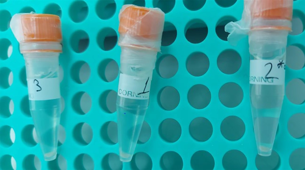
After the growth signal, the colonies were incubated in an oven at 37°C for 2 days. The colonies grown in the MGIT tube were then collected and homogenized thoroughly with a sterile glass bead by vortexing at 2400 rpm. To prepare a 3 - 4 mL Mcf-2 bacterial suspension, all colonies grown in four MGIT cultures were utilized. One milliliter of the Mcf-2 suspension prepared by this method was transferred to a 1.5 mL screw-capped centrifuge tube. The tube was sealed with parafilm and kept at 37°C for 3 days. A loopful of the suspension was then inoculated onto CBA plates to check for contamination. The pellet was obtained by centrifugation at 11,500 rpm for 30 minutes from the inactivated colonies that were incubated at 80°C for 20 minutes (Figure 1). Three pellets were prepared at different times for each method and stored at 4°C until DNA isolation.
3.4. Preparation of Pellets from Direct MGIT Colonies
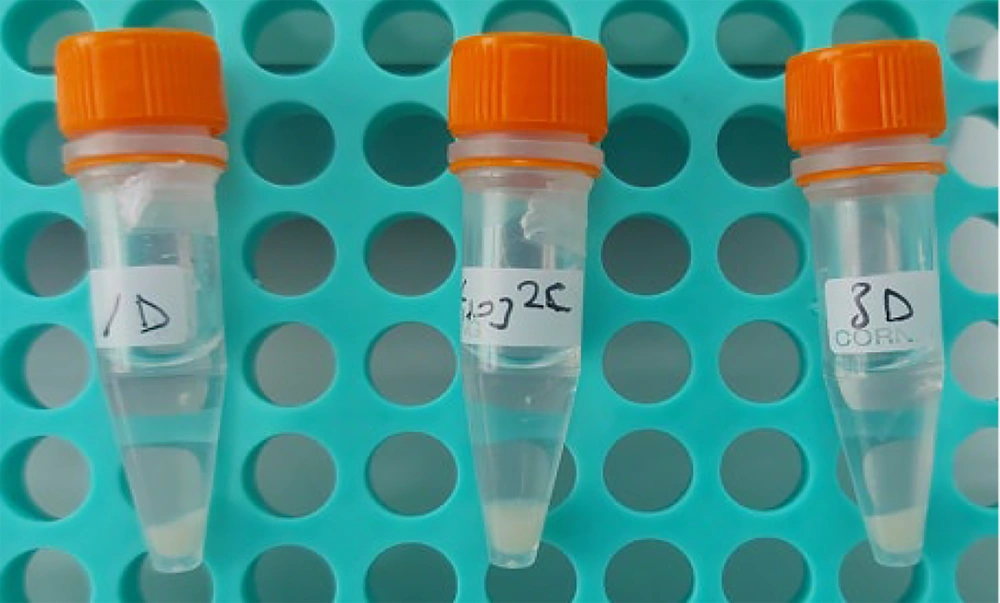
Following the growth signal, the MGIT tubes were incubated at 37°C for 3 - 4 days. A 10 µL sample of the liquid medium was inoculated onto CBA plates and inspected for contamination. For each DNA isolation, all colonies grown in two MGIT cultures were transferred into 1.5 mL screw-capped sterile centrifuge tubes, and the tubes were sealed with parafilm before being incubated at 80°C for 20 minutes. The pellets were obtained by centrifugation at 10,500 rpm for 5 minutes from the inactivated colonies (Figure 2). Three pellets were prepared from MGIT tubes cultured on different days for each isolation method. The pellets were distributed to the methods in numbered groups by a blind person who was unaware of the methods and groups to prevent bias.
3.5. DNA Extraction Methods
Genomic DNA was isolated using four different extraction methods: The CTAB method and three spin column methods: (i) the GeneJET Genomic DNA Purification Kit (GeneJET-Genomic DNA-PK) (Thermo Scientific, K0721); (ii) the Quick-DNA Fecal/Soil Microbe Kit (Quick-DNA-Fecal/Soil-MK) (ZymoResearch, D6010); and (iii) the Genematrix Tissue & Bacterial DNA Purification Kit (Genematrix-Tissue/Bacterial-DNA-PK) (EURx, E3551-0S). All final DNA preparations were made in a 30 µL volume. The CTAB method followed the protocol published by Jagatia and Cantillon with modifications (10). Changes were made to the duration of intervention with (i) Proteinase K (20 mg/mL) in a 65°C water bath, ranging from 30 minutes to 1 hour; (ii) CTAB-NaCl solution incubation time at 65°C in a water bath, also ranging from 30 minutes to 1 hour; and (iii) an extended incubation time at 80°C for 1 hour after forming a homogenate with 24:1 chloroform-isoamyl alcohol-ice-cold isopropanol.
Spin column DNA extraction methods were performed according to the manufacturer's instructions with modifications. For the GeneJET-Genomic DNA-PK, the lysis time was extended to 3 hours at 37°C after adding 200 µL of lysis buffer (20 mM Tris, mM EDTA, 1.2% Triton X-100, and 20 mg/mL lysozyme enzyme) to the pellet. The remaining steps, including washing and DNA elution, were performed as instructed. For the Quick-DNA-Fecal/Soil-MK, after adding 600 µL of bashing beads to the pellet, the solution was transferred to a bead lysis tube and vortexed for 40 minutes at 2 400 rpm. Following vortexing, the bashing bead tube was heated at 80°C for 20 minutes. Modifications were also made to the elution step of DNA by heating the elution buffer to 65°C for 30 minutes before addition, and elution was performed by adding 30 µL of elution buffer to the column and holding it for 2 minutes at room temperature. For the Genematrix-Tissue/Bacterial-DNA-PK, modifications included extending the lysis time with BL solution and Proteinase K to 2 hours and 30 minutes and 1 hour and 30 minutes, respectively. Additionally, the incubation temperature and duration with SOLT were adjusted to 70°C for 10 minutes. The second part of the procedure started with modifying the volume of SOLT by adding 300 µL instead of 200 µL.
Before commencing DNA extraction methods, one of the pellets from the Mcf-2 bacterial suspension and all pellets formed from the MGIT culture were mechanically crushed by mixing with a pipette tip for 2 - 3 minutes. Chemicals and solutions in the extraction protocols were mixed using a pipette tip or by hand inversion to prevent DNA breakage.
3.6. Assessment of Genomic DNA Quality
Concentration, purity, and integrity of DNA were determined as quality control parameters for NGS and WGS analysis. Concentration was measured using the Qubit High Sensitivity DNA kit via a fluorometric assay. DNA purity was assessed using a NanoDrop 2 000 UV-Vis Spectrophotometer by measuring absorbance ratios at 260/280 nm as an indicator of protein/RNA contamination (optimal range 1.8 - 2) and at 260/230 nm as an indicator of solvent/salt contamination (optimal range 2 - 2.2). Both values served as criteria for DNA quality assessment (11). DNA integrity was assessed for pellet DNA formed from colonies of the MGIT culture using a numeric value called the DNA integrity number (DIN), measured by the Agilent 2 200 Tapestation system.
4. Results
4.1. Concentrations of DNA
The concentrations of DNA extracted from pellets formed by the 1 mL Mcf-2 pellet and MGIT culture colonies were measured in triplicate for each method (Table 1). The average DNA quantity extracted from the 1 mL Mcf-2 pellet was 5.52 ng/µL with the CTAB method, 8.38 ng/µL with the GeneJET-Genomic DNA-PK, 19.7 ng/µL with the Quick-DNA-Fecal/Soil-MK, and 4.98 ng/µL with the Genematrix-Tissue/Bacterial-DNA-PK. Mechanical disruption of the pellet with pipette tips increased DNA quantity by approximately 2 ng/µL in the CTAB method, 5 ng/µL in the GeneJET-Genomic DNA-PK, and 16 ng/µL in the Quick-DNA-Fecal/Soil-MK, while it did not affect the quantity in the Genematrix-Tissue/Bacterial-DNA-PK. Using pellets from colonies obtained from two MGIT culture tubes increased DNA quantity for extraction methods, except for the Genematrix-Tissue/Bacterial-DNA-PK due to the higher bacterial load. Among these methods, the Quick-DNA-Fecal/Soil-MK provided the highest amount of DNA, with an average concentration of 85 ng/mL. Additionally, this commercial kit performed best in terms of DNA concentration extracted from the 1 mL Mcf-2 turbidity pellet.
Abbreviation: CTAB, cetyltrimethylammonium bromide.
a Mechanically homogenized by pipette tips before starting DNA extraction.
4.2. Purity of DNA
The highest purity of DNA (ranging from 1.6 to 1.9) in terms of protein contamination was obtained with the Quick-DNA-Fecal/Soil-MK. The absorbance value at 260/230 nm was generally low for all extraction methods and starting materials, except for the colonial pellet DNA from MGIT culture extracted by the Quick-DNA-Fecal/Soil-MK (Table 2).
| Extraction Methods | Pellet of 1 mL Mcf-2 suspensions (260/280 - 260/230) | Colonial pellet from the MGIT Culture (260/280 - 260/230) | ||||
|---|---|---|---|---|---|---|
| 1 | 2 | 3 | 1 | 2 | 3 | |
| CTAB | 1.3 - 0.81 | 1.1 - 0.95 | 1.2 - 0.62 | 1.4 - 0.70 | 1.4 - 0.65 | 1.50 - 1.14 |
| GeneJET-Genomic DNA-PK | 1.1 - 0.66 | 1.6 - 1.23 | 1.4 - 0.56 | 1.5 - 0.86 | 1.5 - 0.84 | - |
| Quick-DNA-Fecal/Soil-MK | 1.8 - 0.62 | 1.6 - 0.51 | 1.6 - 0.36 | 1.9 - 1.3 | 1.9 - 1.61 | 1.9 - 1.58 |
| Genematrix-Tissue/Bacterial-DNA-PK | 1.1 - 0.93 | 1.1 - 0.78 | 1.1 - 0.79 | 1.1 - 0.41 | 1.1 - 0.66 | 1.2 - 0.34 |
Abbreviation: CTAB, cetyltrimethylammonium bromide.
4.3. Integrity of DNA
DNA integrity was evaluated based on the DIN value, which indicates the degradation of DNA. The preferred DIN number indicating intact DNA for WGS is a high DIN value (≥ 7) (12). DNA integrity number scores for DNA extracted from MGIT culture colony pellets by the four extraction methods are shown in Table 3. The CTAB method preserved DNA integrity, yielding the highest DIN value of approximately 9.5 in two out of three replicates. The Quick-DNA-Fecal/Soil-MK provided acceptable, but not preferred, DNA quality with a DIN value of almost 6.8 in two out of three replicates. The other two spin column methods yielded DNA with approximately 8 DIN scores in at least two replicates.
| Extraction Methods | DIN Number | ||
|---|---|---|---|
| 1 | 2 | 3 | |
| CTAB | 9.7 | 0.1 | 9.4 |
| GeneJET-Genomic DNA-PK | 0.1 | 8.1 | 8.5 |
| Quick-DNA-Fecal/Soil-MK | 7.5 | 6.8 | 6.9 |
| Genematrix-Tissue/Bacterial-DNA-PK | 8 | 7.8 | 6.2 |
Abbreviations: CTAB, cetyltrimethylammonium bromide; DIN, DNA integrity number.
4.4. Time Requirement
The CTAB method had a long completion time of approximately 28 hours for each sample due to the 24-hour lysis time. DNA isolation with the Quick-DNA-Fecal/Soil-MK was the fastest method, taking approximately 2-3 hours for three samples of M. tuberculosis colonies. The time required for DNA extraction with other commercial kits was approximately 5 - 6 hours.
5. Discussion
Advancements in NGS technologies over the past two decades have significantly facilitated whole-genome analysis (13). In particular, WGS is increasingly preferred in molecular genetics for TB control strategies, as it offers rapid, reliable, and detailed data on parameters such as diagnosis and first and second-line drug resistance simultaneously (14). Ensuring high-quality, contaminant-free DNA is essential for accurate sequencing. Extracting sufficient genomic DNA, particularly for M. tuberculosis WGS, remains challenging because of its complex cell wall (7). The initial DNA amount is not a significant hurdle for Illumina WGS analysis, including library kits like Nextera XT, which require only 1 ng of input DNA (2, 5). However, long-read technologies like ONT, which have the capability to analyze complex genomic loci and large repetitive elements such as the M. tuberculosis genome, demand higher DNA quantities for WGS. Oxford Nanopore Technology library kits typically require at least 400 ng of DNA, necessitating a minimum initial DNA concentration of 40 ng/µL (2, 6).
DNA concentration is influenced by various factors, including sample type, growth phase, initial material amount, extraction method, chemicals, and incubation timing (5, 7). The CTAB method, commonly used for DNA isolation from plants or polysaccharide-rich bacteria, is also favored for high-yield DNA isolation in M. tuberculosis (7, 11, 15). Modifications to the CTAB protocol were made in this study to enhance DNA yield due to the compact MGIT colonies and low pellet quantity. Similarly, other methods were modified. Additionally, the efficiency of the extraction methods was assessed using different initial pellet quantities: A low pellet amount from 1 mL of Mcf-2 pellet and a high pellet amount from MGIT colonies equivalent to around 100 µL of liquid in the centrifuge tube. Among these methods, the Quick-DNA-Fecal/Soil-MK provided the highest DNA yield from both starting materials. Using the colonial pellet, a suitable DNA quantity with an average of 85 ng/µL for long-read WGS analysis was obtained. Modifications, including holding the bashing bead tube at 80°C for 20 minutes and preheating the elution buffer before elution, could be effective in achieving high DNA yields using this method.
DNA purity is also crucial for all NGS technologies, particularly for ONT-based long-read WGS, which relies on the direct passage of DNA through nanopores. Contaminants can obstruct nanopores, reducing flow cell longevity (16). The types of extraction methods, especially those like the CTAB method that include many chemicals and enzymes, significantly influence DNA purity. The Quick-DNA-Fecal/Soil-MK demonstrated higher purity in this study. Modifications such as increasing intervention times and adjusting chloroform isoamyl alcohol and washing steps may also enhance DNA purity.
DNA integrity is another critical factor for successful long-read WGS, as degradation can occur due to various factors, such as repeated freezing and thawing, aged culture materials, or mechanical handling (2, 5, 7). DNA integrity number scores are used to indicate DNA degradation, with lower scores reflecting poorer integrity (12, 14). To preserve DNA integrity, this study employed techniques to avoid pipetting during solution addition and used gentle mixing methods. The most unfavorable DIN score, almost 6.8 or 7, was obtained with the Quick-DNA-Fecal/Soil-MK method. This result may be attributed to the 40 minutes of vortexing of the bead-beating tube included in the method protocol. Modifications, such as decreasing the vortexing time, may be effective in obtaining higher integrity DNA.
Recent studies have examined DNA extraction methods for obtaining high-quality DNA suitable for long-read sequencing in M. tuberculosis and other mycobacteria (11, 15, 16). Bouso and Planet validated six different methods for nontuberculous mycobacteria (NTM), with method 5, which involved early bead-beating in SDS, phenol extraction, and isopropanol precipitation, yielding high molecular weight DNA (51.598 bp), purity (260/280: 1.893, 260/230: 1.947), and quantity (7.263 µg) for Oxford Nanopore sequencing without the need for additional clean-up steps (11).
Elton et al. compared spin-column and precipitation CTAB methods from MGIT liquid cultures and 7H11 agar colonies of M. tuberculosis. They found that DNA yields were higher in 7H11 cultures (resistant isolates: 966 ng, susceptible isolates: 1712 ng) than in MGIT cultures (resistant: 688 ng, susceptible: 1414 ng), with the CTAB method providing better DNA integrity, particularly in MGIT cultures (56,150 bp for MDR-TB, 54,776 bp for DS-TB) (15). Percy et al. optimized a spin-column protocol by testing bead-beating times from 15 to 120 seconds, with DNA concentrations increasing from 25 ng/µL to 45 ng/µL. They observed that DNA integrity declined after 45 - 60 seconds in M. abscessus, while it remained stable for M. tuberculosis up to 60 seconds but decreased at 120 seconds (17).
Short-duration and user-friendly DNA extraction methods are preferred for routine use. In this study, the Quick-DNA-Fecal/Soil-MK provided the highest quality and quantity of DNA in the shortest time, taking a maximum of 2 - 3 hours. However, the inability to conduct comprehensive long-read WGS analysis on DNA samples obtained using the Quick-DNA-Fecal/Soil-MK represents a limitation, hindering a thorough understanding of the complete impact of this method on sequencing reads and outcomes. Another significant limitation of the study is the small number of pellet groups evaluated in each repetition, which may restrict the generalizability of the results and affect statistical analysis.
5.1. Conclusions
Based on three repetitions of the Mcf-2 and colonial pellet extractions, it can be stated that the Quick-DNA Fecal/Soil-MK kit yielded the highest DNA quantity and purity but lower DNA integrity compared to other methods. The kit requires adjustments for optimal results. As a starting material, the pellet composed of colonies from two MGIT cultures, which is equivalent to approximately 100 µL or more in a 1.5 mL microcentrifuge tube, is suitable for long-read WGS with this kit. However, while this study provides guidance on selecting a suitable DNA extraction method for long-read sequencing of M. tuberculosis, a larger sample size would be necessary to generalize the findings and reach a definitive conclusion. For long-read sequencing of challenging bacteria like M. tuberculosis, DNA extraction protocols need to be optimized to balance DNA yield, fragment size, and purity, ensuring efficient sequencing and accurate drug resistance analysis.


