1. Background
Shigellosis is one of the main sources of complication in children with diarrhea in developing countries. The annual number of epidemic cases of Shigella throughout the world is estimated to be 164.7 million, of which 163.2 million occur in developing nations (1). Worldwide, the prevalence of deaths caused by the disease per year is estimated to be 1,100,000, and two-thirds of the patients are children under five years of age (2). Recently, it was estimated that 91 million people worldwide were affected by diarrhea caused by Shigella each year; in Asia, 410 000 children were affected by this disease, who were mostly malnourished and died (3). There are four species of Shigella; Shigella dysenteriae, S. flexneri, S. boydii and S. sonnei; also known as groups A, B, C and D, respectively (4). Shigellosis is one of the major causes of morbidity in children with diarrhea in Iran (5, 6) and recently the World Health Organization has emphasized the need to understand the disease charge and epidemiology in developing countries (7).
Identification, understanding of antibiotic resistance patterns and molecular characterization of genetic elements of Shigella species are important because of both its clinical and epidemiological implications (8). It has been shown that in order to study the epidemiology of this disease, genotyping methods are much better than phenotypic characterization. Analyses of Shigella isolates by various genotyping methods can give us helpful information for outbreak investigations (9, 10), disease transmission pathways (2), disease monitoring (11), and evolutionary studies (12). Several genotyping methods have been developed for Shigella strains, such as plasmid profiling, which was used in our previous study for subtyping of Shigella strains. However, plasmid profiling exhibits a lower level of discriminatory power than pulsed field gel electrophoresis (PFGE) (12-15). However, plasmid profiling is more useful than PFGE in some respects for investigation of the genetic relationships among Shigella strains circulating over a longer time range, and it is also useful for discrimination of certain strains that could not be distinguished by PFGE.
2. Objectives
To compensate for some drawbacks of plasmid profiling in molecular epidemiology of Shigella strains, we used the PFGE method as a complementary test in this study, as it has been proven to be a powerful tool for discriminating Shigella strains in many laboratories (15, 16). We analyzed 82 Shigella isolates recovered from children with diarrhea in Shiraz (Southern Iran) by IpaH and IpaBCD PCR-restriction fragment length polymorphism (RFLP). In parallel, PFGE patterns of total DNA of the S. sonnei isolates were determined to find the clonality among these strains. The data were then compared with the results obtained from our previous study using the plasmid profiling method.
3. Patients and Methods
3.1. Patients and Samples
A total of 82 clinical strains of Shigella spp. isolated from stool samples of 719 patients aged two months to 14 years, were characterized in this study. This study was conducted during 2011, on strains, from stool samples of sporadic diarrheic patients with positive occult blood (OB) test collected in Shiraz, during a period of six months, from April to October 2003. The strains were identified at the serogroup level and characterized using biochemical tests by standard procedures (17). These strains included S. sonnei (n = 61), S. flexneri (n = 16), 3 S. boydii (n = 3) and S. dysenteriae (n = 2).
3.2. PCR- Restriction Fragment Length Polymorphism Analysis
Detection of Ipa genes was performed by amplifying both IpaH and IpaBCD genes by PCR. The primer sequences were previously reported (18, 19) and obtained from TIB MOLBIOL Syntheselabor GmbH (Berlin, Germany). Other enzymes and chemicals were provided by Cinnagen Chemical Company (Tehran, Iran). For DNA extraction, Shigella colonies were suspended in sterile distilled water and disrupted by boiling for three minutes, and aliquots of the suspension were directly used for PCR assays. Amplification was performed in a thermal cycler (Eppendorf, Germany) according to the methods described by Aranda and Faruque (18, 19). Briefly, Thermal cycling parameters for the PCR assays consisted of denaturation at 94°C for two minutes, annealing of primers at 55°C for two minutes, and primer extension at 72°C for three minutes.
Amplification was performed for 35 cycles. Using an appropriate molecular size marker (100-bp DNA ladder, MBI, Fermentas, Lithuania), electrophoresis was performed in 1.5% agarose gel to ascertain expected sizes of the amplicons. PCR amplified IpaH fragments were digested with HinfI, HaeIII and IpaBCD fragments were digested with HinfI, and, pstI restriction enzymes for four hour at 37°C in the appropriate buffer recommended by the supplier (MBI, Fermentas, Lithuania). The digests were analyzed by electrophoresis on 2% agarose gel with 1x tris-acetate-EDTA buffer followed by ethidium bromide staining.
3.3. Pulsed Field Gel Electrophoresis (PFGE)
Genomic DNAs of 41 S. sonnei were restricted by XbaI and analyzed by PFGE according to the procedures explained by Mammina et al. with some modifications (20). These strains were selected based on our previous data about their plasmid and antibiotic sensitivity patterns. Indeed, they had the highest similarities in these two patterns. Thus, PFGE was used to allow greater discrimination between these two strains. Twenty-three and six patterns were revealed according to their plasmid profiles and drug sensitivity, respectively. Shigella isolates were grown in 3 ml brain hearth infusion (BHI) broth at 37°C overnight. The cells were pelleted, washed and resuspended in 1 mL of 75 mM Tris-HCl, 25 mM EDTA. The same volume of 1.2% agarose gel (pulsed-field grade; Bio-Rad, Hercules, CA, USA) was then added to the suspension, mixed completely and poured into the slots of a plastic mold (CHEF DRII; Bio-Rad). The agarose plugs which were obtained in the previous step were transferred into the lysis buffer (50 mM Tris-HCl, pH 7.2, 50 mM EDTA, 1.0% N-lauroylsarcosine and 0.1 mg/mL proteinase K) and cells were lysed for 16-20 hours at 55°C. Subsequently, five times washing for 30 minutes was applied to the agarose plugs using washing buffer (10 mM Tris-HCl, pH 8.0, 1 mM EDTA) while gently shaking and the gels were stored in the same buffer at 4°C.
A 1-mm slice of each agarose plug was equilibrated in 100 μL of restriction buffer for 30 minutes and digested at 37°C for 18 hours with 50 units of XbaI (MBI, Fermentas, Lithuania). After digestion, genomic DNA was separated in 0.9% agarose gel with a Gene NavigatorTM System (Amersham Biosciences, USA). The conditions for electrophoresis were 6 V/cm for 33 hours with the pulse time increasing from 5 to 40 seconds. Lambda ladder PFG marker (BioLabs, New England) was used as the molecular weight standard. Computer-generated PFGE banding patterns were performed by the Photo Capt Mw program.
3.4. Genetic Similarity Among Strains
Similarities among isolates on the basis of PFGE profiles were analyzed by the NTSYS-PC software (Numerical Taxonomy and Multivariate Analysis System ver. 2.02) for dendrogram construction. The matrix of similarity of coefficients was subjected to un-weighted pair-group method analysis (UPGMA) to generate dendrograms using the average linkage procedure.
4. Results
4.1. Prevalence of Shigella Virulence Genes and PCR-RFLP Analysis
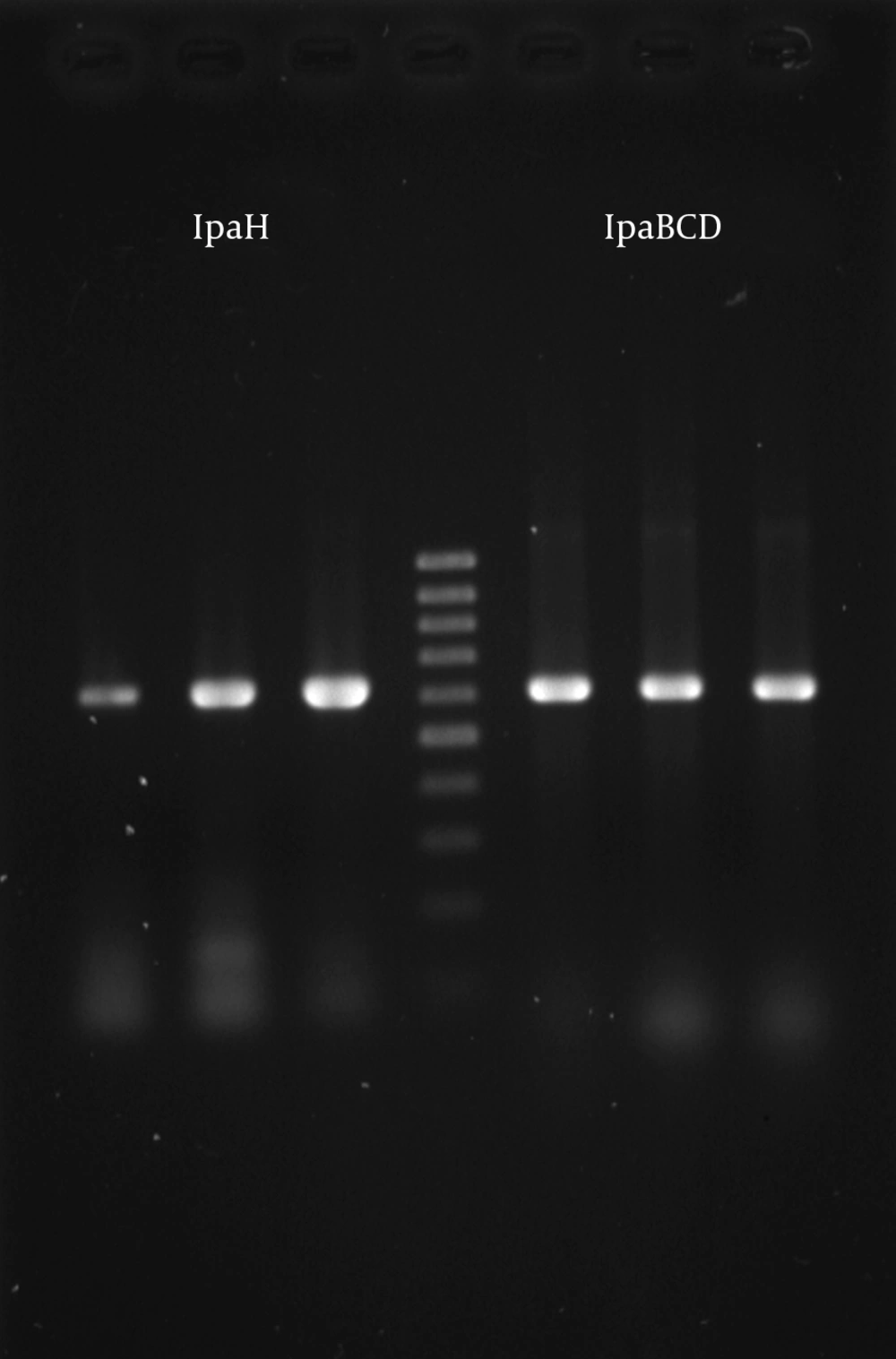
The results of DNA amplification by the PCR method based on primers used in this study showed the presence of a 619-bp fragment for the IpaH gene and a 612-bp fragment for the IpaBCD gene in DNA preparations obtained from all the isolates. However, in two strains of S. sonnei a larger band with molecular weight of 664-bp was detected for the IpaH gene. These data showed that all Shigella isolates carried invasive genes. Following digestion of the amplified fragment of IpaH gene, one homogeneous profile, consisting of bands with molecular sizes of 275, 215 and 129-bp with HinfI and 343, 182 and 94 with HaeIII enzyme, was revealed in all except for two S. sonnei strains with different IpaH bands (Figure 1).
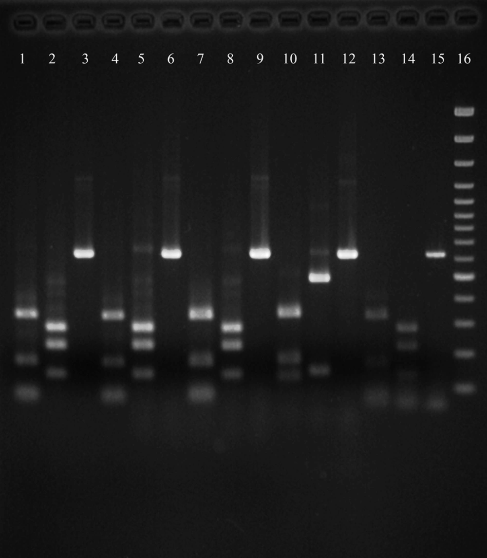
In these two strains, IpaH gene was broken to three bands of 343, 189, 139 and two bands of 505 and 159 base pairs in the presence of HinfI and HaeIII enzymes, respectively. The sequences of the fragments amplified from IpaH genes of these two strains are currently under investigation by the sequencing method in our lab. HinfI, HaeII and pstI digestion of the 612-bp IpaBCD fragment resulted in a strictly homogeneous profile for all Shigella serotypes and strains isolated from the patient IpaBCDs. We obtained three bands of 320, 170 and 122-bp with HinfI, two bands of 469 and 143-bp with HaeII, and two bands of 350 and 262-bp with pstI enzymes digestion, for all the strains.
Lane 1 and 2, IpaH gene product (619-bp) from representative strains; lane 3, IpaH gene product from S.flexneri ATCC 12022 as a positive control; lane 4, DNA molecular size marker (100-bp Ladder); lane 5, IpaBCD gene product (612) from S. flexneri ATCC 12022 as a positive control; lane 6 and 7, IpaBCD gene product form representative strains.
4.2. Pulsed Field Gel Electrophoresis of Total Genome DNA
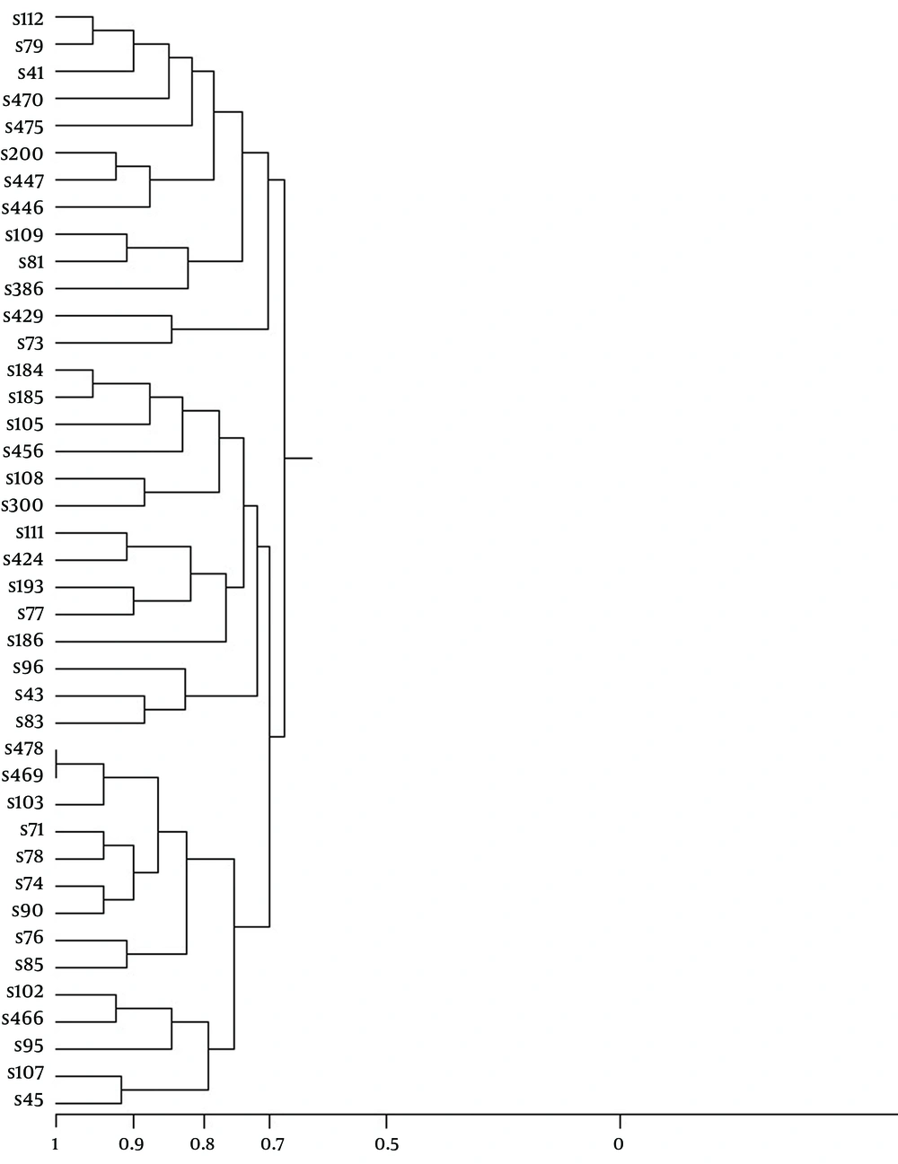
XbaI digestion produced maximum fragments (21 bands) in one and the minimum (10 bands) in another strain of S. sonnei. Analysis of total DNA fragments revealed an average of 15.5 bands (range from 10-21) in each isolate of all the strains. Some representative profiles and patterns from PFGE are shown in Figure 2. The majority of the strains (24.4%) showed 12 bands, and the patterns with 19, 21, 11 or 10 bands were the least frequent. The sizes of the fragments ranged from 4.279 to 630.5 kb in the strains under study. To define the patterns, we considered a PFGE pattern with one or more DNA bands, which were different from the others, as a unique PFGE pattern. In total, 40 restriction patterns were determined among all 41 S. sonnei. Sample patterns were divided into two groups based on the drawn dendrogram (Figure 3). Similarities ranged from 70% to 100%. Organisms were clustered into two groups; the first group included 26 samples, with the highest similarity and the second group included 15 isolates, with the lowest similarity.
Lanes 3, 6, 9, 12, 15, amplified IpaH gene in five representative S. sonnei strains; lanes 1, 4, 7, 10, 13, digested IpaH gene by HinfI; lanes 2, 5, 8, 11, 14, digested IpaH gene by HaeIII; lane 16, DNA molecular size marker (100-bp Ladder); lanes 10, 11, 12 show a S. sonnei strain with different PCR-RFLP patterns.
5. Discussion
The World Health Organization (WHO) Department of Vaccines and Biologicals (V and B) has increasingly emphasized on developing Shigella vaccines, and several candidate vaccines are being tested by clinical trials. However, understanding the disease burden and the molecular epidemiology of Shigella infection in developing countries is essential to establish a new generation of protective vaccines available in the relatively near future (7).
Our data showed that IpaH, a chromosomally located multicopy virulence gene, was stable and present in all strains under study (Figure 1). Therefore, PCR screening of environmental samples for the IpaH gene could be a better indicator of the possible presence of Shigella in comparison with screening for IpaBCD genes, which are present on plasmids. The major virulence genes in Shigella are encoded by accessory genetic elements such as plasmids or by chromosomal genes, which have a bacteriophage origin (21-24). This may indicate that Shigella may lose or acquire clusters of virulence genes as well as other accessory functions. Consequently, a transit between virulent and possible non-virulent forms, which are indistinguishable from normal gut flora, may happen (25-27). On the other hand, in parallel we used PCR-RFLP to analyze the virulence strains of Shigella according to the variability of their IpaH and IpaBCD genes.
The results showed that all Shigella serotypes and the strains isolated from the patients had one homogeneous profile indicating that these two virulence genes are strictly conserved (Figure 1). Although they may be suitable indicators of the possible presence of Shigella strains, it seems that they are not reliable enough for molecular epidemiological studies in comparison with plasmid profiling, because the conserved sequences are not suitable for these purposes. However, we found two S. sonnei strains with different IpaH bands (Figure 2). In these two strains IpaH gene was broken to three bands of 343, 189, 139 and two bands of 505 and 159 base pairs in the presence of HinfI and HaeIII enzymes, respectively. This is the first report of S. sonnei with a larger band of IpaH with molecular weight of 664-bp. The sequences of the fragments amplified from IpaH genes of these two strains are under investigation by sequencing methods at our lab.
In our previous study we found that S. sonnei is the major species causing shigellosis in Shiraz, Iran (6). As S. sonnei contains only one serovar, the development of a serological typing schema is difficult for this species. The typing systems, which are based on the phenotypic properties of the microorganism, have some disadvantages or limitations. (22, 28-31) More recently, sensitive and reproducible molecular markers, including those used in ribotyping (5, 8, 32), plasmid profiling, and pulsed-field gel electrophoresis (PFGE) (33, 34), have been applied with success, for S. sonnei and other microorganisms (31).
In our previous study we showed that a traditional method like antibiogram analysis was not a useful epidemiological marker, due to poor reproducibility, insufficient discriminatory power and also affected by physiological factors (6). Accordingly, we found that comparing plasmid profiles is more useful to assess possible relatedness of individual clinical isolates for epidemiological studies. Based on such data, plasmid profiles distinguished more strains than antimicrobial susceptibility patterns (6). The data indicated that shigellosis in patients seen in Shiraz, is caused by a large number of clones which could not be differentiated by antimicrobial susceptibility patterns. This was similar to the results of studies done in Bangladesh (23). However, it was in contrary with the data obtained in the developed world, where one or a few clones account for Shigellosis in a community (35). However, one disadvantage of plasmid profiling is that during the preparation of the samples for plasmid extraction some plasmids could be lost due to the fact that large plasmids (> 15-kb) are usually unstable and consequently, only small plasmids below the band of chromosomal DNA on the gel are suitable for analysis. Moreover, plasmid low discriminatory power is further increased by restriction endonuclease digestion (21, 30). Therefore, in this study we used PFGE to confirm the relationship among S. sonnei strains during outbreaks according to their total DNA contents.
As shown in Figure 4, we observed 40 pulsotype patterns based on the PFGE for 41 S. sonnei strains, while in our previous study plasmid profiling and antibiotic susceptibility test defined only 23 and six patterns, respectively. Therefore, PFGE was more reliable for genotyping of the strains under study. Liu et al. found seven distinct macrorestriction patterns among the 20 clinical isolates of S. sonnei using PFGE and ERIC-PCR methods, while their rRNA gene (rDNA) restriction fragment length polymorphisms with EcoRI or HincII gave the same single profile (31). They suggested that PFGE and ERIC-PCR have the highest discriminatory power for the differentiation of strains of S. sonnei in comparison with plasmid profile analysis, REAP, and ribotyping (31). However, limited diversity in the strains of Shigella spp. has been reported previously in Ireland (36), India (37) and Japan (38).
Clustering of our 41 isolates by XbaI PFGE patterns revealed 11 clusters at a genetic distance of ≥ 90% within the clusters consisting of 26 isolates. The lowest similarity among the strains was defined as a genetic distance of 70% for 27 strains (Figure 3). These data showed that S. sonnei clinical isolates under study could have a clonal relationship. The best description for such similarities is that the isolates representing the outbreak strains could be the recent progeny of a single (or common) precursor. On the other hand the high diversity of PFGE patterns (40 patterns in 41 strains) observed in this study is not surprising because numerous samples were collected from different patients in the community.
In the present study, 10-21 bands with the molecular weight of 4.279-630.5kb were observed in the PFGE profiles of the strains. Liu et al. reported 20 fragments; their sizes ranged from 32.4 to 582 kb, in molecular typing of Taiwan S. sonnei isolates (31). These different results indicate the high diversity of S. sonnei strains in different geographic areas. Random genetic events including point mutations, insertion and deletion of DNA can alter PFGE profiles. These events are evident by the presence or absence of the determined bands (39, 40). However, technical difficulties of laboratory methods can be considered as another reason for such observed discrepancies. They also depend on other factors such as individual person, laboratory set up and equipment, reagents, interruptions or distractions and unknown reasons.
To our knowledge, this is the first report that has applied the PFGE genotyping method to study the molecular epidemiology of S. sonnei infection in Shiraz, Iran. This report revealed the reliability of PFGE for molecular epidemiology of Shigella strains isolated from the population of children and that the strains under study could be epidemically related. It seems that an alternative subtyping method is needed for the study of clonality and global transmission of S. sonnei. However, due to high diversity of the PFGE patterns we could not find a correlation between these patterns and antibiogram profiles. Consequently, obtaining additional information, such as the use of supplementary epidemiological analysis or a second strain typing method is recommended. Here, we also reported for the first time, two strains of S. sonnei with a different PCR-RFLP pattern for IpaH gene.


