1. Background
Toxoplasma gondii and Neospora spp. are obligate, intracellular, coccidian protozoan parasites belonging to the Apicomplexa phylum and are closely related tissue-dwelling protozoan parasites with similarity in morphological and biological characteristics, infecting almost all warm-blooded animals (1, 2). Antibodies of these parasites have been found in the sera of many species of domestic and wild animals worldwide (3-6). Both parasites have indirect life cycles. Definitive hosts of T. gondii are cats and other felids (7). Mainly dogs and perhaps other canids are the definitive hosts of N. caninum, but the definitive host of N. hughesi is still unknown (8, 9). These parasites have a wide range of intermediate hosts including sheep, goat, cattle, horse, bison, camel, pig, and deer (10-13). However, horse can serve as the potential intermediate host of these parasites and can be infected following ingestion of sporulated oocysts via contaminated feed or water, as well as via vertical transmission from mother to fetus through the placenta, which is an alternative route (14, 15). Both parasites are related to Coccidia, which is reported to cause encephalitis in horses (7). Toxoplasmosis is one of the most common parasitic zoonoses worldwide (16). Antibodies against T. gondii were detected in horses in the Czech Republic (17), Turkey (18, 19), Egypt (20), Sweden (21), and Switzerland (22). Toxoplasmosis is a common infection that rarely evolves to clinical disease (23), but may cause fever, ataxia, retinal degeneration, abortion, and severe encephalomyelitis (24).
N. caninum and N. hughesi were first described in dogs in Norway in 1984 in the brain and spinal cord of an adult horse in California, USA, respectively (25, 26). In 1990, the first serological evidence of N. caninum infection in a horse was described (27). Antibodies against Neospora sp. in equine populations have been reported in many parts of the world such as the United States (28), South Korea (29) Iran (30), and Brazil (31). In Europe, antibodies against N. caninum have been reported in horses in France (32), Italy (33), Sweden (34), Turkey (35) and the Czech Republic (36). Horses can be infected by N. caninum and N. hughesi. Although exposure to Neospora spp. in horses seems to be common, clinical diseases are rare. The clinical disease is characterized by abortion, neonatal diseases, and neurological findings of severe encephalomyelitis (14). N. caninum and N. hughesi are related to abortions and neurological diseases in horses (31).
2. Objectives
Few data has been reported about the prevalence of T. gondii and N. caninum in Arab horses in Iran. The current research was conducted to investigate the seroprevalence of T. gondii and Neospora spp. infections among Arab horses, the most popular horse species in Khuzestan province, southwest of Iran, using modified agglutination test (MAT).
3. Materials and Methods
3.1. Study Area
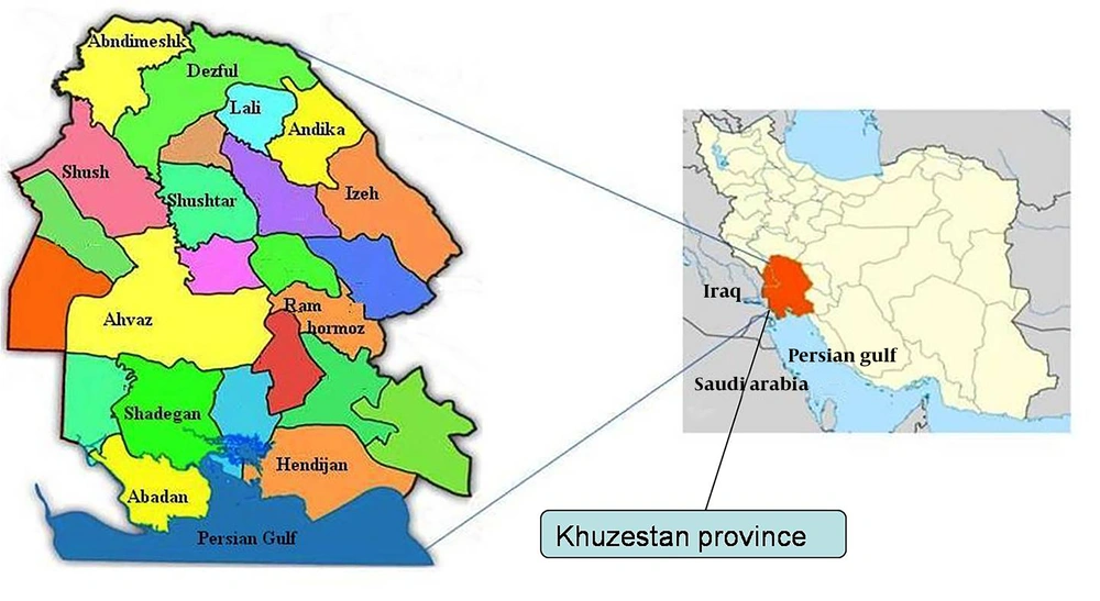
The study area, Khuzestan province, covers approximately 65000 km2 and is located in southwest of Iran (31º 3´ N, 48º 7´ E) (Figure 1). The climate of this area is generally hot and occasionally humid. Summertime temperatures routinely exceed 50°C. Khuzestan province is known to master the hottest temperatures on record for a populated city anywhere in the world.
3.2. Collection of Sera
From October 2009 to March 2011, blood samples (5 to 10 mL) were collected from jugular veins of 235 Arab horses of different ages and sexes. The samples were from 12 cities (Table 1) of Khuzestan province, southwest of Iran (Figure 1). According to the information taken from owners and the physical examinations performed by veterinarians on age and gender of horses, they were categorized into three age groups (≤ 2, 2-10, and ≥ 10 years old). The blood samples were centrifuged 10 minutes at 3000 rpm and serum was removed and stored at -20°C until used.
| City | Samples, No. | Males, No. | Females, No. | T. gondii (+) | Neospora spp. (+) | ||
|---|---|---|---|---|---|---|---|
| Male | Female | Male | Female | ||||
| Abadan | 31 | 16 | 15 | 4 | 7 | 2 | 2 |
| Ahvaz | 58 | 27 | 31 | 21 | 12 | 7 | 11 |
| Ramhormoz | 15 | 9 | 6 | 3 | 5 | 1 | 1 |
| Shush | 18 | 9 | 9 | 4 | 2 | 1 | 4 |
| Shushtar | 15 | 10 | 5 | 4 | 5 | 2 | 2 |
| Dezfol | 24 | 13 | 11 | 6 | 4 | 1 | 2 |
| Lali | 10 | 6 | 4 | 2 | 1 | - | 1 |
| Andika | 10 | 3 | 7 | 3 | 1 | 1 | 1 |
| Andimeshk | 15 | 7 | 8 | 3 | 3 | - | - |
| Hendijan | 10 | 6 | 4 | 3 | 4 | 1 | - |
| Izeh | 13 | 8 | 5 | 5 | 3 | 2 | - |
| Schadegan | 16 | 8 | 8 | 5 | 4 | 2 | 3 |
| Total | 235 | 122 | 113 | 63 | 51 | 20 | 27 |
The Number of Samples From Different Sites in Khuzestan Province and Their Serological Status Against Toxoplasma gondii and Neospora caninum
3.3. Parasites
The T. gondii and N. caninum antigens used in MAT and N-MAT were prepared from the RH strain of T. gondii and the NC-1 strain of N. caninum tachyzoites, harvested from cells grown in mice in the Pasteur Institute of Tehran and the Razi Institute of Shiraz, Iran, respectively.
3.4. Toxoplasma gondii Serology
T. gondii antibodies were investigated using modified agglutination test, as described by Desmonts and Remington (37) and Dubey and Desmonts (38). The sera were diluted two folds (1:20 to 1:320) with phosphate buffered saline containing 0.2 M 2-mercaptoethanol and 50 µL of each dilution was put in a well of 96 U-bottom ELISA plates. Thereafter, 50 µL of the whole formalin-preserved T. gondii tachyzoites were added to each serum dilution. The wells were then mixed thoroughly by pipetting up and down several times, covered, and then incubated at 37°C overnight. The test was considered positive when a layer of agglutinated parasites was formed in wells at dilutions of 1:20 or higher. Positive and negative controls were included in each test.
3.5. Neospora spp. Serology
The N-MAT was used for detection of antibodies to Neospora spp. in horses, described by Romand et al. (39), which is very similar to MAT which is used for detection of toxoplasmosis in sera, except that Neospora tachyzoites were used as antigens. Briefly, the sera were diluted with phosphate buffer saline (pH = 7.2) containing 2-Mercaptoethanol (2-ME) and screened with 1:40 and 1:80 dilutions. Positive sera at 1:80 dilutions were subsequently submitted to serial dilutions (1:160 to 1:640) (29).
3.6. Statistical Analysis
Differences in antibody prevalence of horses infected with T. gondii and Neospora spp. with regard to gender, age and geographical regions were analyzed using chi-square test and were calculated with SPSS (version 16.0). P value < 0.05 was considered statistically significant.
4. Results
All the equines remained clinically normal and there was no evidence of abortion. The serological results are summarized in Tables 1, 2 and 3. The horses’ sera were collected from different cities of Khuzestan province (Table 1). Statistical analysis showed that some factors like age affected the prevalence of T. gondii infection. In this study, we divided all the horses into three groups based on age, to 53 (≤ 2 years old), 121 (2-10 years old), and 61 (≥ 10 years old) horses. The T. gondii seroprevalence in adult horses (≥ 10 years old) was significantly higher than other age groups (63%: 38/61). The results showed that T. gondii seroprevalence in horses increased with age. Toxoplasma antibody with MAT in horses in the age group of 2-10 was 49% (59/114) and was 33% in the age group of ≤ 2 (17/53). However, we could not find any correlation regarding antibodies and age between the three groups. We found 31 (13/2%) samples that showed antibodies for both of these parasites in at least the 1:20 titer.
| Titers | |||||
|---|---|---|---|---|---|
| 1:20 | 1:40 | 1:80 | 1: 160 | 1:320 | |
| Male | 51/122 (42) | 7/122 (5.8) | 2/122 (1.7) | 1/122 (0.8) | 2/122 (1.7) |
| Female | 33/113 (30) | 12/113 (10.7) | 2/113 (1.8) | 3/113 (2.7) | 1/113 (0.9) |
| Total | 84/235 (35.8) | 19/235 (8.1) | 4/235 (1.7) | 4/235 (1.7) | 3/235 (1.3) |
Modified Agglutination Test Titers for T. gondii in Sera From 235 Arab Horses of Khuzestan Province a
| Titers | |||
|---|---|---|---|
| 1:40 | 1:80 | 1:160 | |
| Male | 17/122 (14) | 2/122 (1.7) | 1/122 (0.8) |
| Female | 22/113 (20) | 3/113 (2.7) | 2/113 (1.8) |
| Total | 39/235 (16.6) | 5/235 (2.2) | 3/235 (1.3) |
4.1. Toxoplasma gondii
Antibodies to T. gondii were detected in 114 (48.5%) Arab horses. The majority of the positive sera had titers ≤ 1:20 (Table 2). The seroprevalence in horses varied from 1.8% to 28% between regions. No differences in seroprevalence were found between male and female horses. Antibodies to T. gondii were found in 84 (35.8%) of 235 horses with titers of 1:20, 19 (8.1%) with titers 1:40, 4 (1.7%) with titers of 1:80, 4 (1.7%) with titers of 1:160, and 3 (1.3%) with titers of 1:320. MAT was repeated twice in this step.
4.2. Neospora spp.
Antibodies to Neospora spp. were detected in 47 (20%) among Arab horses. Sera were examined by the developed Neospora agglutination test (NAT) as described by Romand et al. (39) NAT is similar to MAT for T. gondii except that tachyzoites of Neospora spp. are used instead T. gondii tachyzoites. Antibody titers to Neospora spp. in examined horses are shown in Table 3. At 1:40 dilution, the prevalence of antibodies to Neospora spp. was 14% for males and 20% for females, while at 1:80 and 1:160 dilutions prevalence was 1.7% and 0.8% for males and 2.7% and 1.8% for females respectively.
5. Discussion
This is the first report of T. gondii and Neospora spp. infections in Arab horses in southwest of Iran. Results of this investigation indicated that 114 (48.6%) of 235 Arab horses had been exposed to T. gondii and antibodies to Neospora spp. were detected in 47 (20.5%) of them. MAT is considered as one of the highly specific and sensitive tests for detection of T. gondii and Neospora spp. in animals, especially in horses (23, 40). Although antibodies to T. gondii have been reported in horses by MAT, ELISA or IFAT from many countries, there is no clear evidence that this protozoan causes clinical diseases in horses (23).We did not find any significant differences between the seroprevalence of T. gondii and Neospora spp. and horses gender. Dubey et al. the first time detected tachyzoites of N. caninum in a fetus lung (27) and N. hughesi tachyzoites were isolated from an adult horse in the US (8). Horizontal transmission of Neospora spp. in horses appears to be a major mode of transmission (41). Because of serological cross reactivity between N. caninum and N. hughesi, we could not confirm which species of Neospora infected Arab horses using MAT. The seroprevalence of T. gondii in older horses indicated more prevalence rates, which was in agreement with Boughattas’s study in Tunisia (42). In a recent Iranian survey in northeast of Iran on Neospora in horses, they detected Neospora antibodies in 30% of horses using N-MAT (43). The serological results of this study indicated that 47 of 235 (20%) horses were exposed to Neospora spp. which was comparable to those reported from the northwest, north, south and west of Iran as 28%, 30%, 32% and 40.8%, respectively (30, 43-45). Our results were comparable to those reported from the US, France and Italy (27, 33, 46). According to hajialilo’s study in Qazvin, Iran, 71.2% of sport horses were seropositive for T. gondii (47).
The prevalence of Neospora spp. antibodies in cattle in the southeast of Iran was 12.6% (48) and in water buffaloes in the southwest of Iran it was 37% (49). This results imply that exposure to this parasite is common in south of Iran. Dogs could be one of the main definitive host in this region. According to the study of Hosseininejad et al. the prevalence of N. caninum in dogs from west and central parts of Iran was 26.8% (50). Another study in west of Iran (Hamedan province) indicated that the prevalence of N caninum in dogs was 27% (51). Further research on the epidemiological and molecular evidence for identification of Neospora strains is required. This study was the first investigation on T. gondii and Neospora spp. in Arab horses of Khuzestan province, Iran, and indicates that there is exposure to these parasites in this region. Therefore, designing control strategies including restriction of the existent stray cats and dogs in farm hoses as well as follow up and treatment of the owned dogs is recommended and further studies such as molecular and sequencing methods are needed to distinguish N. caninum from N. hughesi.
