1. Background
Cholera is an important infectious diarrheal disease and sometimes causes death in humans. This disease is both epidemic and endemic in different regions of the world, especially in the countries that lack proper sanitary management. Pandemics that are caused by this bacterium have severely affected many countries in multiple continents over a span of many years (1). Vibrio cholerae is a natural inhabitant of aquatic environments and the toxigenic strains are causative agents of Cholera disease. These bacteria are rod-like, Gram-negative, facultative anaerobe and motile (2, 3).
Pathogenicity of Vibrio spp. is depended on the expression of some virulence factors such as Cholerae Toxin (CT) that is facilitated by the Toxin-Coregulated Pilus (TCP). These two genes are encoded by the ctx and tcp genes, respectively (4). Another virulence factor is the Outer-Membrane Protein W (OmpW) that is involved in stimulating the immune response via induction of protective immunity. It also plays an important role in bacterial pathogenesis by increasing the adaptability of pathogenic strains and is encoded by OmpW gene (5).
Different studies showed that toxigenic V. cholerae that caused epidemic and pandemic potential forms of cholerae diarrhea are O1 or O139 sero-groups. V. cholerae O1 and O139 serogroups are positive for ctx, tcp and OmpW genes (6).
Traditionally, identification of Vibrio spp. is consisted as isolation on the selective blood agar medium, followed by biochemical and serological testing (7). However, Polymerase Chain Reaction (PCR) is a rapid and versatile in vitro method for identification of Vibrio spp. and it amplifies defined target DNA sequences that are present within a source of DNA. Also, this method is designed to permit selective amplification of a specific targeted DNA sequence within a heterogeneous collection of DNA sequences (8). Enterobacterial Repetitive Intergenic Consensus sequence (ERIC)-PCR is a method for interspecies profiling of several bacteria such as Bacillus anthracis and B. cereus, Enterobacter sakazakii, Lactobacillus, Listeria monocytogenes, Salmonella enteritidis and it provides discriminative DNA patterns for these bacteria (9). Also, Repetitive Extragenic Palindromic (REP)-PCR is a technique that has been used to study epidemiological relationships among V. cholerae isolates and DNA fingerprints production. It is used to discriminate among bacterial species or strains of the same species. This technique includes the application of oligonucleotide primers based on families of short and highly conserved extragenic repetitive sequences (9, 10).
Different studies showed molecular characterization of V. cholerae in different samples; Waturangi et al. (9) in Indonesia, with examining 75 isolates of V. cholerae from ice samples and using the REP- and ERIC-PCR methods showed that fingerprinting profiles of V. cholerae isolated from ice samples were very diverse. In another study, Alishahi et al. (11) in Iran, by using multiplex PCR on 72 V. cholerae samples of stool showed that all of them carried toxR and ompU genes, but ctxA gene was detected in only 61 isolates. Also, Naha et al. (12) in India, in 180 diarrheal stool samples detected 247 V. cholerae O1 and by using PCR detected rstR, rtxC and tcpA genes (12).
2. Objectives
V. cholerae O1 and O139 are causative agents of cholerae epidemics and endemics. Therefore, molecular detection and identification of V. cholerae strains are important. The REP and ERIC-PCR are fast, simple and valuable methods for molecular typing, genomic fingerprinting and proving of relationships within and among V. cholerae strains, which can be used in epidemiological studies of this microorganism infections (13, 14). Given the importance of the items listed above, this study aimed to describe molecular characterization of isolated V. cholerae from clinical samples by REP and ERIC-PCR methods in Kurdistan Province, Iran.
3. Materials and Methods
3.1. Samples
Forty-eight clinical specimens that suspected for cholerae were considered (under the health center, Kurdistan Province, Iran). Stool samples were collected from these clinical specimens in different cities of Kurdistan Province, Iran, summer 2013. Samples were collected using sterile rectal swabs.
3.2. Bacterial Cultures
Collected samples were transferred to Cary-Blair transport medium (Merck, Germany). Alkaline peptone water was used for enrichment (Merck, Germany) and then bacteria were isolated on Thiosulphate Citrate Bile Salts Sucrose (TCBS) agar plates (Merck, Germany).
3.3. Biochemical and Agglutination Characterization
All bacterial isolates were screened by the biochemical test including oxidase reaction, motility status on Sulfur-Indole-Motility (SIM) agar (Merck, Germany), Triple Sugar Iron (TSI) agar (Merck, Germany) for sucrose, mannose and arabinose fermentation tests, Kligler Iron Agar (KIA) (Merck, Germany) for glucose fermentation, lactose fermentation, sulfur reduction and Methyl Red Voges-Proskauer test (MRVP) (Merck, Germany). Agglutination test in the presence of polyvalent O1 and O139 antisera was used for confirmation of V. cholerae strains (Mast Diagnostics Ltd., Bootle, Mersey side, UK). Positive samples (O1 and O139) were placed in a Luria-Bertani (LB) broth (Merck, Germany) with 30% glycerol at -80°C for other tests (11).
3.4. DNA Extraction
For DNA extraction, screened isolates were cultured in a LB broth for 24 hours at 37°C; DNA genome of each sample was purified using a DNA extraction kit according to manufacturer’s instructions. Afterwards, 500 μl Thioglycollate broth (Merck, Germany) and 2 × 10⁹ (10 - 20 mg) bacterial cells were added to 2ml tubes, then they were vortexed. In other step, tubes were centrifuged at 4.000 rpm for 5 minutes. Then 100 μl of prelysis buffer and 10 μl Ribotinase were added to the tubes and they were incubated at 55°C for 30 minutes. Then 100 μl of each samples were added to a sterile 1.5 or 2 polypropylene tubes. After this stage, 400 μl of lysis buffer was added to tubes and they was vortexed at max speed for 20 seconds, then 300 μl precipitation solutions was added to them and they vortexed at max speed for 5 seconds. Solution was transferred to a spin column with a collection tube. Tubes were centrifuged at 12.100 × g, 13.000 rpm for 1 minute. Spin column was placed in a new collection tube and 400 μl wash buffer 1 was added to tubes. Then they were centrifuged at 12.100 × g, 13.000 rpm for 1 minute. After centrifuge, spin column was placed in a new collection tube and wash buffer 2 was added to it and it was centrifuged at 12.100 × g, 13.000 rpm for 1 minute (wash buffer 2 stage was repeated). Column was carefully transferred to a new 1.5 ml tube. Afterwards, 50 μl of 65°C preheated elution buffer was poured in the center of column and then it was incubated for 3 - 5 minutes at 65°C. This solution was centrifuged at 12.100 × g, 13.000 rpm for 1 minute (According to the manufacturer’s instructions, (CinnaGene, Iran).
3.5. Polymerase Chain Reaction Assay
Uniplex PCR assays were performed for tcpA, ctxA and OmpW genes in a reaction volume of 26 μl; 26 μl of the reaction mixture contained the following reagents:
Mastermix: 12 μl, MgCl2: 25 μl, Distilled Water (DWs): 8.75 μl, primer (pr) Reverse (R): 1 μl, primer Forward (F): 1 μl and 3 μl DNA. The cycling profile for ctxA and OmpW was as follows: initial denaturation was at 94°C for 5 minutes, and 35 cycles of denaturation at 94°C for 1 minute, annealing at 59°C for 1 minute, extension at 72°C for 1 minute and final extension at 72°C for 16 minutes. The cycling profile for tcpA was as follows: initial denaturation at 94°C for 5 minutes, 35 cycles of denaturation at 94°C for 1 minute, annealing at 61°C for 1 minute, extension at 72°C for 1 minute and final extension at 72°C for 16 minutes. The PCR products were electrophoresed at100 v for approximately 1 hour on a 1.5% agarose gel in 0.5- TAE. The PCR primer set that was used: pr ctxA-R: (5' - CAAGCACCCCAAAATGAACT-3'), pr ctxA-F: (5' - TTGTTAGGCACGATGATGGA-3'), pr tcpA-R: (5' - GACTAAGGCTGCGCAAAATC-3'), pr tcpA-F: (5' - CCCCTACGCTTGTAACCAAA-3'), pr ompW-R: (5' - GCATCTGCACCTGCTTTGTA-3') and pr OmpW-F: (5' – ACTTGACGACTCATGGGGAC-3') (Cinna Clon, Iran) (11).
3.6. Enterobacterial Repetitive Intergenic Consensus Polymerase Chain Reaction Assay
The amplification protocols that were employed for ERIC-PCR was as follows: 1 cycle of 95°C for 5 minutes, 35 cycles of 92°C for 45 seconds, 52°C for 1 minute and 70°C for 10 minutes with a final extension step of 70°C for 10 minutes. The PCR products were electrophoresed at 100V for approximately 1 hour on a 1.5% agarose gel (Merck, Germany) in 0.5-TAE (Tris-acetate- EDTA) (Merck, Germany). The PCR primer set that was used as follows: pr R: (5'-ATGTAAGCTCCTGGGGATTCAC-3') and pr F: (5' - AAGTAAGTGACTGGGGTGAGCC-3') (Cinna Clon, Iran) (9).
3.7. Repetitive Extragenic Palindromic Polymerase Chain Reaction Assay
The amplification protocols that were employed for REP-PCR was as follows: 1 cycle of 94°C for 5 minutes, 35 cycles of 94°C for 1 minute, 61°C for 1 minute and 72°C for 1 minute with a final extension step of 72°C for 16 minutes. Then PCR products were electrophoresed at100 v for approximately 1 hour on a 1.5% agarose gel in 0.5-TAE. The PCR primer set used: pr R: (5' – IIIICGICATCIGGC-3') and pr F: (5' - ICGICTTATCIGGCCTA-3') (Cinna Clon, Iran) (9).
3.8. Composed Matrix of 0 and 1
In the using of molecular markers, schemas are bands that are produced with a primer. After electrophoresis, the presence or absence of bands were determined with the numbers 0 and 1 for the isolates, respectively. For data analysis, a 0 and 1 similarity matrix was drawn. Detection of bands was performed by manual editing. After the similarity matrix formation, data were analyzed using the software NTSYS-pc ver 2. 02 and cluster analysis was performed (14).
4. Results
4.1. Biochemical Tests and Bacterial Culture Results
Biochemical tests and bacterial culture results showed that of 48 clinical specimen samples that suspected for cholera, 39 (81.2%) samples were positive for V. cholerae.
4.2. Polymerase Chain Reaction Assay Results
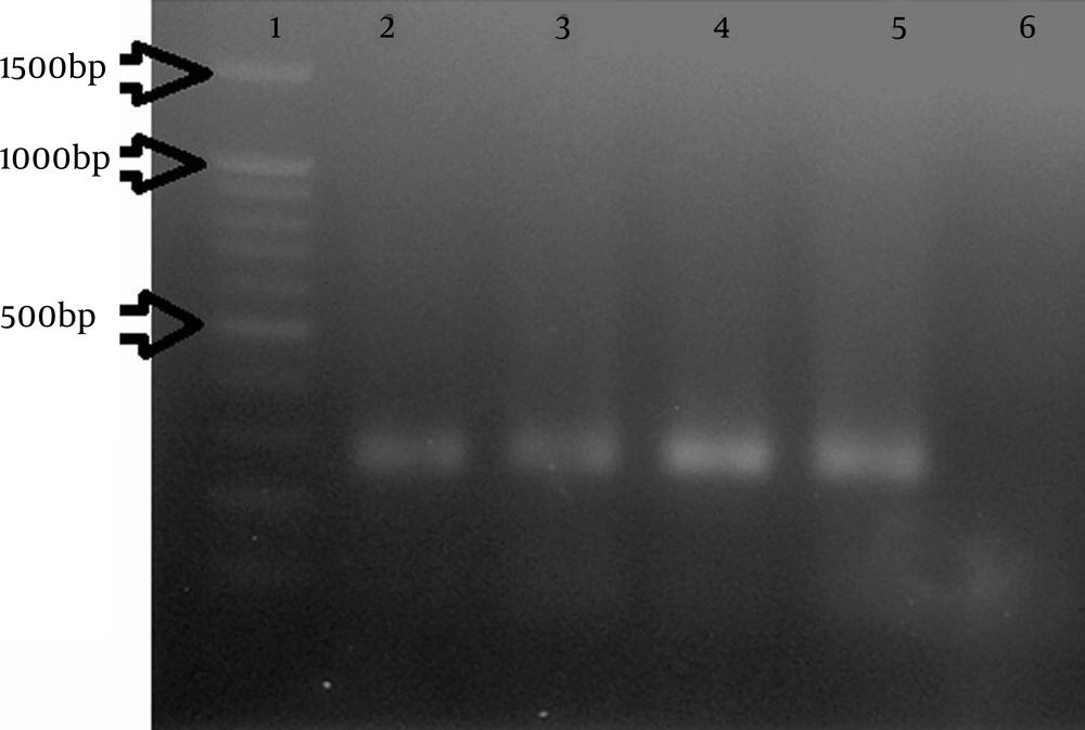
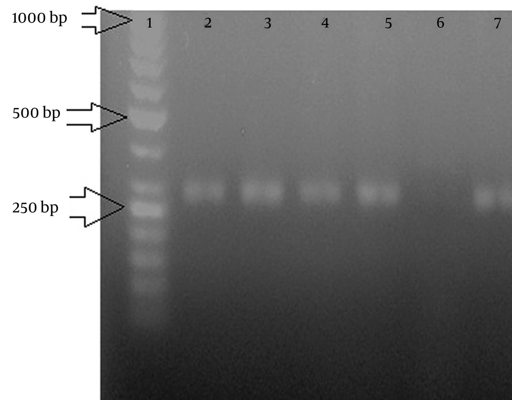
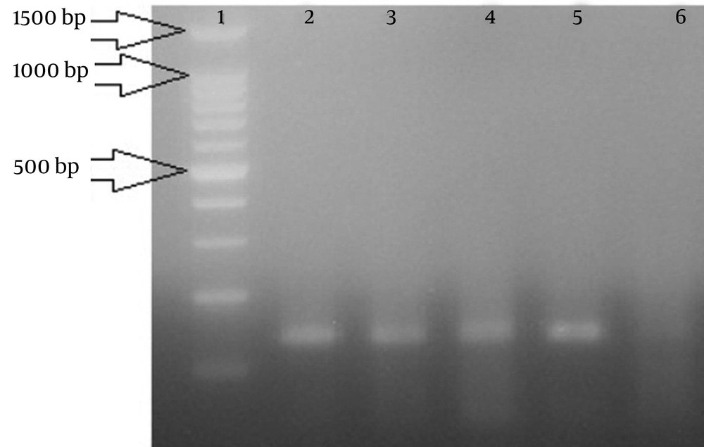
The PCR assay results showed that of 39 V. cholerae positive samples, ctxA was present in 35 (89.7%) (Figure 1), tcpA was present in 34 (87.1%) (Figure 2) and OmpW was present in 37 (94.8%) samples (Figure 3). These results showed specificity for all tested V. cholerae strains. The nucleotide sequence of the tcpA, ctxA and OmpW genes had revealed an amplicon of 287-252 and 132 Base Pair (bp).
4.3. Repetitive Extragenic Palindromic Polymerase Chain Reaction Results With Enterobacterial Repetitive Intergenic Consensus Primer
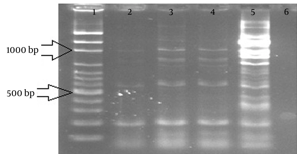
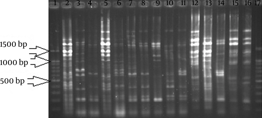
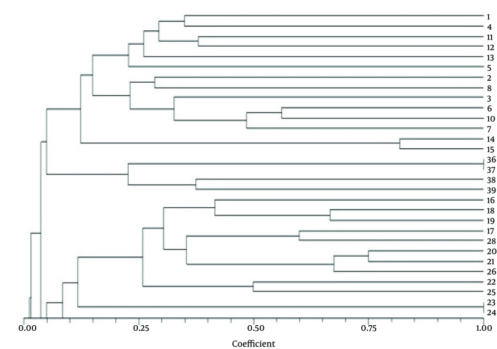
The results achieved by REP-PCR with the ERIC primer showed that strains were divided into 10 groups. Similarity coefficient between these groups was 1% to 51%. Most strains were placed in group 4, which had 30% similarity coefficient (Figures 4 and 6).
4.4. Repetitive Extragenic Palindromic Polymerase Chain Reaction Results With the Repetitive Extragenic Palindromic Primer
The results that obtained from the REP-PCR method with the REP primer showed that strains were divided into 13 groups and similarity coefficient between these groups was 1% to 50%. Maximum strains were placed in group 1 and similarity coefficient was 22% (Figures 5 and 6).
5. Discussion
V. cholerae causes one of the most potent diarrheal diseases in the world. Toxigenic Vibrio spp. expresses virulence factors of different genes such as ctxA, stn, OmpW and tcpA (15, 16).
Goel and Jiang (17) in India showed that out of 114 suspected clinical cases for V. cholerae in different locations from 2004 to 2007, 100% were positive for V. cholerae O1 strains. According to biochemical tests and bacterial cultures in our study, out of 48 clinical specimens that suspected for cholera, 39 (81.2%) were positive. These results indicate that a large number of samples were unfortunately positive for this bacteria. However, in our study 9 (18.7%) samples were not positive for V. cholerae. Different factors such as poor economics, inadequate sanitation, poor hygienic practices, poor nutritional status and low immunity may be effective on V. cholerae rate that was isolated from patients in different regions (15).
Sharma et al. (16) in India using the PCR method reported that out of 115 environmental isolates of Vibrio, ctxA gene was isolated from 14 (13%) and OmpW gene existed in 100% of the samples. The PCR results in our research showed that from 39 V. cholerae isolates, 35 (89.7%), 34 (87.1%) and 37 (94.8%) were positive for ctxA, tcpA and OmpW genes, respectively. These toxin genes are mobile among isolates and they spread in environment. It is possible that a different mechanism of gene transfer such as horizontal gene transfer between serotypes and genetic changes cause V. cholerae transmission in different geographic areas (16, 18).
Maleki et al. (19) by taking advantage of PCR, isolated ctxA, zot, ace and tcpA genes of 39 V. cholerae strains from the summer epidemic in Iran. These genes were present in 89.7%, 84.6%, 100% and 100% of the isolates, respectively. Maleki’s study results (19) are very close to our obtained results. Sampling in our study was conducted in summer (late August and early September). Virulence genotype and phenotype of these pathogens that are associated with infection may differ according to climatic changes (20). Some studies showed seasonal patterns of V. cholerae transmission. Larger number of these transmissions occurred in summer (from June to September) compared to winter (21). Consumption of untreated water and uncooked seafood in summer is another epidemiologic evidence of V. cholerae transmission (22).
Pourshafie et al. (23) in Iran studying 50 V. cholerae O1 serotype Inaba isolates that were collected during several cholerae outbreaks during the summer by PCR method showed that 100%, 98% and 98% carried the ctx, zot and ace genes, respectively. Also, in the Pourshafie’s study strains were genotyped using Randomly Amplified Polymorphic DNA (RAPD), Pulsed-Field Gel Electrophoresis (PFGE) and ribotyping techniques. In our study a PCR-based technique was used for detection of toxigenic genes of V. cholerae O1 and O139 from different cities of Kurdistan Province in Iran. V. cholerae is a well-defined species on the basis of biochemical tests and commercial biochemical identification systems, but these traditional methods are time- consuming, laborious or not always accurate. Also, these methods are not able to specify bacterial strains. So, many researchers use PCR techniques that are fast, have specificity and accuracy for identification of the organism (16, 24). Since strain typing and determination of relationships among V. cholerae strains are important processes for diagnosis, treatment and epidemiological investigations of this bacterium, molecular methods for genotyping and gene analysis are applied. Polymerase chain reaction is able to differentiate biotypes and also detect virulence factors (16, 18, 25).
Goel et al. (26) in India using ERIC- PCR, revealed similar DNA patterns during the outbreak in Chennai City. Moreover, the ERIC sequence is one of the most informative and discriminative methods for the analysis of V. cholerae diversity (9). Ten strain groups were obtained using ERIC-PCR in our results and it classified the tested strains into 4 groups with 30% similarity coefficient. These groups were also highly homogenous.
Taneja et al. (27) by PFGE, REP-PCR and ribotyping studied genetic characteristics of V. cholerae isolates during sporadic and outbreak cases. In Taneja’s study, the REP-PCR divided all clinical isolates into four major profiles. Also, the REP-PCR method with REP primer in our study showed that strains were divided into 13 groups with 1% to 50% similarity coefficient. Group 1 had maximum strains with 22% similarity coefficient, also groups were highly homogenous. Different studies showed that REP-PCR is a less informative and discriminative method compared to other applied methods for the analysis of V. cholerae diversity. However, it is a good technique to differentiate clinical toxigenic isolates from nontoxigenic environmental isolates (9, 27).
In our results, extracted DNA of V. cholerae using the REP-PCR method showed different fingerprints that had a variety of sizes between 150 to 5400 bp. Also, the results of our study showed that V. cholerae that isolated from patients created different fingerprint patterns in REP-PCR, so, these results showed different origins for the infection and the fact that it was spreading. In cluster analysis of REP-PCR results, isolates were classified into 13 categories. In ERIC-PCR analysis, isolates were sorted in 10 categories. These results showed marker similarity between the isolates that were obtained from ERIC-PCR was more than REP-PCR. In the dendrogram of REP-PCR results, 6 samples had 22% similarity. In ERIC-PCR, most strains belonged to group 4, which had 30% similarity. The comparison of two primers used in this test revealed that results of REP primer had 1% - 50% similarity between selected isolates. Therefore, the results that belonged to REP-PCR, ERIC-PCR and cluster groups were similar. In conclusion, considering the prevalence of V. cholerae outbreaks in different countries, detection of reservoir of V. cholerae and appropriate methods to prevent these outbreaks should be considered.
