1. Background
Bovine viral diarrhea (BVD) is one of the most important diseases of cattle, worldwide (1). Bovine viral diarrhea virus (BVDV) causes immunosuppression, and reproductive, digestive and respiratory disorders in cattle. Clinical conditions can most often be from subclinical to severe hemorrhagic (2). Bovine Viral Diarrhea Virus causes significant economic losses in the cattle industry. Bovine fetuses infected with BVDV between days 30 and 125 of gestation can develop immune tolerance against the virus and will be born persistently infected (PI) (3). Cattle infected persistently, as a continuing source of the virus and the main factor in transmission of the disease between and among herds, are the main source of BVDV and a primary factor in the epidemiology of the disease (4). Persistently Infected animals are virus positive and antibody negative when they are infected with a sole genotype or subtype of BVDV (5). These animals can be identified by the combined use of serological and virological tests (6). Detection and elimination of infected animals, especially PI animals, is essential to control and eradicate BVDV infections (7).
Bovine Viral Diarrhea Virus is a member of the Pestivirus genus in the Flaviviridae family. Among viral proteins, Erns (previously E0 or gp48), E2 structural proteins and the non-structural NS3 protein have been identified as the principal immunogens of BVDV (8). Envelope glycoprotein Erns, encoding a dsRNase, that is capable of apoptosis and Interferon synthesis, is efficiently inhibited by the dsRNase (9). Bovine viral diarrhea virus particles present the Erns in multiple copies on their envelope. Moreover, large amounts of a soluble form of Erns are shed in the medium by BVDV-infected cells (5, 10). This finding and the fact that Erns is genetically and antigenically conserved among different isolates (11) have permitted the use of Erns as a useful marker for the identification of PI animals. During the recent years, control programs aiming at eradicating BVD, have consequently attracted increasing interest, compared to BVD management by vaccination (12).
Many European countries are conducting a program for BVDV control and eradication, and the first goal of this program is the detection and elimination of PI animals (13). In Iran, the prevalence of BVDV antibodies in adult cattle is around 25% (14, 15). It is therefore desirable to have a rapid, sensitive and reliable means of identifying infected animals for control and eradication of BVD. Virus isolation, RT-PCR and most often antigen detection are the basis for the diagnosis of persistent infection (16). For this purpose, commercial antigen capture enzyme linked immunosorbent assay (AC-ELISA) kits, exploiting Erns-specific monoclonal antibodies (MAbs) were designed and used for identification of PI animals, which constitute 1% - 2% of cattle populations in endemic areas (5). Until now, Erns-specific monoclonal antibodies (MAbs) have been produced following immunization with the whole virus.
2. Objectives
The main objective of this study was to produce monoclonal antibody (MAb) for the first time against recombinant Erns antigen of BVDV, which was produced in an efficient bacterial expression system, to design a local AC-ELISA for detecting PI animals in the future.
3. Materials and Methods
3.1. Purification of Maltose-Binding Protein-Erns Fusion Protein
Recombinant MBP-Erns protein in pMalc2x expression vector, under the control of the lac promoter in Escherichia coli BL-21 strain had been previously produced and expressed in our laboratory (17). After expression, the bacterial pellet was resuspended in column buffer and sonicated to release the bacterial proteins. Purification of the expressed protein (MBP-Erns) from the supernatant of the sonicated bacteria was carried out on a column of maltose-affinity chromatography based amylose resin, according to the manufacturer’s instructions (18). In order achieve this in the first step, the purification was performed based on MBP’s affinity to amylase. Then in the second step, the MBP-Erns protein was detached from amylose resin by using 10 mM maltose solution as a competitor of amylase. Maltose-binding protein is a fusion partner of about 42 kDa (without expression of the alpha fragment of the beta galactosidase) encoded by pMAL-c2X plasmid vector at the N-terminus part of recombinant proteins. The MBP molecule plus the alpha fragment of the beta galactosidase with an approximate weight of 50 kDa is present in bacteria containing only pMAL-c2X. Several studies have shown that MBP is a soluble protein and can even solubilize fused recombinant proteins (19, 20). As solubility of the recombinant protein was enhanced by MBP at the beginning of the recombinant molecule, the purification processes were done without any need of treatment with chemical substances like urea. Finally, the recombinant protein (MBP-Erns) in collected samples was examined by sodium dodecyl sulfate polyacrylamide gel electrophoresis (SDS-PAGE).
3.2. Immunization
BALB/c mice are usually chosen as the source of immune spleen cells, because the myeloma cells used for fusion are of BALB/c origin. For this purpose, four to six-week-old BALB/c mice (obtained from the Razi Vaccine and Serum Research Institute, Iran) were immunized by intraperitoneal injection of 100 µg of purified MBP-Erns on days 0, 15 and 34. The first injection was with complete freund’s adjuvant (CFA). The Second and third injections were performed using incomplete freund’s adjuvant (IFA), in order to stimulate a good immune response. The mice were tail-bled, and the serum was assayed for antibody activity by an indirect ELISA on day 45, after the first injection. Mice with the highest titer of anti-Erns antibodies by indirect ELISA were selected and three days before the fusion, a booster injection of MBP-Erns without adjuvant was performed and their spleens were removed for fusion (21, 22).
3.3. Preparation of Myeloma and Mouse Feeder Cells
SP2/0 murine myeloma cell line is a good fusion partner for immune spleen cells because of its good growth rate, efficiency of hybridoma production after fusion and because it doesn’t synthesize or secrete any immunoglobulin chains. The SP2/0 cells were cultured with 8-azaguanine to ensure their sensitivity to hypoxanthine aminopterin thymidine (HAT) selection medium, used after cell fusion. About 1 × 10⁷ SP2/0 cells in the logarithmic phase with viability of more than 95% were used for fusion. Mouse peritoneal cells (feeder cells), most of which are macrophages, are an effective source of soluble growth factors for hybridoma cells. For Preparation of feeder cells, adult BALB/c mice were killed and 8 mL of 0/34 M chilled sucrose solution was injected peritoneally, entering directly at the base of the sternum and rest tip of the needle over the liver. After a gentle massage of the abdomen, the fluid was withdrawn and viable cells were counted and diluted with HAT medium (GIBCO, Grand Island, NY) to 1 × 10⁵ feeder cells/mL. This cell suspension was added to the 60 inner wells of 96-well plates, 24 hours before fusion (22).
3.4. Hybridoma Production
For hybridoma production, 1 × 10⁷ SP2/0 myeloma cells in the logarithmic phase were fused with 1 × 10⁸ spleen cells from the immunized mice using polyethylene glycol (Sigma, USA), as the fusing agent. The cells in the fusion mix were resuspended in 35 mL of HAT medium and incubated at least 30 minutes in a CO2 incubator. Then, 100 µL of the fusion mixture were distributed to the 60 inner wells containing feeder cells in 96-well plates and incubated five days in a CO2 incubator. After five days, 100 µL of HAT medium was added to each well and replaced with fresh HAT medium every other day (22). Aminopterin in the HAT medium blocks the de novo pathway. Hence, unfused myeloma cells die, as they cannot produce nucleotides by de novo or salvage pathway. Unfused B cells (spleen cells) die as they have a short lifespan. In this way, only the B cell-myeloma hybrids survive. These cells produce antibodies (a property of B cells) and are immortal (a property of myeloma cells) (21).
3.5. Detection of Anti-Erns Antibody Secreting Hybrids
Two weeks after fusion, when hybrid cells reached 10% to 50% confluence, culture supernatants were screened by indirect ELISA. Enzyme immunoassay (Nunc, Roskilde, Denmark) plates were coated with 50 µL recombinant MBP-Erns per well at a concentration of 2 µg/mL in coating buffer (0.5 M NaHO3/Na2CO3, pH 9.3) at 4°C, overnight (MBP-Erns ELISA). Also, recombinant MBP was coated concurrently and separately (MBP ELISA). The unspecific bindings were blocked with 0.05% (v/v) phosphate buffered saline tween-20 (PBST) containing 5% skim milk powder, after incubation for two hours at 37°C and three washes with PBST. The supernatant of hybridoma was incubated for one hour at 37°C, with sera (1:2000 in PBST containing 2% skim milk) from non-immunized and immunized mice as negative and positive controls, respectively, and also a supernatant of other hybridoma as a negative control. Next, plates were washed three times with PBST. The bound antibodies were detected with goat anti-mouse IgG peroxidase (Sigma, USA) diluted 1:2000 in PBST containing 2% skim milk buffer, after incubation for one hour at 37°C. Finally, tetramethylbenzidine (TMB) peroxidase substrate solution was added and left for 10 minutes at room temperature in the dark. The reaction was stopped by stop solution (1M H2SO4) and the absorbance was read at 450 nm using the Dynatec-MR 5000 ELISA reader. Positive hybrids in MBP-Erns ELISA, which didn’t react with recombinant MBP in MBP ELISA, were considered as positive (22).
3.6. Isolation and Cloning of Hybridoma Cells
Several cloning rounds were carried out until more than 90% of the wells containing single clones were positive for antibody production, indicating that the cells were identical and had the same origin. For this purpose, positive clones were expanded in 24-well plates containing feeder cell and were grown overnight in a CO2 incubator. The positive hybrids were then cloned by limiting dilution (8 cells/mL) in hypoxanthine thymidine (HT) medium (HAT without aminopterin) and distributing 100 µL of the dilution in 96-well plates. The single clones with the highest anti-Erns antibody titers using indirect ELISA were sub-cloned at least three times until all sub-cloned supernatants were positive for antibody production. After three rounds of cloning, positive cultures were grown to larger volumes and frozen in liquid N2, as soon as possible, in fetal bovine serum (GIBCO, Grand Island, NY) containing 10% of DMSO (22).
3.7. Reactivity of MAbs to Recombinant and Natural Antigens by Western Blotting
The stability of antibody secretion in the positive clones after three rounds of cloning was monitored by ELISA and the reactivity of the anti-Erns MAbs to recombinant and natural Erns was evaluated by Western blotting. At first, recombinant Erns antigen was electrophoresed on 10% vertical SDS-polyacrylamide gel and the protein band was transferred to a nitrocellulose membrane. Following transfer, the nitrocellulose membrane was cut into strips and blocked with PBST containing 5% skim milk, after two hours of incubation at 37°C and three washes with PBST. The strips were then incubated for one hour with the supernatant of each positive hybridoma, separately. Concurrently, the supernatant of other hybridoma as the negative control and also sera (1:2000) from non-immunized and immunized mice were used as negative and positive controls, respectively. Finally, the strips were washed again as above and incubated for one hour with goat anti-mouse IgG peroxidase diluted to 1:2000 in PBST containing 2% skim milk. After washing, the strips were developed by 4-chloro 1-naphtol (Sigma, USA) and H2O2 as the substrate (22). For reactivity of MAbs to natural Erns antigen, BVD virus propagated in bovine turbinate (BT) cells and the culture medium was centrifuged and the precipitate was suspended in 200 μL of SDS-PAGE sample buffer and 200 μL PBS. This sample as the natural Ag with MBP and recombinant nucleoprotein of influenza virus as the marker in a separate well were electrophoresed in 10% SDS-PAGE. At the completion of electrophoresis, the gel related to markers was cut and stained with coomassie blue and the rest gel was evaluated by Western blotting, following the same procedures as before (22).
4. Results
4.1. Expression and Purification of MBP-Erns Fusion Protein
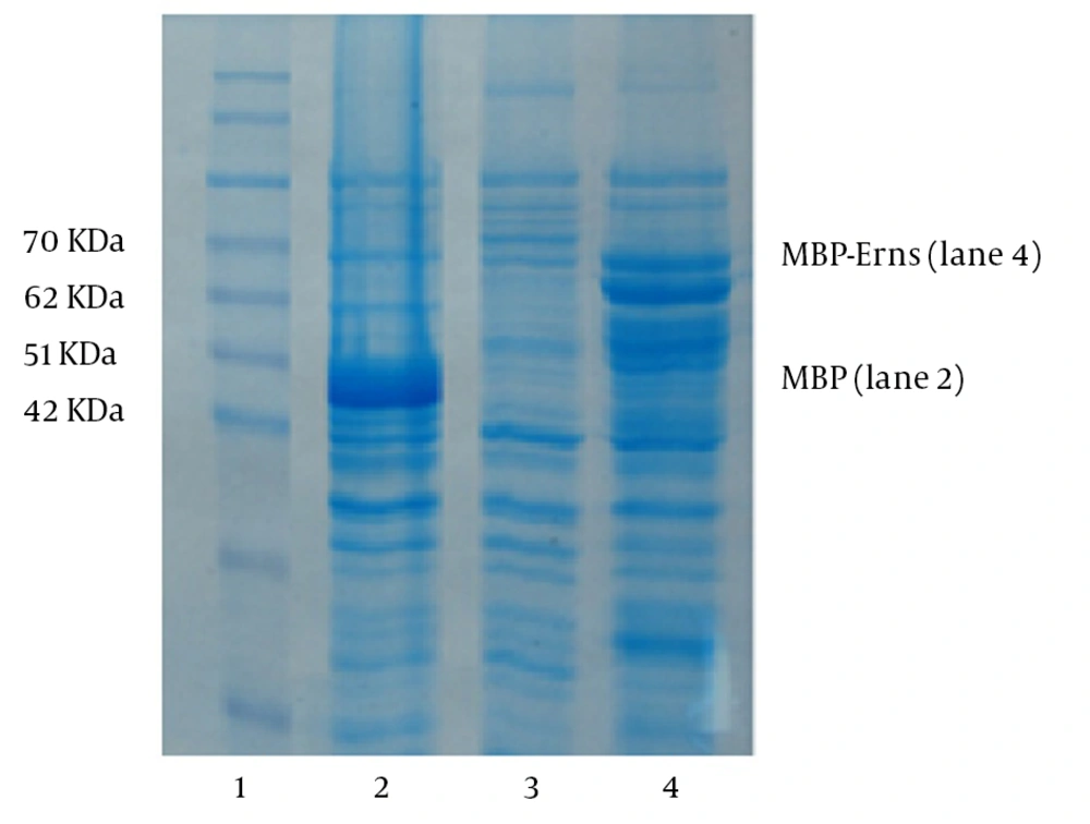
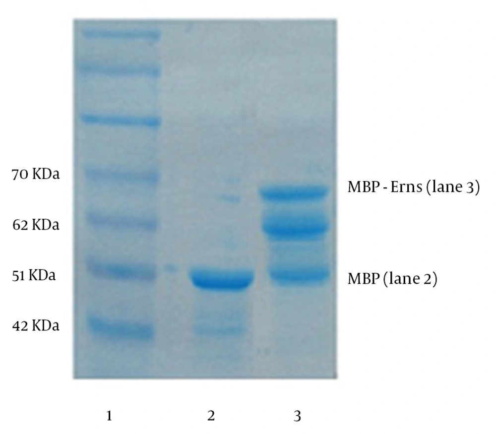
Expression of recombinant MBP-Erns protein in pMalc2x expression vector in E. coli BL-21 strain was induced by adding IPTG (isopropyl-beta-D-thiogalactopyranoside) to the bacterial culture based the instruction of pMalc2x expression system (18). Protein profiles of induced and non-induced bacteria by SDS-PAGE revealed that at least two polypeptides of 62 and 66 KDa were expressed in the induced bacteria (Figure 1). Considering molecular weights of 42 and 24 kDa for MBP and Erns respectively, MBP-Erns fusion protein was estimated to have an approximate molecular weight of 66 kDa. The protein expression was confirmed by Western blotting, using BVDV antibody positive bovine serum. After purification of the expressed protein (MBP-Erns) from the supernatant of the sonicated bacteria on a column of amylose resin and analysis by SDS-PAGE, a major protein band of 66 kDa and multiple smaller bands indicated that the MBP-Erns fusion protein was subjected, in some extent, to proteolytic cleavage (s) in the bacterial host (Figure 2). Therefore, all of the expressed and purified polypeptides seemed be related to the MBP-Erns fusion protein.
The molecular weight marker is shown in Lane 1. Lane 2 indicates a control colony (containing only pMAL-c2X) expressing MBP (50 KDa). Lanes 3 and 4 belong to bacteria expressing the MBP-Erns fusion protein before and after induction by IPTG, respectively. The expression of a protein of about 66 kDa, corresponding to the predicted molecular weight of MBP-Erns is shown in lane 4.
4.2. Hybridoma Production and Isolation
After immunizing mice with the recombinant MBP-Erns, anti-Erns antibody in the serum of mice was detected by indirect ELISA on day 45 and the absorbance was read at 450 nm using the ELISA reader. The optical density (OD) values of immunized sera (1:2000 dilution) were from 1.54 to 1.72 compared with the OD value of non-immunized serum (negative control), which was 0.08. Spleen cells of the immunized mouse, with the highest titer of anti-Erns antibody, were fused with SP2/0 myeloma cells using polyethylene glycol (PEG). The cells in the fusion mix were resuspended in HAT medium and distributed in 96-well plates. Hybrids were selected by their ability to grow in HAT medium and when hybrid cells reached 10% to 50% confluence, culture supernatants of primary clones were screened by MBP-Erns ELISA and MBP ELISA. Positive hybrids in MBP-Erns ELISA, which didn’t react in MBP ELISA were considered as positive. On the basis of primary screening, anti-Erns antibody in the supernatant of eight clones was identified with OD value from 0.2 to 1.1 (average 0.44). The positive clones with the highest titer of anti-Erns antibody were cloned and screened by indirect ELISA three times. The OD values of ELISA were higher, ranging from 1.2 to 2.7 in the second and third rounds of cloning with no reaction towards MBP. However, anti-Erns antibody production was stopped in two clones during cloning, probably due to somatic mutations or deletions of chromosomes.
4.3. Reactivity of MAbs to Recombinant and Natural Antigens
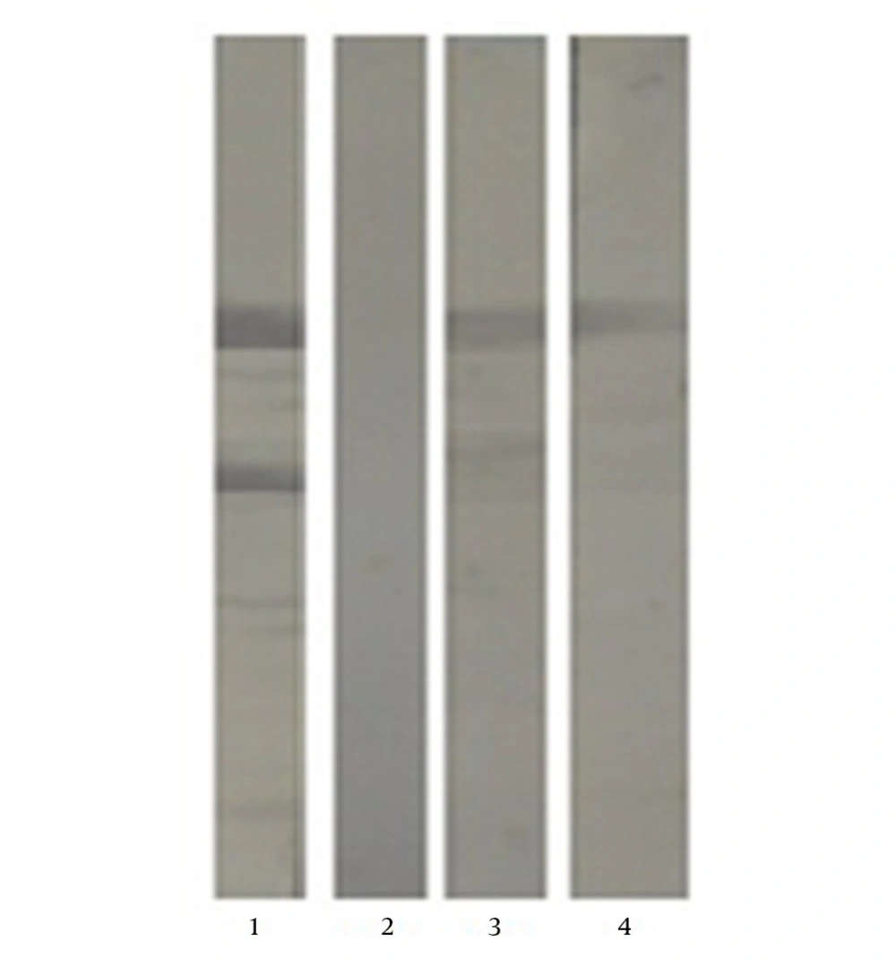
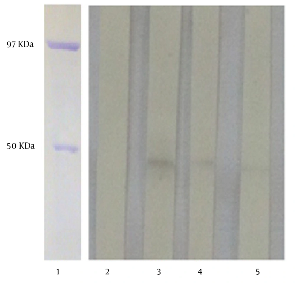
The positive clones after three rounds of cloning were selected and the reactivity of the MAbs with recombinant Erns antigen was screened and established by Western blotting (Figure 3). Also, the specificity and reactivity of the MAbs with the natural form of the Erns antigen, as shown in Figure 4, were examined and confirmed with BVD virus infected cells, as the source of the virus in Western blotting. In the Western blot analysis, anti-Erns MAb reacted with a protein of BVDV that was lower than the 50 kDa marker, which was compatible with the approximate weight of natural Erns (48 kDa) of BVDV.
Lanes 1 shows the purified nucleoprotein of the influenza virus (97 KDa) and purified MBP (50 KDa) from the control colony containing only pMAL-c2X as the protein weight marker. Lanes 2 shows the supernatant of an unrelated hybridoma. Lanes 3 - 5 represent the supernatant of subclones containing anti-Erns MAbs.
5. Discussion
Among viral structural proteins, the Erns glycoprotein has been shown to be a useful marker for identification of PI animals, since it is genetically and antigenically conserved among different isolates (6, 23, 24). To characterize the immune response of cattle to BVDV glycoprotein Erns, Kwang et al., (1992) produced a recombinant insoluble erns-glutathione-s-transferase (GST) fusion protein in E. coli. They found that antibodies to Erns were present in cattle, following vaccination with killed or modified-live viruses, as well as natural infection (25). The BVDV particle presents the 48-kDa glycoprotein (Erns) in multiple copies on its envelope, thus allowing binding and detecting antibodies raise against identical or adjacent epitopes (26). The Erns remains both intracellular and is secreted from infected cells, and can thus be detected both in situ and in serum. Also, Erns appears to be less influenced by inhibition of maternal antibodies in young PI calves (5). Furthermore, PI animals are highly infectious yet often do not manifest their carrier status through obvious clinical symptoms and antibody detection (27).
Identification of these animals is by virus isolation, RT-PCR and detection of viral antigens by immunohistochemistry and AC-ELISA (3). The ELISA is more suited for screening of large series of samples. So far, several BVDV-specific ELISAs have been developed for detection of both antibodies and viral antigens (5). In one research, mAb ELISA was compared with existing ELISAs, which rely on polyclonal antibodies (pAbs) for detecting captured antigens. The mAb detection ELISA was more sensitive than pAb detection ELISAs (28). After BVDV-specific Mabs became available, several laboratory assays reported BVDV detection based on anti BVDV MAbs. Animal sera, whole blood, organ and ear notch tissue samples can be used for BVDV diagnosis. Bedekovic et al. (2011) described an Indirect immunofluorescence (IF) method based on a pool of BVDV-specific monoclonal antibodies (VLA, Weibridge, UK) using ear notch tissue samples, for diagnosis of PI cattle. Compared with the RT-PCR assay, the IF assay had a sensitivity and specificity of 100% and is a good alternative to RT-PCR and antigen ELISA with high speed and accuracy (4).
An anti-gp53 (D89) MAb was used in an indirect fluorescence antibody (IFA) procedure on frozen tissue sections and cell culture. The IFA was a suitable diagnostic assay for detection of BVDV. However, combination of D89 with another BVDV MAb may improve the ability to detect all strains of BVDV in tissues (29). Also, one study reported on an indirect immunoperoxidase assay, which used anti-gp53 and anti-NS3 MAbs for detection of BVDV antigens in CNS (30). The utilization of microparticle immunoagglutination assay has been investigated using coated microparticles with anti-BVDV monoclonal antibodies for BVDV detection. The microparticle immunoagglutination was more sensitive than RT-PCR to detect the virus in the shortest time (31). Several AC-ELISA were developed for rapid detection of BVDV, based on either polyclonal Antibodies and MAbs specific for one or more viral antigens following immunization with the whole virus (5, 26).
A research by Mignon et al., (1992) developed an AC-ELISA using anti-gp48 (Erns) and anti-NS3 MAbs for detecting BVDV antigens in blood samples with sensitivity and specificity of 100%. They proved that the AC-ELISA is a good candidate for replacing virus isolation as a reference method for BVDV antigen detection in PI animals (26). On the other hand, the traditional method of virus isolation is protracted, labor intensive and expensive (28). Current testing strategies to detect PI calves rely heavily on immunohistochemistry and a commercially available AC-ELISA assay. These viral assays often depend on monoclonal antibodies, which target the Erns glycoprotein of BVDV (32). The Erns specific monoclonal antibodies are valuable tools in AC-ELISA for identification of PI animals (5). Mars and Van Maanen (2005) showed that the Erns antigen ELISA had 99% sensitivity and 99.5% specificity as compared to RT-PCR (33). In this regard, a commercial, easy to use AC-ELISA kit based on anti-Erns MAbs is available for detection of viral antigens in serum as well as whole blood and even skin biopsies of PI animals (3).
To summarize, in the present study, for the first time, large amounts of immunologically-active recombinant Erns protein in an expression system under the control of the strong promoter (lac) of vector pMalc2x were used to produce anti-Erns MAbs by the cell fusion assay. Anti-Erns MAbs were screened by indirect ELISA and the reactivity of the MAbs with recombinant and natural antigen was established by Western blotting. The economic impact of BVDV infections has led a number of countries in Europe to start eradication or control programs. However, in both cases, the primary step is identification and elimination of PI animals (13). Based on our results, it appears that Erns recombinant antigen and the specific monoclonal antibodies produced against it may be suitable for developing BVDV laboratory diagnostic assays, especially AC-ELISA.


