1. Background
Despite the progress that has been made in medical science, infectious pathogens remain an important cause of morbidity and mortality (1). Bacteria attach to contact surfaces and survive in the form of microbial communities known as biofilms (2, 3). A microorganism’s ability to remain on various surfaces and to make biofilm is responsible for many chronic diseases that show extremely high resistance to antimicrobial drugs (4, 5).
For this reason, researchers are now investigating strategies other than antibiotic therapy to deal with resistant pathogens (6). Ever since the era of ancient medicine, herbs and their accessory compounds have been known to display varying degrees of antimicrobial efficiency. Extensive research has been done on many of these plants to investigate their antimicrobial activities (7).
Euphorbia is a genus of the Euphorbiaceae family, many of whose members are known as spurges. This family is found in temperate zones worldwide, like Africa and America (8), and is very applicable today in the area of medicine, with a number of studies highlighting its valuable antimicrobial and antioxidant properties (9, 10).
2. Objectives
In the present study, the antimicrobial activity of E. hebecarpa is estimated for the first time, and its antibacterial action on the free-cell and biofilm structures of Staphylococcus aureus, Bacillus cereus, Streptococcus pneumonia, Pseudomonas aeruginosa, Escherichia coli, and Klebsiella pneumonia is evaluated.
3. Materials and Methods
3.1. Collection, Identification, and Extract Preparation
A plant of E. hebecarpa was collected from Kerman, Iran in April, 2012. After collection, the whole plant was rinsed and air dried for two weeks, and was then ground to a fine powder by means of an electric blender (Bosch, Germany). The powder was saved in a dark box for further use.
Ten grams (10 g) of plant material was macerated in a 100 mL mixture of different solvents. A total of 80% ethanol (Pars Chemic Co., Kerman, Iran) and 96% methanol (Pars Chemic Co., Kerman, Iran) was used for the preparation of the ethanol extract and the methanol extract. The extracts were macerated for 30 hours at 38°C by shaking, and then purified using Whatman No. 1 filter paper (MA, USA). Subsequently, the solution was filter sterilized with 0.22 µm of mixed cellulose ester membranes (MilliporeTM; MA, USA). The final extracts were stored at 4°C until needed for use.
3.2. Test Microorganisms and Culture Conditions
The selected bacteria used in our study included six species, three of which were Gram-positive (S. aureus, B. cereus, and S. pneumonia) and three of which were Gram-negative (P. aeruginosa, E. coli, and K. pneumoniae). The tested microbial species were clinical isolates provided by the faculty of Medicine in Kerman University of Medical Sciences. All of these bacteria were isolated from hospitalized patients. The test microorganisms were maintained in NB/glycerol (20%) at -80°C. Nutrient agar (NA, Merck, Germany) containing Luria-Bertani (LB, Merck, Germany) was used to activate S. pneumonia, while nutrient agar was used for the other bacteria. The Mueller-Hinton agar (MHA, Merck, Germany) medium was applied for the disk diffusion assay, and a nutrient broth (NB, Merck, Germany) was selected for the minimum inhibitory concentration (MIC). The minimum bactericidal concentration (MBC) of the tested bacteria was applied in the Mueller-Hinton agar medium (11). A tryptic soy broth (TSB, Merck, Germany) medium was used for the anti-biofilm assay. For S. pneumonia in all assessments, the medium was enriched by increasing the Luria broth (LB).
3.3. Standard Drugs Used for the Antimicrobial Assay
Ciprofloxacin (Sigma, USA) (2 mg/mL) was used as a reference antibiotic against bacterial species.
3.4. Inhibition Zone Determination by Disk Diffusion Assay
The antimicrobial activities of the crude extracts were assessed using the Bauer-Kirby disk diffusion method (12). A stock culture of test bacteria was cultured in a nutrient broth (NB) medium at 37°C for 18 hours. The final cellular concentration was adjusted to 108 colony-forming units (CFU)/mL with reference to the McFarland turbidometer (13). The optical density at 600 nm of suspense bacteria was then adjusted to 0.13 (VarianCary50, America) (14), and a five hundred microliter (500 µL) quantity of bacterial suspension was added to each petri dish containing the Mueller-Hinton agar and spread over the agar.
In the next step, sterile 6 mm blank paper disks (Padtan Teb Inc., Tehran, Iran) were saturated with filter-sterilized plant extract at the prepared concentration (100 mg/L for about two hours and allowed to dry at 37°C for five hours (15). Two disks macerated with the same volume of ethanol and methanol were used as a negative control, and ciprofloxacin (2 mg/mL) was used as a positive control. Each of the disks was placed on top of the agar layer and the petri dishes were then incubated at 37°C for 18 hours. The diameter of the growth inhibition zone (mm) surrounding the disks showed the inhibitory effect.
3.5. Determination of MIC and MBC
The MICs and MBCs were evaluated using a macrobroth dilution method, as recommended by the clinical and laboratory standards institute (11), with NB as the test medium. The overnight cultures of the microorganisms were diluted to yield a cell concentration equal to 5 × 10⁵ cfu/mL. Various concentrations of serial two-fold dilution extract (0.05 - 50 mg/mL) were contaminated with equivalent volumes of bacterial suspension. These solutions were prepared by dissolving extracts of stock concentration (100 mg/mL) in a sterile culture medium (NB). The bacteria were then incubated at 37°C for 18 hours, and the lowest concentration that inhibited cells was determined as the MIC. Negative controls (bacteria + NB), positive controls (bacteria + NB + ciprofloxacin), vehicle controls (bacteria + NB + solvent), and media controls (NB) were included.
The MBC was determined by spreading 150 µL on an MHA plate from a sample showing no visible growth and incubating this for a further 18 hours at 37°C.
3.6. Biofilm Formation Inhibition Assay
Biofilm formation was measured as described by O’Toole and Kolter (16) with some modifications. At first, crude plant extracts were added to the sterile culture medium in three concentrations (12.5 - 50mg/mL), and 100 µL of each concentration was then added into the wells of a sterile 96-well polystyrene microtiter plate. The overnight cultures of each bacterial species were diluted to an OD600nm equal to 0.2 in fresh TSB. Subsequently, 100 µL of these suspensions was added to the wells and incubated at 37°C for 24 hours. Negative controls (cells + TSB + sterile distilled water), positive controls (cells + TSB + ciprofloxacin), extract controls (TSB + extract concentration), and media controls (TSB) were included.
The stabilized biofilm mass was calculated using crystal violet staining (17). First, the wells were aspirated. Then, to remove the non-adherent cells, each of the wells was washed with sterile PBS three times. In the next step, 150 µL of 96% methanol was distributed in each well in order to fix the adherent cells, and left for 15 minutes. After this time elapsed, the 96% methanol was removed and 200 µL of 1% crystal violet (Gram color-staining set for microscopy; Merck, Germany) was added to each well for 20 minutes. Following the staining step, the washing procedure with sterile water was repeated and the plates were air dried at room temperature. To re-solubilize the dye bonded to the biofilms, 160 µL of 33% glacial acetic acid (Merck, Germany) was added to each well and the optical density of the content was measured using a microtiter plate reader (ELX-800, Biotec, India) at 630 nm. The following formula was applied to evaluate biofilm inhibition:

3.7. Inhibition of a Preformed Biofilm
To produce biofilms, 100 µL of stationary-phase bacterial cultures in TSB medium were added aseptically to the wells of a 96-well polystyrene microtiter plate and incubated at 37°C for 24 hours. After biofilm formation, the medium was aspirated gently, and non-adherent cells were removed by washing the biofilms three times with sterile PBS. To determine extract efficiency, each extract concentration (12.5 - 50 mg/mL) was released in the wells and incubated for 24 hours at 37°C (18). The control wells were the same as those described in section 3 - 6. Further inhibition of a preformed biofilm was analyzed using crystal violet staining. In this way, the percentages of the reduced biofilm structures were calculated.
3.8. Assessment of Biofilm Metabolic Activity
The anti-metabolic activity of E. hebecarpa extracts on stabilized biofilm was quantified as reported by Ramage and Lopez-Ribot (19). At first, the attached biofilms were washed twice with PBS. Each well was then contaminated with extracts (12.5 - 50 mg/mL) and the microplate was incubated for 24 hours at 37°C. Subsequently, 50 µL of a triphenyl tetrazolium chloride (TTC, Merck, Germany) solution was added to each well, allowing the occurrence of a reaction in the dark at 37°C over three hours. Finally, absorbance was measured at 490 nm. The control wells were the same as those described in section 3 - 6, and the percentages of reduced biofilm metabolic activity were determined.
3.9. Statistical Analysis
The differences for individual parameters between the control and treated groups were tested by analysis of variance (ANOVA) using SPSS version 18.0 for Windows. Differences were considered significant in the case of a P value less than 0.01. All experiments were performed in triplicate and repeated three times.
4. Results
4.1. Inhibitory Effects of E. hebecarpa Extracts Against Planktonic Forms of Bacteria
The zone of inhibition (ZOI) for methanolic and ethanolic extracts of E. hebecarpa is shown in Table 1, and the MIC and MBC of these extracts are illustrated in Table 2. The largest ZOI was observed for K. pneumonia, while no ZOI was in evidence for the methanolic and ethanolic extracts of this plant in the case of S. pneumonia. Likewise, there was no ZOI for the methanolic extract in the case of E. coli (Table 1).
| Bacteria | Methanolic Extract | Ethanolic Extract | Ciprofloxacin | Solvent Control |
|---|---|---|---|---|
| Staphylococcus aureus | 15.67 ± 0.6 | 14 ± 0.8 | 23 ± 0.6 | - |
| Bacillus cereus | 11.33 ± 0.9 | 15.67 ± 0.5 | 21 ± 0.4 | - |
| Streptococcus pneumonia | - | - | 9.4 ± 0.1 | - |
| Pseudomonas aeruginosa | 19 ± 0.6 | 22.23 ± 0.7 | 24.6 ± 0.3 | - |
| Escherichia coli | - | 9.67 ± 1.1 | 22.6 ± 0.7 | - |
| Klebsiella pneumoniae | 20 ± 0.9 | 25.67 ± 0.5 | 12 ± 0.9 | - |
Antibacterial Activity of E. hebecarpa Alcoholic Extracts Against Test Microorganisms Using the Disk Diffusion Method (Zone of Inhibition in mm)a
| Bacteria | MIC Methanolic extract, mg/mL | MIC Ethanolic extract, mg/mL | MBC Methanolic extract, mg/mL | MBC Ethanolic extract, mg/mL |
|---|---|---|---|---|
| Staphylococcus aureus | 0.78 | 0.19 | 1.56 | 0.39 |
| Bacillus cereus | 0.19 | 0.09 | 0.39 | 0.39 |
| Streptococcus pneumonia | 0.39 | 0.19 | 1.56 | 0.78 |
| Pseudomonas aeruginosa | 0.78 | 0.39 | 1.56 | 0.78 |
| Escherichia coli | 0.39 | 0.39 | 3.12 | 1.56 |
| Klebsiella pneumoniae | 0.39 | 0.19 | 0.78 | 0.78 |
E. hebecarpa MIC and MBC Values of the Test Bacteria
The results of this study reveal that the inhibitory effects of E. hebecarpa extracts in a broth medium are much greater than in a solid medium. In addition, it was found that extracts with concentrations much lower than the concentration used in preparing the disk (0.09 - 0.78 mg/mL) were able to inhibit all microorganisms under investigation. Of all the pathogenic bacteria, B. cereus showed the greatest sensitivity to E. hebecarpa extracts in the broth medium, although P. aeruginosa required the highest concentration of this plant extract for inhibition to take effect.
4.2. Inhibitory Effects of E. hebecarpa Extracts in Preventing Biofilm Structures
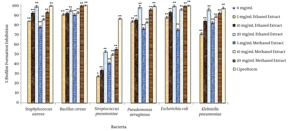
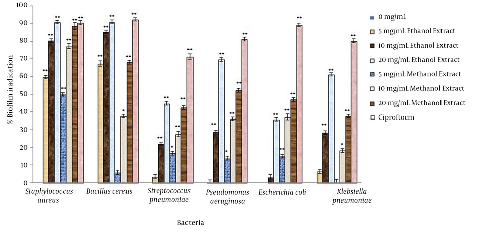
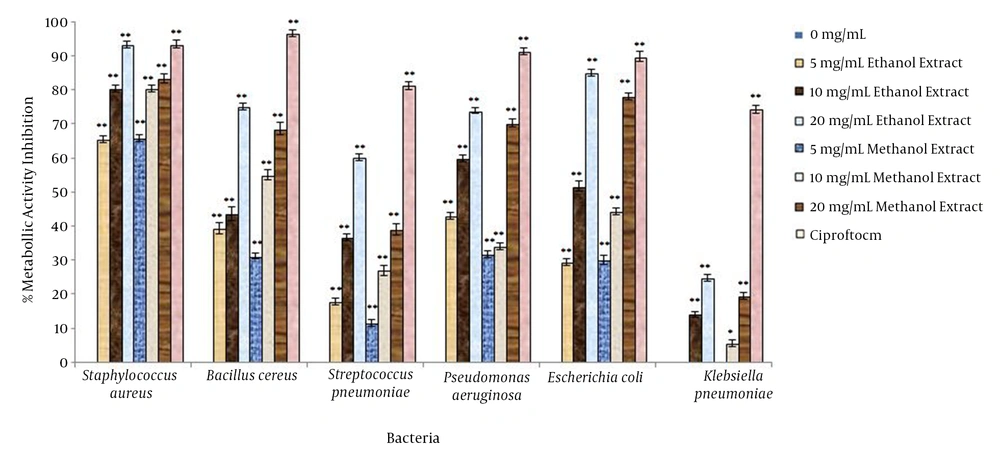
The inhibitory efficiency of each concentration of E. hebecarpa extracts on preventing biofilm formation, demolishing biofilm structures, and inhibiting the metabolic activity of biofilms are shown in Figures 1 - 3. According to the F value of ANOVA analysis on the tested data, it was confirmed that the inhibitory efficiency of the E. hebecarpa extract was significant at 0.01 (P < 0.01).
Based on an examination performed on the biofilm structures, it was concluded that the type of bacteria and the concentration of extracts were significant at 0.01 (P < 0.01), but the type of solvent used in the extraction process was only significant for demolishing the biofilm structures (P = 0.004); in fact, it was found that the inhibition of biofilm formation and metabolic activity in treatment using extracts of E. hebecarpa was independent from the type of solvent used (P = 0.09).
The maximum inhibitory effects of E. hebecarpa extracts on biofilm formation were observed on B. cereus (92.91%) and E. coli (91.2%), although these extracts had low efficiency in terms of inhibiting the biofilm formation of S. pneumonia (43.06%). Regarding the demolition of biofilm structures, the efficiency of the E. hebecarpa ethanolic extract on B. cereus and K. pneumoniae was significantly greater than that of the methanolic types, while on the biofilm of E. coli, the methanolic type was more efficient than the ethanolic type. In the treatment using E. hebecarpa extracts, S. aureus biofilm formation was susceptible (74.49%) but E. coli biofilm formation (23.02%) and K. pneumoniae biofilm formation (25.38%) had a resistant biofilm structure for all the tested bacteria. The metabolic activity of the bacteria in biofilms treated with the E. hebecarpa extract dramatically decreased, with the greatest reduction observed in S. aureus (78.21%) and the lowest reduction observed in K. pneumoniae (10.71%).
5. Discussion
Indiscriminate use of antibiotics in recent decades has caused the emergence and spread of drug-resistant strains of bacteria that make it difficult to fight infections. One of the main causes of drug resistance is the placement of microorganisms in biofilm structures. This prevents the penetration of antimicrobial compounds and also inhibits the proper function of these compounds. Hence, ascertaining new ways to deal with pathogens, especially in biofilm form, is essential (20, 21). Investigation in this context is especially focused on biological derivatives, because the biological nature of these compounds involves reduced side effects compared to conventional chemical agents. Of the biological derivatives available, herbs are considered a popular and suitable option for dealing with pathogenic microorganisms (22, 23).
In this study, the antimicrobial properties of a new species of Euphorbia (E. hebecapra) were investigated against six pathogenic bacteria. A disk diffusion test showed that E. hebecapra extracts have high capability in terms of preventing the growth of selected bacteria. As shown in Tables 1 and 2, ethanolic extracts inhibit planktonic forms of bacteria more effectively than methanolic extracts, with the exception of the disk diffusion method, where a methanolic extract of E. hebecarpa used on S. aureus was found to be more effective than the ethanolic extract. In a disk diffusion test, K. pneumoniae was the most sensitive bacterium, while the inhibitory effect of this plant extract on E. coli was weak. The E. hebecapra extracts had no inhibitory effect on S. pneumonia. However, in the liquid medium that was used for the MIC test, these extracts had an inhibitory effect on the growth of this bacterium. In addition, the extracts had notable preventive effects on the other microorganisms.
Since the inhibitory effects of E. hebecapra on all tested bacteria in a broth medium were higher than in a solid medium, it can be concluded that the active compounds of the extracts, like many other herbal extracts, have a lower diffusion level in a solid medium and therefore display optimum inhibitory effects in such a medium. Thus, a higher concentration of extract is required compared to the broth medium.
The E. hebecapra extracts were found to be efficient in dealing with biofilm structures. Their preventive effect correlated directly with concentration and, except for the demolition test of the biofilm, the inhibitory effect of each extract was independent from the type of solvent used. The ability of the E. hebecapra ethanolic extract to inhibit biofilm formation was greater than its ability to demolish the biofilm or prevent metabolic activity of the microbial cells in the biofilm structure. Thus, it can be concluded that E. hebecapra extracts contain some components that interact with the biofilm formation of bacteria, but these extracts have a low ability to deal with immobilized biofilm structures.
In other studies, the therapeutic properties of some species of Euphorbia have been confirmed, but these studies were mainly carried out on planktonic forms. For example, Abubakar (24) confirmed the inhibitory effects of E. hirta methanolic and aqueous extracts on pathogenic bacteria such as E. coli, P. mirabilis, S. dysenteriae, and K. pneumoniae. In that study, Abubakar revealed that the antibacterial activity of the plant material is enhanced under acidic conditions and at elevated temperatures. Furthermore, the MIC and MBC values in his study ranged from 25 to 100 mg/mL.
Kamba and Hassan (25) examined the ability of various parts of E. balsamifera to combat some pathogenic bacteria, and their results showed that the extracts of this plant have suitable inhibitory effects on S. typhimorium, P. aeruginosa, K. pneumoniae, E. coli, and Candida albicans, and that there are no differences between the antimicrobial properties of the stems, roots, and leaves. Concentration-dependent inhibition of bacterial growth was visually confirmed by these researchers. In their study, the MIC for the tested bacteria was 5.0 - 6.0 mg/mL, and the MBC ranged between 4.5 and 6.0 mg/mL. The differences between these results and our observations are probably due to the diverse chemical substances in different Euphorbia species, and especially the different extraction methods used. In effect, this indicates that the extraction method plays an important role in the presence of the active principles in the extract and influences the extract’s antimicrobial activities. The results of the present study show that if extraction is carried out using conventional maceration, the inhibitory effect of the extracts is more than that concentrate the extract. As a result, it was predicted that the active compounds of this plant are very volatile and extraction was therefore performed using the maceration method.
A study carried out by Nashikkar et al. (26) showed that ethanolic and chloroformic extracts of E. trigona caused a 70% decrease in swarming of P. aeruginosa and P. mirabilis. Also, these extracts decreased biofilm formation by 50%. In this research, while the bacterial growth rate was not adversely affected, a remarkable reduction in virulence factor production was observed. For example, the urease activity of P. mirabilis and rhamnolipid production by P. aeruginosa decreased significantly in the case of treatment with extracts.
5.1. Conclusion
According to the results of this research and other studies that were performed on different species of Euphorbia, it can be concluded that the antimicrobial potential of this plant is confirmed and its extractions are suitable for combating pathogenic microorganisms. Since the ethanolic extract of E. hebecapra used in this study showed suitable inhibitory effects on planktonic forms and the biofilm structures of pathogenic bacteria, it can be suggested that these extracts be used as antibacterial agents against pathogenic microorganisms, and particularly for biofilm structures.


