1. Background
Human Papillomavirus (HPV) infection is one of the most common sexually transmitted infections (1, 2). The infection rate will increase a few years after the begining of sexual activities and it is estimated that approximately 80% of sexually active adult’s lifetime have been infected with at least 1 HPV (3). Although most infections are transient and do not cause disease, some of them can persist for several years and lead to the development of high-grade precancerous lesions or an invasive cervical (4). Cervical cancer is recognized as the second most common malignancy of female reproductive system in Iran, which devotes itself 8.8% of all cancers in women and also near 7.5% of leading causes of cancer deaths (5, 6). Furthermore, cervical cancer age-standardized incidence rate is 2.2 per 100,000 Iranian women per year (7, 8).
Human Papillomavirus genotypes are classified as high-risk HPV (HR-HPV) and low-risk HPV (LR-HPV) groups according to their potential to cause cancer (9). The most common LR-HPV types are HPV-6 and HPV-11 that commonly cause benign warts. HR-HPV are associated with the development of cancers and are found in more than 99% of all cervical carcinomas so HPV as a marker for the early detection of cervical cancer and precancerous. HR-HPV types are including types of 16, 18, 31, 33, 35, 39, 45, 51, 52, 56, 58, 59, 66 and 68 (10-12).
Human Papillomavirus-16 and HPV-18 are consistently 2 most prevalent types reported worldwide. The high frequency of these 2 types of HPV in cervical cancer and other HPV related diseases has made scientists provide HPV vaccines (13). It has been demonstrated that vaccines prevent almost 70-80% of the invasive cervical cancer in the individuals worldwide who are not infected with this virus (14). For this purpose, comprehensive studies in different regions of the world have been done due to varied distribution of HPV genotypes in populations (15). In addition, other evidences have proved that the HPV infection is involved in other tumors such as anogenital, head, neck, and probably breast cancers (16-18).
According to evidence revealed, human Papillomavirus and HPV genotypes prevalence have been shown to diversity among populations according to HPV genotyping protocols employed, age categories, region, and the type of population. Unfortunately, HPV prevalence data is not yet available for the total population of Iran. Therefore, Epidemiological studies are necessary for evaluating the risk of cancer in women who are found to be infected with types of HPVs in different population and vaccination programs.
2. Objectives
The aim of this study was performed to provide the data related to HPV infection types and prevalence in Kermanshah, Iran (A city located in west Iran).
3. Methods
In this cross sectional study, a total of 87 samples, including 61 cervical samples, 24 genital warts in women, and 2 skin biopsy in men were collected from pathology laboratories of Kermanshah, in the period of 2014 to March 2015. The inclusion criteria included the presence of genital warts, uterine cervix microscopically suspicious for HPV, Pap smear result suspicious for HPV, HPV positive partner, no symptoms, and a desire to screening for sexually transmitted infections. Cervical swaps, genital, and skin biopsy were obtained from each participant using an Ayres spatula or Cytobrush. Demographic data and patient’s medical history was collected in a questionnaire.
DNA of HPV specimens were extracted using a genomics extraction kit (G-spin Total DNA Extraction kit (Intron Biotechnology, Seongnam, Korea)) according to the instructions of the manufacturer. The presence of HPV and genotyping was identified using a HPV Easy-Typing kit (Autoimmune Diagnostika GmbH, Strassberg, Germany). Briefly, PCR were performed out in a total volume of 25 μL containing 15 μL working master mix, 2.5 μL 10xbuffer, 2.5 mM MgCl2, 1 unit Taq DNA polymerase, and 100 ng of DNA sample. The PCR reactions were carried out by Initial denaturation at 95°C - 3 minutes and 10 cycles consisting 96°C - 10 seconds, 60°C - 20 seconds, and 26 cycles consisting denaturation at 95°C - 10 seconds, annealing at 55°C - 15 seconds, and extension at 72°C for 15 seconds. The final step was extended to 3 minutes at 72°C.
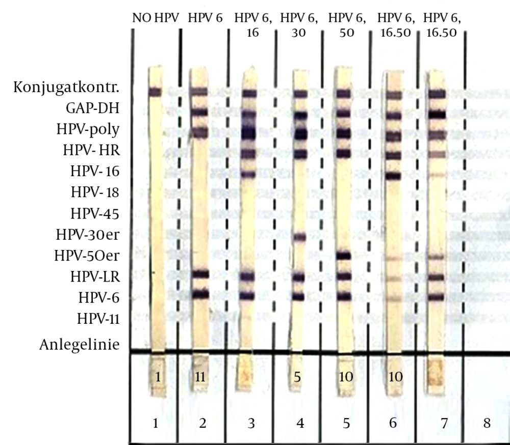
To analyze the PCR products, 5 μL of each PCR product was ran in a 2% agarose gel. The HPV-fragment was 180 bp. After amplification, DNA Bands were stained by ethidium bromide and visualized under UV gel documentation. Human Papillomavirus kit was performed in this assay according to rever-hybridsizaion, however, it is a sensitive method to detect and differentiate infections with relevant HPV genotypes at suitable times and costs. With the aid of HPV Easy-Typing kit, high-risk and low-risk genotypes, and also HPV types of 16, 18, 45, 6, 11, 30s (31, 33, 35, 39), and 50s (51, 52, 53, 56, 58, 59) genotypes could be detected by manufacture protocol.
Eventually Data were analyzed by SPSS software (version 16.0) using Pearson and chi-square tests to determine the frequencies of HPV positivity, age, and HPV genotypes. P <0.05 was considered as the statistically significant.
4. Results
Human Papillomavirus was detected in 54 specimens (62.1%) of our HPV (Figure 1). Among the 87 enrolled patients, 48 suffered from inflammation, 24 from genital warts, 5 from cervical cancer, and 10 normal that 52% (28/54), 37% (20/54), 9.3% (5/54) and 1.8% (1/54) were HPV-positive cases, respectively (Table 1). The patients were 18 to 63 years old and their mean age was 40 ± 12.3 years. Patients in the age group of 35 - 45 years had high HPV infection.
The HR-HPV types were the most frequent ones that have been detected. Among the 54 HPV that were positive, 61.1% (33) and 64.8% (35) of them have shown the presence of HR-HPV and LR- HPV, respectively. Furthermore, HPV-6 was major a genotype, which was found in 64.8% of the HR and LR types.
Type 16 and 18 were the frequently observed ones in the patients (26%), followed by type 50s (24.7%), 45, 30s, and 11 (1.8% from each). 22 samples (40.7% of all HPV positive samples) were infected with multiple HPV types, which included at least 1 HR-HPV. Of all HPV co-infection, HR types were related to LR-HPV types. We isolated co-infection in HPV 6 with 4 different strains of HPV 50s, 16, 18, and 30s and the distribution of these co-infections were found as 54.5%, 40.9%, 9.1%, and 4.5%, respectively.
Furthermore, the most prevalent co-infection samples were HPV 50s/6 (54.5%) and HPV 16/6 (40.9%). The prevalence of HPV infection in cervical cancer was 100% (5/5) included HPV 16 (20%), HPV 18 (20%), and HPV co-infection of 50s/6 (60%). The major genotype of HPV infection in inflammation, genital wart, cervical cancer were obtained 6 (35.7), 50s/6 (30%), 50s/6 (60%), respectively. In addition to the above outcomes, it was understood that 13 types of HPV genotypes were single and multiple infections (Table 2). Statistical analysis showed that there was no significant association between HPV genotypes, the age of patients and different groups of the study.
| HPV Genotypes | Type of HPV Genotypes, % | Normal, % | Inflammation, % | Genital Warts, % | Cervical Cancer, % | |
|---|---|---|---|---|---|---|
| High risk | 16 | 5 (9.2) | 0 | 4 (14.3) | 0 | 1 (20) |
| 18 | 11 (20.4) | 0 | 7 (25) | 3 (15) | 1 (20) | |
| 50s | 1 (1.8) | 0 | 1 (3.6) | 0 | 0 | |
| 30s | 0 | 0 | 0 | 0 | 0 | |
| 45 | 1 (1.8) | 0 | 1 (3.6) | 0 | 0 | |
| Low risk | 6 | 14 (25.9) | 0 | 10 (35.7) | 4 (20) | 0 |
| 11 | 0 | 0 | 0 | 0 | 0 | |
| Multiple HPV types | 16/6 | 6 (11.1) | 1 (100) | 2 (7.2) | 3 (15) | 0 |
| 18/6 | 2 (3.7) | 0 | 0 | 2 (10) | 0 | |
| 18/11 | 1 (1.8) | 0 | 0 | 1 (10) | 0 | |
| 50s/6 | 10 (18.5) | 0 | 1 (3.6) | 6 (30) | 3 (60) | |
| 16/50s/6 | 2 (3.7) | 0 | 1 (3.6) | 1 (10) | 0 | |
| 16/30s/6 | 1 (1.8) | 0 | 1(3.6) | 0 | 0 | |
| Total | 54 (100) | 1 (1.8) | 28 (52) | 20 (37) | 5 (9.2) |
The Prevalence of number Human Papillomavirus Genotypes and Multiple Human Papillomavirus Genotypes (n = 87)a
5. Discussion
Human Papillomavirus can be considered as the main risk factor for the persistent infection with HR-HPV. Also, this virus plays a major role in the pathogenesis of cervical cancer (19-21). Thus, it is required to identify the HPV infection and its genotyping description the in clinical issue. Among 87 examined samples in our study, 62.1% of them have shown positive signs for the presence of HPV. Human Papillomavirus infection in inflammation, genital wart, cervical cancer and normal was observed 52% (28/54), 37% (20/54), 9.3% (5/54), and 1.8% (1/54), respectively. However, this result was a relatively high report, which is consistent with the previous surveys done in Asian countries (22, 23). The prevalence of HPV strains is different in various geographical parts of Iran (5.5 to 60%), although our statistical result was higher than the maximum of this range (22, 24, 25). Based on the study of Khorasanizadeh, the mortality to incidence ratio was 42% in Iran (26). Furthermore, other studies conducted in the United States demonstrated that the prevalence of HPV was in range of 2.9% to 80.8% (27, 28).
The prevalence of HPV in sub-Saharan African regions, Latin American and Caribbean regions, Eastern Europe, and South-eastern Asia is 24%, 16%, 14.2%, and 14%, respectively, while the prevalence of HPV in our study is higher than these statistics (29). On the other hand, the prevalence of HPV is low in European countries, North America, and Japan (30). The distribution of HR-HPV positive isolates has been reported to be about 14.7%, 19.2%, and 40.0% in Nigeria, Uganda, and Mozambique, respectively (31-33). In contrast, the frequency of HR-HPV in developed countries located in the Western Europe range from 9.4% to 12.1 (34); however, HR-HPV frequency announced by our investigation is much higher than this range (61.1%). The heterogeneity in the intra-country and intra-region studies was due to the selected variables such as age and population type. Also, HPV prevalence varied according to the HPV testing method used.
Throughout the study of specimens, we identified 4 major HPV genotypes 6, 16, 18, and 50s were the most common high risk observed types. Other prevalent types were HPV 45, 30s, 11 (1.8% from each). Recent studies done in the other parts of Iran, revealed that HPV 16 (41%) and 18 (20%) were the most common HR-HPV genotypes according to Daneshvar et al. and the other study revealed that the most common HR-HPV genotypes obtained were HPV 16 (33%) and 53 (9%) (35, 36). Moreover, in agreement with the other studies done in several Asian countries, our results showed that HPV 16 and 18, HPV 50s was mostly presented in the cervical samples (13, 23, 37, 38).
Likewise, we detected multiple HPV genotypes infection in 40.7% of HPV-positive samples. This finding was higher than that announced by Alsbeih et al. and lower than that declared by Sun et al. (39, 40). The most common combination of multiple HPV infections in our finding was HPV 50s/6 followed by HPV 16/6. Infection with multiple HPV genotypes has been associated with the increased risk of HPV persistence. Human Papillomavirus vaccine is not used for people, while this virus has high prevalence and it can be prevented through a vaccination program held by the organization of health in Iran. Consequently, based on our finding, it could be claimed that vaccine-targeted HPV6, HPV16, HPV18, and HPV50s can be useful to detect HPV genotypes in this region.
Generally, a wide variety of diseases is caused by specific HPV types. For instance, HPV 6 or 11 cause 90% of genital warts and HPV types 16, 18, 31, 33, and 35 are occasionally found as co-infections with these diseases (41). In our study, HPV types 6, and HPV co-infection of 50/6 were presented highly in genital warts. This outcome is important since it can be associated with high-grade intraepithelial neoplasia (41).
Finally, it can be concluded that the variations among the obtained results may be due to the differences in populations under study, geographic localizations, differences in sampling approaches or methods, and the primers used in PCR. In Iran, only some individuals with cervical cancer are observed, however, the number of them is increasing gradually due to the absence of mass screening programs and vaccination as a routine primary prevention method. These data provide beneficial information regarding the epidemiology of the HPV infection that can guide future applications of screening and prevention of this precancerous disease in this region of Iran. Therefore it seem to pay serious attention of public health organizations in diagnosis and their identification in order to treat them and evaluating the efficiency of HPV vaccines for the prevention of vaccine-targeted HPV genotypes. In conclusion, the prevalence of the HPV infection was relatively high. The most frequently detected genotypes were HPV6, HPV16, HPV18, and HPV50s, respectively.
