1. Background
Dermatophytes, yeasts, and non-dermatophyte moulds are known as causative agents of cutaneous fungal infections (1, 2). It is estimated that they affect more than 20-25% of people all over the world (3). Skin and nails are the favored locations for colonization, invasion, and finally disease development by these pathogens (4). Candida species are mostly responsible for cutaneous fungal infections known as cutaneous candidiasis. They frequently are recovered from different cases. Candida species contain pathogens that exist as normal flora of the body of healthy humans, particularly in oral cavity, intestinal tract, and on the skin. They are able to develop a range of diseases including non-life-threatening to life-threatening invasive diseases (5, 6).
Despite, Candida albicans has been reported as the most important causative agent of this family, other species such as C. parapsilosis, C. tropicalis, C. guilliermondii, C. famata, and C. krusei also are isolated from clinical samples. This diversity for Candida species has been frequently observed in prior studies. However, many comprehensive studies have shown the significant growth of the infections related to other non-Albicans Candida species (NACs) (7-10).
The opportunistic nature of Candida species indicates that the patient’s natural immune system is seriously impaired. This is because complete immune system secures the occupation of the potential growth sites of the pathogens by non-harmful micro flora that as a protective barrier limits the colonization and development of mycotic infection (11). In addition, the occurrence of candidiasis could be affected by some predisposing factors such as geographical location, type of job, presence of underlying diseases (diabetes, nutrient deficiency complications, and immune deficiency diseases), applying some therapies (long period antibiotic therapy, cancer therapy, etc.), site of infection (lesion), and age (12, 13). Indeed, these factors may have close associations with the infection development.
More importantly, a diversity would be possible in the distribution of Candida species not only between two neighboring countries, but also in different provinces and areas of a big country such as Iran. Thus, providing critical understanding about the pattern of the distribution of Candida species might be extremely beneficial for recruiting protective and control strategies and devising effective antifungal therapies.
2. Objectives
The aim of the present study was to gain insights into the molecular analysis of Candida species. To this end, this work focused on illustrating the diversity and distribution of Candida species in patients suffering from cutaneous candidiasis in Tehran, Iran. In addition, we assessed the role of several predisposing factors in the induction and development of the disease.
3. Methods
3.1. Study Population
In this study, 3000 samples obtained from patients with suspected candidiasis referring to Razi hospital in Tehran were examined from March 2014 to 2015. A questionnaire containing demographic specifications and clinical signs was designed and completed for each individual.
3.2. Mycological Examination
Sampling area was decontaminated with 70% ethanol. The fungal samples were obtained by scraping skin and different parts of nails. Then, each individual sample was divided into two pieces. One piece was recruited for direct microscopic examination using 10% potassium hydroxide solution (%10 KOH) to find any possible trace of Candida elements, such as pseudohyphae and yeast cells. The other piece was cultured on Sabouraud dextrose agar (SDA) (Difco, Detroit, MI, USA) with 0.005% chloramphenicol (Difco, Detroit, MI, USA). All plates were incubated at 30°C for 48 to 72 hours. Yeast isolates were maintained for molecular analysis.
3.3. Molecular Examination
3.3.1. DNA Extraction
Candida DNA extraction was performed according to a procedure previously described by Mirhendi et al. (14) using phenol-chloroform technique. DNA concentration and purity were determined by measurements at wavelengths of 260 and 280 nm, respectively.
3.3.2. PCR Conditions
To amplify the internal spacer region (ITS1- 5.8S - ITS2) of the yeast rRNA genes, two universal primers (including ITS1 [5′-TCCGTAGGTGAACCTGCGG-3′] and ITS4 [5′-TCCTCCGCTTATTGATATGC-3′]) (Bioneer, South Korea) were used (15). The PCR reaction was performed in the final volume of 100ul containing 0.2 mM each deoxynucleoside triphosphate (Roche, mannhiem, Germany), 1.5 mM magnesium chloride (Roche, mannhiem, Germany), 0.5 mM each primer, Taq buffer, and 2.5 U of Taq polymerase (GeNet Bio, Korea), and 1 mg extracted DNA from each individual plate as template. Thermal cycle reaction was carried out by a thermal cycler (Biorad, United states). It was composed of a pre-denaturation at 94°C for 5 minutes followed by 35 cycles of denaturation at 94°C for 30 seconds, annealing at 56°C for 45 seconds, and a final extension step at 72°C for 7 minutes. To assess the presence and the length of the PCR amplicons, electrophoresis was accomplished in 1.5 % agarose gel (Roche, mannhiem, Germany) stained with SYBR green. Subsequently, gels were visualized under ultraviolet transillumination.
3.3.3. RFLP Analysis
Msp1 enzyme (Fermentas Life Sciences, Lithuania) was used to digest PCR products. Briefly, 10 L of all amplicons, 0.5 μL of enzyme, 1.5 μL of 10 × Buffer Tango, and 3 μL of nuclease-free water were mixed. The reaction mixture was incubated at 37°C for 2 hours. To visualize the migration profile of digested products, electrophoresis was accomplished exactly as explained above.
4. Results
Out of 3000 subjects referring to the mycology lab of RAZI hospital from March 2014 to 2015, 290 (9.66%) patients were identified with cutaneous candidiasis, including 118 (40.7%) men and 172 (59.3%) women.
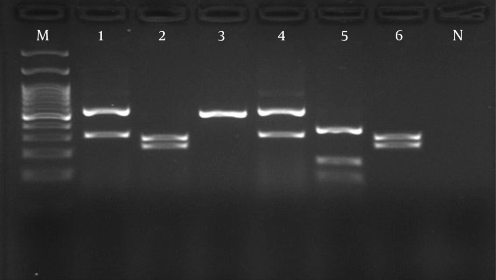
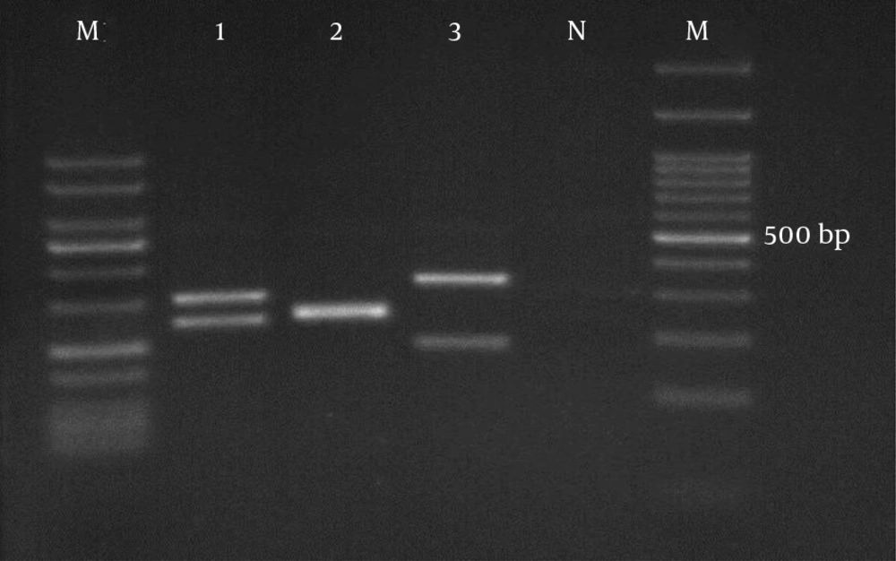
Molecular genotypic identification of Candida species was done by using the PCR-RFLP method. C. albicans showed the highest prevalence (n = 132, 45.5 %) among all species, followed by C. parapsilosis (n = 77, 26.5%), C. tropicalis (n = 37, 12.7 %), C. glabrata (n = 22, 7.5 %), C. krusei (n = 16, 5.5 %), and C. guilliermondii (n = 6, 2%) (Figures 1 and 2).
Due to the opportunistic nature of Candida species, some predisposing factors such as site of infection (lesion), age, occupation, and underlying disease were also considered in this survey. Candida infection symptoms are various and closely associated with the location of the infection. Our results revealed that the most commonly affected location was nails (n = 165, 56.7%), while the least commonly affected site was face (n= 2, 0.6%). In addition, C. albicans and C. parapsilosis predominantly were isolated from different sites of infection (Table 1).
The lowest prevalence rate was observed in the 11 - 20 age group (n = 7, 2.4%), whereas the most predominantly affected age ranges were 41 - 50 and 51 - 60 (n = 63, 21.7% and n = 65, 22.4%, respectively) (Table 2). Among several job categories that were considered in this survey, the majority of positive samples were related to the women working at home (housewives) (n = 135, 46.5%) (Table 3). Many prior studies confirmed the high prevalence of candidiasis among people with an underlying disease (16-18). As mentioned in Table 4, out of 73 individuals that simultaneously suffered from another disease, people with diabetic disorder were mostly susceptible to Candida infection. They showed the highest prevalence than the patients with other underlying diseases (n = 41, 56.1%).
| Candid Species | Nail | Groin | Axillar | Interdigital of Foot | Interdigital of Hand | Face | Total |
|---|---|---|---|---|---|---|---|
| C. albicans | 70 (42.4) | 27 (48.2) | 14 (56) | 11 47.8 | 9 (47.3) | 1 (50) | 132 (45.5) |
| C. parapsilosis | 48 (29) | 15 (26.8) | 5 (20) | 5 (21.7) | 4 (21) | 0 | 77 ( 26.5) |
| C .tropicalis | 22 (13.3) | 7 (12.5) | 2 (8) | 3 (13) | 3 (15.8) | 0 | 37 (12.7) |
| C. glabrata | 12 (7.27) | 4 (7.14) | 2 (8) | 1 (4.3) | 2 (10.5) | 1 (50) | 22 (7.6) |
| C. krusei | 9 (5.4) | 2 (3.57) | 2 (8) | 2 (8.6) | 1 (5.2) | 0 | 16 ( 5.5) |
| C. guilliermondii | 4 (2.42) | 1 (1.8) | 0 | 1 (4.3) | 0 | 0 | 6 (2) |
| Total | 165 (100) | 56 (100) | 25 (100) | 23 (100) | 19 (100) | 2 (100) | 290 (100) |
Distribution of Candida Species in Regard to the Site of Infection in Patients Referring to the Mycology Lab of RAZI Hospital From March 2014 to 2015a
| Disease | No. (%) |
|---|---|
| Psoriasis | 9 (12.3) |
| Pemphigus | 7 (9.58) |
| Heart failure | 4 (5.47) |
| Diabetes | 41 (56.1) |
| Basal-cell carcinoma | 1 (1.3) |
| Hypertension | 4 (5.47) |
| Lung disease | 1 (1.3) |
| Thalassemia major | 1 (1.3) |
| Lichen planus | 1 (1.3) |
| liver cirrhosis | 1 ( 1.3) |
| Leukemia | 1 (1.3) |
| MF | 1 (1.3) |
| Chemically injured skin | 1 (1.3) |
| Total | 73 (100) |
Prevalence of Positive Samples Among People With Underlying Diseases
5. Discussion
Candidiasis is one of the well-known fungal opportunistic diseases induced by Candida species. They could develop both cutaneous and systemic infections (16). Due to the opportunistic nature of the disease, establishing appropriate and constant monitoring programs to increase knowledge about prevalence, strains distribution, and changing pattern of Candida species are of great importance. These data will help us design disease prevention strategies and prescribe effective antifungal therapies (16, 19).
This study was developed to have a comprehensive baseline about the prevalence of cutaneous candidiasis infections and identification of predominant Candida strains that induce the disease. In addition, the effects of several predisposing factors on the disease were investigated. Cutaneous candidiasis prevalence has been subjected by many researchers in several parts of Iran. Its prevalence is estimated roughly between 6 and 60% (2, 20, 21). In this study, the prevalence of cutaneous candidiasis was determined as 9.6% among 3000 clinical specimens that referred to the mycological department of Razi hospital from March 2014 to 2015. In addition, women were slightly more affected than men. The significant reduction of Candida infection prevalence could be attributed to the development of life conditions, personal hygiene, effective anti-fungal therapies, and education level.
Many epidemiological studies revealed a considerable increase in infections induced by C. albicans and non-C. albicans (7-10). To investigate the diversity of Candida pathogenic species, PCR-RFLP technique was applied. MSP1 endonuclease enzyme was used to distinguish Candida strains. It enables to differentiate several Candida strains such as C. albicans, C. tropicalis, C. glabrata, C. krusei, and C. guilliermondii (22-26). However, this enzyme could not discriminate between C. dubliniensis and C. albicans (27). Our findings revealed that C. albicans still has the highest prevalence (45.5 %) among the other species, followed by C. parapsilosis (26.5%), C. tropicalis (12.7 %), C. glabrata (7.5 %), C. krusei (5.5 %), and C. guilliermondii (2%). These results are in agreement with those of prior studies conducted in Iran (16, 27, 28). Furthermore, both C. albicans and C. parapsilosis are common Candida species that are found in nail and intertriginous area (22, 28).
Candida colonization and disease development is closely correlated with several predisposing factors (12, 13, 29). In this study, some of these factors such as infection location, age, occupation, and underlying disease were deliberated. Onychomycosis, regardless the nature of causative agent, is a well-known nail disorder. Candida strains are known as the important pathological agents of yeast onychomycosis. Similar to other studies conducted in other regions of the world, our study confirmed that onychomycosis was the principal clinical presentation (56.9%) (30-32). Moreover, this form of infection has been found more often in female than male (33, 34). This trend could be attributed to their activities because most of the affected females were working at home (defined as housewife in Table 3).
Candida strains also are the common cause of intertriginous infections. Wearing tight and synthetic underclothing, excessive sweating, poor hygiene, immune system suppression due to taking drugs that suppress the immune system and chemotherapy, and also metabolic disorders are conditions that can lead to Candida overgrowth and finally Candida skin infection. This form of disease may be observed in several locations such as mouth, interdigital, nail, axillar, and groin (35). As was expected, the first highest prevalence was related to onychomycosis and based on the data obtained in this study, the second highest prevalence belonged to groin candidiasis (19.6%).
Fungal infections can occur in both younger and older patients. However, it has been found that older patients are mostly at risk of the disease. This is because aging is associated with changing physiological functions and the elderly are most probably affected by particular medical care such as chemotherapeutic and immunosuppressive drugs for cancer and immunosuppressive therapies, respectively. These agents can make them susceptible to the disease (36). Our data illustrated that Candida infections significantly affected patients aged 40 - 70 years. These data were in agreement with those of previous studies, indicating that the prevalence of fungal infections rises in patients over 65. However, defining an exact age cut-off point is still impossible (18, 36-38).
As mentioned in the results, the present study also tried to display a small scheme of Candida infection distribution among people with different occupations. Among the several job categories shown in Table 3, women working at home (housewives) mostly were susceptible to Candida infection. It could be due to the repeated exposure of their hands to water during house chores such as washing dishes and doing the laundry. Therefore, it seems there is a correlation between site of infection (clinical manifestation) and type of patient activity (job). Furthermore, underlying diseases more often are responsible for immune response performance reduction that makes the patients prone to pathogen colonization and consequently getting the infection. In the current study, patients with diabetes, psoriasis, and pemphigus displayed the highest prevalence of cutaneous candidiasis as 56.1%, 12.3%, and 9.58 %, respectively.
In conclusion, this study showed a correlation between the occurrence of Candida infection and increasing the age of the population in our country. In addition, we could not ignore the critical role of underlying diseases such as metabolic disorders (diabetes) and cancer chemotherapy in the infection. Although our study just focused on samples from Tehran city and we had not access to the samples of other regions of Iran, the obtained data illustrated a small scheme of cutaneous Candida infection distribution and revealed the effects of some important predisposing factors on the induction and development of the infection. Among different Candida strains that are circulating in human population, C. albicans is still the major strain of Candida putting people at risk. Collectively, it seems necessary to establish constant and routine monitoring programs for identification of Candida infection in people, especially patients suffering from other diseases. Such programs could help us with early diagnosis and application of appropriate medical care.

