1. Background
Hepatitis E virus (HEV) causes fulminant hepatitis E infection in immunosuppressed individuals, especially in high-endemicity regions of the world (1). The main route of HEV transmission is the oral-fecal through drinking water contamination. Hepatitis E virus, as a non-enveloped RNA positive sense with 7.2 kb in length, comprises 3 open reading frames among which open reading frame 2 (ORF2) containing 660 amino acids (aa) encodes capsid protein (2). The capsid protein is responsible for host interaction, specific immune response, and genotyping determination. According to the nucleotide sequences of ORF1, ORF2, and ORF3 regions, HEV has been classified into 5 genotypes. All genotypes belong to one serotype (3). Given the lack of specific treatment for HEV, immunization has been considered as an effective option for the prevention of HEV infection. Since no cell culture system has been adopted for the cultivation of killed and attenuated HEV vaccines, the development of subunit vaccines by using recombinant proteins seems to be a good hypothesis.
Virus-like particles (VLPs) by maintaining the complete structure of virus particles have multiple epitopes on their surface and they are considered as effective types of subunit vaccines. Regarding the absence of genetic materials, VLPs do not have the capability to reverse, recombinate, and re-assort, thus they do not pose a challenge for the vaccine administration (3, 4). The Escherichia coli and Baculovirus expression systems (BES) are frequently used for the HEV VLP vaccine (3, 5). Deletion at C-terminal 52 aa and N-terminal 111 aa has been shown to be necessary for the VLP formation in Sf9 cells (6). The developed HEV VLP vaccine in E. coli was designed by truncated ORF2 (368 - 606) (1, 7).
The developed VLP vaccines have the capability to induce the mucosal immunity; however, vaccine researchers usually use adjuvants to increase their efficacy. Adjuvants have been shown to induce long-term immunity and enhance the efficacy of vaccines (8). Aluminum derivatives such as aluminum hydroxide and aluminum phosphate have been widely used as adjuvants in developing vaccines (9, 10). The truncated nonstructural protein (NSP4) (112 - 175) from simian (SA11C13) and the full-length of NSP4 of attenuated porcine Rotavirus (OSU-a strain) have been reported to act as non-toxic adjuvants (11). The NSP4 contains 175 amino acids. Nonstructural protein 4 domains play important roles in morphogenesis, pathogenesis, and immune response that are consequently used in Rotavirus vaccine construction (12, 13).
2. Objectives
In previous studies, the VLP vaccine of truncated ORF2 was constructed in BES and E. coli and aluminum hydroxide was used as an adjuvant (13). In order to design a new mucosal vaccine and adjuvant with more ability to stimulate the mucosal surface, we fused Rotavirus NSP4 (OSU-a strain) at C-terminal of the truncated ORF2 (112 - 607) and evaluated the expression of truncated ORF2-NSP4 in BES and E. coli. The VLP formation of this fusion protein was also investigated in prokaryotic and eukaryotic systems.
3. Methods
3.1. Ethics Statement
All experiments were carried out in accordance with the regulations set by ethics committee of Ahvaz Jundishapur University of Medical Sciences (IR.AJUMS.REC.1393-153).
3.2. The Construction of Truncated ORF2-NSP4 Recombinant Plasmids in the Eukaryotic Expression System
The construction of a recombinant baculovirus containing the capsid protein gene (112 - 607) fused with Rotavirus NSP4 was described previously (14). The Sf9 cells (IBRC; Tehran, Iran) were infected with recombinant baculovirus at a multiplicity of infection of 5 for the ORF2-NSP4 protein production and incubated at 26.5°C for 7 days, then cultured in about 1 liter. The supernatant was concentrated by Amicon Ultra-4 centrifugal filter (Millipore, USA). The yield of protein was checked by Nanodrop (Thermoscientific, USA).
3.3. Evaluation of Protein Expression
After extraction of protein from the Sf9 cells, their total recovery and integrity were determined by sodium dodecyl sulphate–polyacrylamide gel electrophoresis (SDS-PAGE). The western blot was carried out by anti-ORF2 primary antibody (rabbit polyclonal, dilution 1:250; Biorbyte; England) and secondary polyclonal antibody (Goat anti-rabbit IgG conjugated with HRP) to evaluate the expression of protein. Finally, labelled proteins were detected using a chemiluminescence (Thermoscientific, USA) western blotting system. The ultrafiltrated samples with Amicon 30 kDa Ultra-4 centrifugal filter Unit (EMD Millipore, USA) were purified by ultracentrifugation (Vision Scientific Co., ltd; Daejeon, South Korea).
3.4. The Construction of the Truncated ORF2-NSP4 Recombinant Plasmids in the Prokaryotic Expression System
The truncated ORF2-NSP4 gene was subcloned into pET28a (containing His. Tag coding sequence) by double digestion with BamHI/SalI (Thermoscientific, USA) and transformed into E. coli DH5α. Then, the selected colonies were cultured on Luria Bertani medium (LB) (Merck, Germany) agar plate containing kanamycin (25mg/L) (Sigma, USA). The cloned gene was confirmed by single and double digestion and PCR using T7 promotor primers (T7 forward: 5’-TAA TAC GAC TCA CTA TAG GG-3’ and T7; reverse: 5’-TAGTTATTGCTCAGCGGTGG-3’).
3.5. The Determination of Protein Solubility
To determine the protein solubility, the bacterial cultures were induced using 1mM concentrations of IPTG and incubated in 37°C for 6 hours. The bacterial pellet was lysed according to Qiagen protocol (The QIA expressionist TM, Hilden, Germany). The expressed protein was analyzed by SDS-PAGE and western blot.
3.6. The Solubilization of E. Coli Recombinant Protein from Inclusion Bodies
In order to solubilize the ORF2-NSP4 protein, the cell was lysed with lysis solution according to the protocol (15). The protein samples were folded by dilution method and concentrated using Amicon 30 kDa (USA).
3.7. The Purification of Recombinant Protein
The purification by Ni2+ chelate affinity chromatography (Qiagen, Germany) under denaturant conditions was carried out according to the manufacturer’s protocol (The QIA expressionist TM, Hilden, Germany). The purity of the protein was checked by SDS-PAGE. The yield of protein was evaluated by Nanodrop (Thermoscientific, USA).
3.8. Electron Microscopy
Three microliters of filtrate truncated ORF2-NSP4 were placed on carbon coated grids and stained with 2% uranyl acetate. The prepared grids were checked by transmission electron microscopy (Philips, Germany, CM120 model).
4. Results
4.1. The Production of Recombinant Truncated ORF2-NSP4 Protein
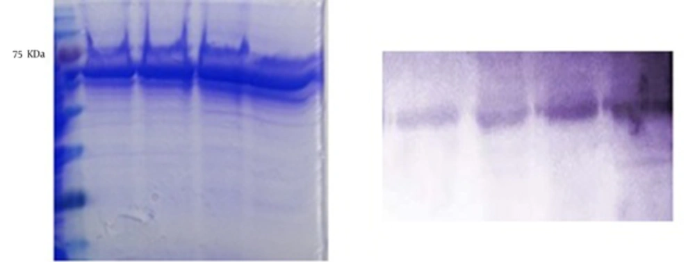
As shown in Figure 1, the SDS-PAGE analysis showed that ORF2-NSP4 protein band appeared on 75 KDa. The western blot analysis confirmed the SDS-PAGE results. The yield of protein was about 2 mg/L.
4.2. The Subcloning of the Truncated ORF2-NSP4 Gene in pET28a Plasmid
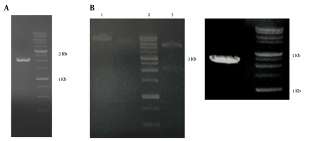
The truncated ORF2-NSP4 gene was double digested, purified, and inserted in linearized pET28a. The subcloned gene was confirmed by single, double digestion, and PCR. The size of truncated pET28a-ORF2-NSP4 recombinant plasmid was 7390 bp (Figure 2A and B).
A, the truncated ORF2-NSP4 gene double digested with BamHI and SalI, 2000 bp. B, left, Lane 1, the truncated ORF2-NSP4 single digestion with BamHI; Lane 2, ladder 1 kb; Lane 3, The truncated ORF2-NSP4 double digestion with BamHI and SalI, two bands appeared at 2000 bp and 5369 bp; right, PCR with T7 promotor primers, the ORF2-NSP4 band appeared at 2300 bp.
4.3. The Determination of Protein Solubility
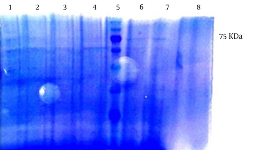
In order to determine the solubility of protein, the recombinant pET28a- ORF2-NSP4 protein was transformed in E. coli BL21 and then lysed by lysis buffer. The SDS-PAGE and western blot analysis showed that many target proteins appeared on precipitant. These results indicated that ORF2-NSP4 proteins formed inclusion bodies and the proteins precipitated (Figure 3A).
A, the determination of solubility of the ORF2-NSP4 protein by SDS-PAGE. Lane 1, supernatant; and Lane 2, precipitant. The 75 KDa ORF2-NSP4 bond appeared with more concentration in the precipitant. B, the SDS-PAGE analysis of solubilized inclusion bodies of the ORF2-NSP4 protein. Lane 1, the pre induction of the ORF2-NSP4 protein. Lane 2, supernatant of ORF2-NSP4 protein. Lane 3 - 9, washing steps with urea 4M. C, the western blot analysis of solubilized inclusion bodies of the ORF2-NSP4 protein. Lane 1, the pre induction of the ORF2-NSP4 protein. Lane 2, the supernatant of the ORF2-NSP4 protein. Lane 3 - 8, washing steps with urea 4M.
4.4. The Solubilization of Inclusion Bodies Target Protein
The protein was lysed under denaturing condition several times and protein bands were appeared in supernatant of washing steps 3, 4, 5, and 6 (Figure 3A and B).
4.5. The Purification of the ORF2-NSP4 Protein
The target protein was expressed and purified by Ni2+ chelate affinity chromatography. The expression and purification of the truncated ORF2-NSP4 protein were confirmed by SDS-PAGE. The SDS-PAGE analysis showed a protein band at about 75 kDa, which was in agreement with the expected molecular weight. SDS-PAGE of the purified ORF2-NSP4 protein showed that the target protein appeared in the elute of elution buffer (pH 4.5). The purity of the protein was above 80%. The yield of the purified protein was about 1 mg/L (Figure 4).
4.6. Electron Microscopy
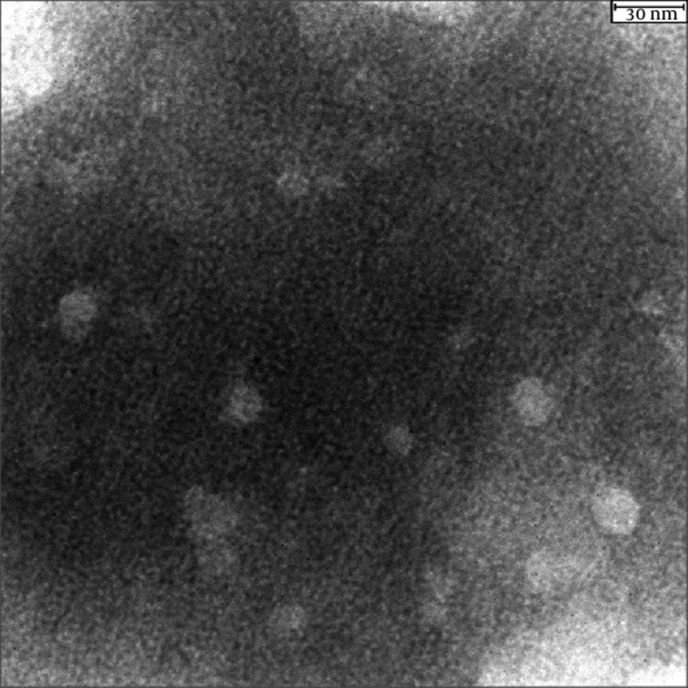
As shown in Figure 5, the results of electron microscopy showed that VLP of ORF2-NSP4 was successfully constructed in the eukaryotic system while not developed in the prokaryotic system.
5. Discussion
In the present study, the attenuated form of Rotavirus NSP4 was fused to the truncated ORF2 (112 - 607). The protein was successfully assembled, and then it could form VLP in the eukaryotic expression system (BES). Because of their immunogenicity, the truncated and full length of ORF2 capsid proteins of hepatitis E virus has been used in many expression systems for vaccine development. The hepatitis E virus (HEV; 112 - 607) and HEV (368 - 606) vaccines developed VLP in BES and bacterial expression systems, respectively (1, 7). The resulting VLP has 23.7 nm diameter with T = 1 icosahedral symmetry and consists of 60 copies of truncated ORF2 while the native virion has 27 nm diameter and T = 3 icosahedral symmetry (16).
The Baculovirus expression system acts as a highly efficient insect system for the HEV VLP vaccine development because of 1, its ability to produce large amounts of recombinant proteins by maintaining capsid protein structure; 2, minimizing the risk of culture contaminant pathogens; 3, having a limited host range causing no threat to vaccinated persons; 4, easy inactivation by simple chemical materials; 5, easy purification due to its accumulation in cytoplasm; 6, being suitable for large-scale vaccine production; and 7, ability to post-translational modification (3, 5, 17). In spite of the higher safety of subunit vaccines, their efficacy compared to full pathogen-based inactivated or live attenuated vaccines is lower. Therefore, higher or booster doses and in some cases the co-administration of adjuvants are required (5).
Until now, two HEV VLP vaccine candidates, baculovirus 56 KDa protein (Novavax) in phase I/II trials and E. coli HEV 239 protein in phase II/III trials (Hecolin), have been administrated by alum adjuvant (7). Eosinophilia, sterile abscesses, and myofascitis are some reported side effects of alum (18). Therefore, using a new adjuvant with the ability to induce mucosal immune system and lower side effects seems to be necessary. In addition to induction of protective immunity against infectious diseases, the mucosal adjuvants are believed to have a potential to treat immunological diseases and can potentiate the immunity of mucosal barriers against all pathogens by preventing attachment, colonization, penetration, and replication in the gastrointestinal mucosa (19). Previous studies have indicated that mutant forms of Cholera (mCT) and heat-labile toxin (mLT) without toxicity, while retaining adjuvant properties, induce the mucosal immunization. The NSP4 of Rotavirus could induce the mucosal immunization in Rotavirus VLP vaccine (11).
The continuous cell culture passage of virulent strain of NSP4 (OSU-v) can produce an attenuated strain (OSU-a) with the lowest pathogenicity (20). Previous studies have shown that the insertion of a peptide at C-terminal of HEV not only affects self-assembling of VLP formation, but also helps the produced VLP be used as carrier for other antigens (21, 22). Our results in agreement with previous reports showed that the OSU-a strain of Rotavirus NSP4 was successfully linked to C-terminal of the truncated ORF2 to assemble to VLP. It has been reported that HEV (368 - 606) VLPs formed in the bacterial expression system did not have a homogenous structure, as they lacked post-translational modification (1). Our study investigated the development of HEV ORF2 (112 - 607)-NSP4 VLP vaccine in bacterial and BES expression systems showing that VLP of truncated ORF2-NSP4 was formed in BES, but not in the bacterial expression system. According to the non-toxicity of ORF2-NSP4 protein to E. coli, as shown by the present results, the unsuccessful formation of VLP in E. coli may be resulted from 1, Unrefolding of protein after urea treatment; 2, Low concentration of protein after purification, and/or 3, No self-assembling of recombinant ORF2-NSP4 protein as a result of the insufficient post translational modification.
6. Conclusions
Regarding the established role of Virus like particle in the induction of mucosal immune response by oral administration (21, 23) and the protective effect of anti-NSP4 antibody, the truncated ORF2-NSP4 was developed as a recombinant candidate vaccine to protect against both HEV and Rotavirus. Moreover, the adjuavancity of NSP4 could increase the efficacy of ORF2 (11, 12). In this study, the fusion of NSP4 to the truncated ORF2 was successfully assembled to VLP in BES. However, further studies need to be performed to evaluate the VLP development in E. coli and the immunity of the truncated ORF2-NSP4 protein as a potential candidate vaccine against HEV and Rotavirus.


