1. Background
Staphylococcus aureus has different virulence factors such as enterotoxins that could act as superantigens. These superantigens could stimulate T lymphocytes and trigger a cytokine storm (1). In addition, these enterotoxins are among the most common causes of food poisoning (2, 3) as well as chronic diseases (4, 5) such as septic shock and arthritis (6). However, antibiotic resistance and production of toxins have made S. aureus an opportunistic pathogen (7).
One of the most important enterotoxins that are produced by S. aureus is staphylococcus enterotoxin E (SEE) (8). This superantigen is one of the classical superantigens, which has the most similarity with staphylococcal enterotoxin A (SEA), and the lowest prevalence among others (9). Some reports stated that 84% similarity exists between SEE, SEA, and staphylococcal enterotoxin P (SEP) with respect to their genome sequences (10). The existence of S. aureus enterotoxins in patient’s blood has not been considered. However, in the case of septic arthritis, staphylococcal species strain with antibiotic resistance were isolated from synovial fluid of rheumatoid arthritis (RA) patients (11).
Recent studies were shown that the molecular fingerprinting of a collection of 94 S. aureus isolates from patients with osteomyelitis and methicillin-resistant was significantly higher among others that were not resistant (12). The results of another study showed that superantigens as virulence factors are effective in staphylococcal infections (13). The role of staphylococcal superantigens as virulence factors in some chronic diseases such as RA and nonseptic arthritis has been reported (6). Thus, the purpose of this study was to assay the staphylococcal enterotoxin E in RA patients’ blood.
2. Objectives
This study aimed to assess staphylococcal enterotoxin E in the blood of patients with rheumatoid arthritis.
3. Patients and Methods
S. aureus strain, which contained the entE gene and producing enterotoxin E isolated from the clinical samples and were characterized by Gen Bank reference (M21319.1) as the standard strain (14).
3.1. Blood Sample Collection
From April 2011 to August 2013, a total of 83 patients with RA were referred to laboratory by rheumatologists. Inclusion and exclusion criteria of the RA patients were based on ACR 2010 criterion (15). This project was approved by Ethics Committee of Baqiyatallah University of Medical Sciences on November 29, 2011 with Code No: 24 Paragraph 28. Sampling has been carried out based on Microbial Standard Protocol and by perfect observance of aseptic conditions. Thus, after disinfecting the sampling site, 5 to 10 mL blood were taken by syringe. Then, 3 - 5 mL of the blood was inoculated into Castaneda medium (Baharafshan Co, Tehran, Iran) and has been incubated at 37°C for 48 hours. Next, 2mL of the collected blood has been added to sterile CBC tube and the remained was added to clotting tube.
3.2. Blood Culture
After inoculating Castaneda medium for 48 hours incubation at 37°C, 1 mL of Castaneda medium was aspirated and subcultured on blood agar plate (CONDA. SA). After incubation for 24 hours and at 37°C, the isolated bacterial strains have been studied by using colony characteristics, Gram stain, catalase, oxidase, motility, sugar fermentation, DNase (Merck Germany), coagulase tests, and susceptibility to bacitracin and novobiocin.
3.3. DNA Extraction from Bacteria
For performing the PCR method, the bacterial standard strain was inoculated into the 5 mL LB broth medium (Merck Germany) for 24 hours incubation at 37°C. The bacterial suspensions were centrifuged, and cellular sediments were obtained and then the DNA extraction has been done by using modified salting-out method (16).
3.4. DNA Extraction From Blood
In aseptic condition, patient’s blood has been carefully added to CBC tube and was centrifuged (10 min, 7000 × g in 4°C). Then, blood buffy coat was selected and transferred to DNA free sterile tube. Next, each of the blood buffy coat genomes has been separately extracted by CinnaPure DNA Extraction Kit (Cinnagen Co. Iran). Based on the Kit instructions, the amount of 100 μL of blood buffy coat has been added to the Kit’s microtube, then 400 μL of lysis buffer was added to it and vortexed for 20 seconds, then 300 μL of precipitation solution has been added to microtube and again vortexed for 5 seconds. In this stage, all microtube components have been transferred to the column, and centrifuged (1 minute, 5°C, 13000 × g). Then, the column has been transferred to the new microtube and has been washed by 400 µL of washing buffer 1, and again was centrifuged (1 minute, 5°C, 12000 × g). Then, for the second time, the column was transferred to the other new microtube and washed by 400 µL of washing buffer 2, and again was centrifuged (1 minute, 5°C, 12000 × g). After drying and changing the column, the amount of 30 μL of elution buffer was added and centrifuged (5 minutes, 12000 × g), finally the DNA quality and quantity were measured by NanoDrop (Thermo Scientific NanoDrop 2000 Spectrophotometer, USA).
3.5. Primer Design
Using online Gene script software, the primer was designed on the basis of the reference sequence (S. aureus MW2 enterotoxin E gene, access number: M21319.1) and was analyzed by primer3 software. In addition, multiple alignments were carried out by DNASIS MAX trial version. This primer pair was amplified a 693 bp fragment.
3.6. Polymerase Chain Reaction
PCR method has been set up for the diagnosis of enterotoxin E gene, by using the specific primer pairs, which amplified a 693 bp fragment and the standard strain genome. For DNA amplification, the master mix was made in 200-μL microtubes using a 25 μL reaction mixture that contained the 2 μL DNA template, 0.3 U of Taq DNA polymerase, 2.5 μL of 10X PCR buffer as well as 0.16 mM of each dNTPs, 2 mM MgCl2 (all reagents were from Cinnagen Co, Iran), 10 pmol of the primer pair (synthesized by Cinnagen) and double-distilled water, to a final volume of 25 μL. All amplifications were carried out in a thermal cycler (Bio-Rad, C1000) with initial denaturation at 95°C for 3 minutes followed by 35 cycles of denaturation at 94°C for 30 seconds, primer annealing at 61°C for 40 seconds and extension at 72°C for 40 seconds, followed by a final extension at 72°C for 5 minutes. The amplified PCR products have been electrophoresed in a 1.5% agarose gel for 45 minutes, and then stained with ethidium bromide for 20 minutes (0.05 mg/mL; Sigma Aldrich). The gels were photographed under ultraviolet light using Gel Doc (Bio-Rad Universal Hood II, USA). Also, molecular size markers (50 and 100 bp) have been included in each agarose gel. Then, all sample buffy coat genomes have been assayed by the optimization PCR.
3.7. Enzyme-Linked Immunosorbent Assay
The indirect Enzyme-Linked Immunosorbent Assay (ELISA) was used for the validation of the enterotoxin E existence in patients’ blood samples. First, patients’ plasma (after that CBC tube has been centrifuged in 10 minutes with 7000 × g) was taken out and assessed by ELISA with the following procedure. Based on previous report (17), 50 µL of patients’ plasma have been mixed with 50 µL of phosphate-buffered saline as diluents. Then, they were added to the defined wells of ELISA plates that were previously coated with polyclonal monospecific antibody against staphylococcal enterotoxin E (Rb PAb staphylococcus enterotoxins E 15922 500 lot 713307 Abcam). After one hour incubation and three times washing, 50 µL conjugate antibody was added. Then, enzyme substrate was added, and finally 100 µL stopping solution was added. After 10 minutes of this process, the color changed. Finally, the wells absorption were measured by the ELISA reader device in 450 nm wavelength and then the cut off value has been calculated for each plates.
4. Results
A total of 83 blood samples were assessed patients with RA from April 2011 to August 2013. During this research and after sequential sub cultures, only 5 bacterial species were isolated. Based on the results of biochemical tests, just one case has been detected as S. aureus and others as S. intermedius, as well as three Gram negative bacterial strains: two strains as Psedumonas putida and one strain as P. aeroginosa.
4.1. The Results of DNA Extraction
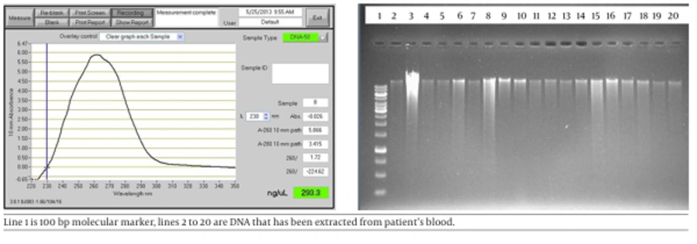
The quality and quantity of the standard strain and patients sample genomes have been examined by both gel electrophoresis (the agarose gel concentration 0.8%, in 100 volt for 45 minutes) and NonoDrop device. Anyhow, both results were acceptable.
4.2. The Results of Primer Design and PCR of the Blood of Patients with Rheumatoid Arthritis
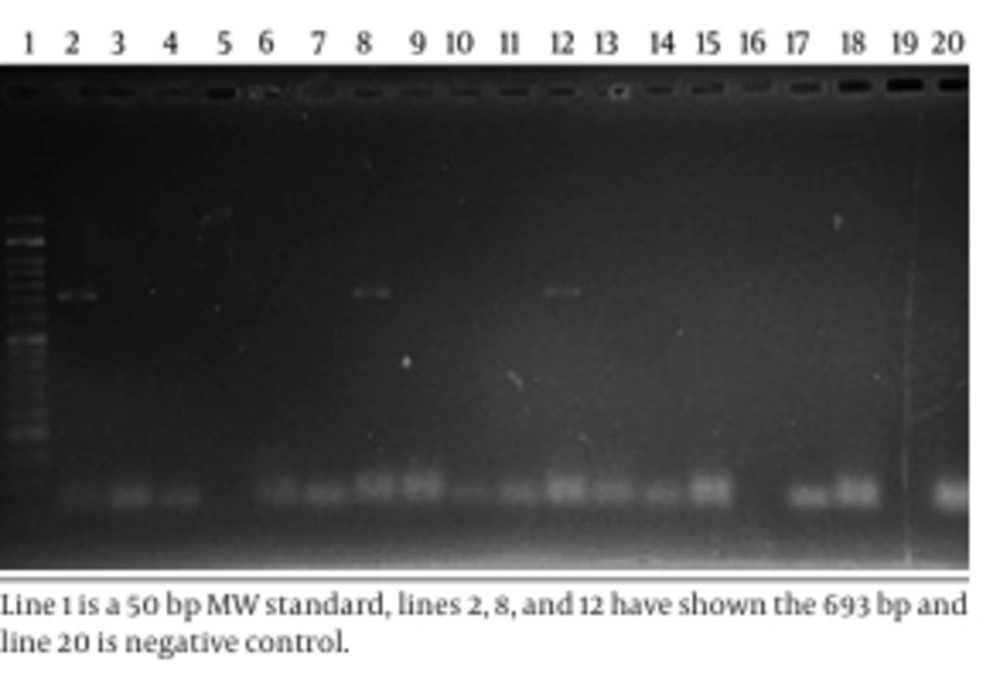
The results of primer designing were as follows: F- gtagc GGATCC agc gaa gaa ata aat gaa a and R- gcgcg AAGCTT tca agt tgt gta taa ata c, which were amplified at 693 bp amplicons as PCR products. After some examinations based on the standard strain genome and Master Mix components, PCR has been set up and applied as a positive control for each test. The results of the samples’ PCR with amplifier primer of a 693 bp fragment suggested positive in 13.25% (Figure 2). Of samples, there was a part of entE gene. Figure 2 shows the result of PCR product electrophoresis with amplifier primer of a 693 bp fragment for a number of samples.
4.3. The Results of Enzyme-Linked Immunosorbent Assay (ELISA)
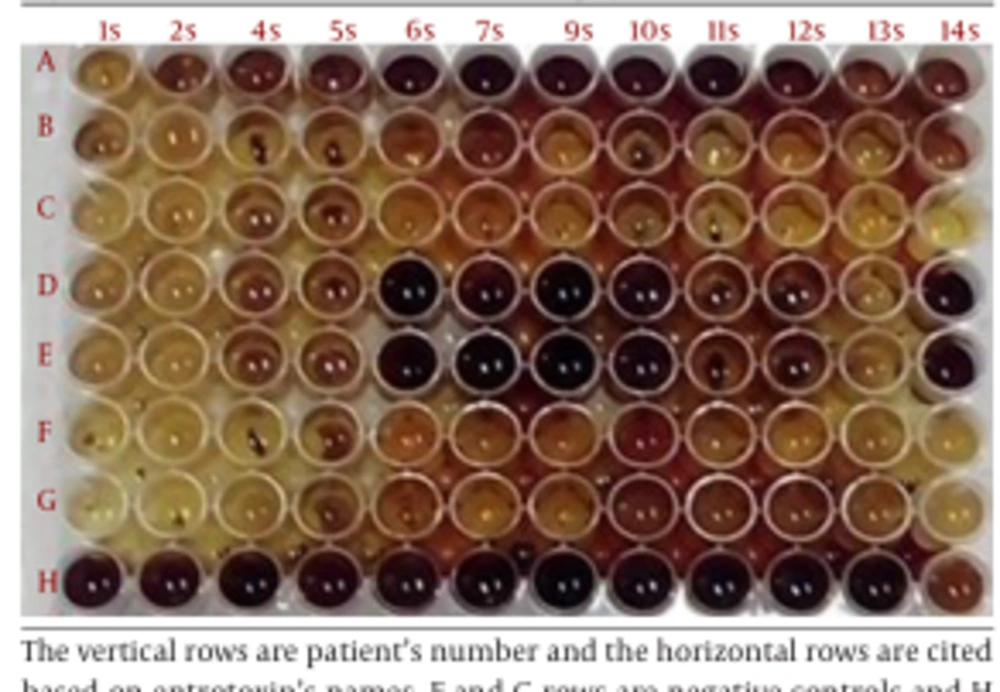
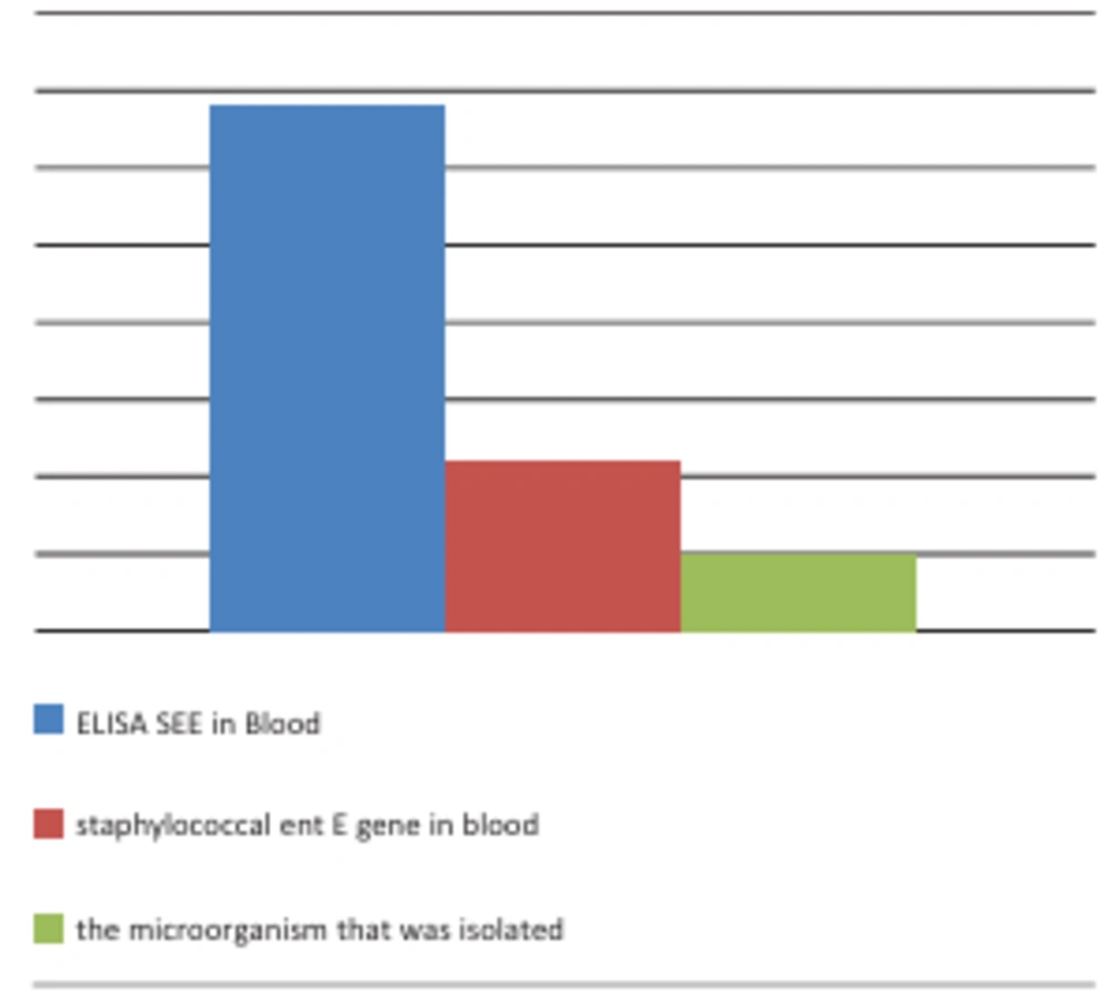
The results of Enzyme-Linked Immunosorbent Assay ELISA plates indicated that 40.96% of RA boold (plasma) were positive for staphylococcal enterotoxin E (Figure 3). The comparative results of PCR, ELISA, and bacterial culture were illustrated in Figure 4.
5. Discussion
The exact etiology of RA is not determined yet; nevertheless, all scientists have recognized it as an inflammatory reaction. To clarify the disease etiology, many researches were conducted. For example, Sioud et al. designed and conducted a study on TcR V α in IL-2R + T cells from 6 patients, which showed restriction in their SF T cell receptor variable region β (TcR V β) gene usage (18). In another study, the correlation between the inflammatory proteins such as IL-1β; IL-6 and fibrinogen as well as Tumor Necrosis Factor alpha (TNF-α) and the number of blood monocytes were demonstrated (19). An experimental model by using intravenous injection of toxic shock syndrome toxin-1 (TSST-1) provides a means to examine the possible role of superantigens in RA and related diseases, and also to analyze the cellular and molecular pathways induced by microbial products (20).
It is reported that the Mycoplasma arthritidis products, named M. arthritidis-T cell mitogen (MAM) and Mycoplasma arthritidis-derived superantigen (MAS), have biological activities in common with superantigens such as MAS, staphylococcal enterotoxin E (SEE), or lipopolysaccharide (LPS). The ability of MAM to induce proinflammatory interleukin 1 beta (IL-1 beta) and tumor necrosis factor alpha (TNF-α) gene expression in the THP-1 monocytic cell line were examined and showed that play an important role in the pathology of arthritis in rodents, which closely resembles human RA (21, 22). The results of one investigation revealed that the level of IgM and IgG antibodies against staphylococcal enterotoxin D (SED) were higher compared to normal cases. The investigators concluded that stimulation of B cells using SED preferentially induced RF plus B cells in normal controls and in patients with seronegative and seropositive RA (23).
In one study, T cells were cultured with SEB in the presence of interferon-γ (IFN-γ)-treated synovial cells. Then, T cell proliferation and activation were assessed by 3H-thymidine incorporation and IL-2 production and showed that activated synovial cells are potent antigen-presenting cells for SEB to T cells, and concluded that that superantigen (SEB) may play a critical role in the pathogenesis of RA through activated synovial cells (24). However, large numbers of studies have been carried out and results are variable with some authors indicating evidence for the effect of uncharacterized superantigens expanding or deleting T cells with particular V beta regions. While, others have suggested that observations of restricted V region usage and limited junctional regions imply that clones of cells have been expanded by antigen (25). An experimental study results revealed that proliferation and interleukin-2 (IL-2) production was dependent on the dose and type of staphylococcal enterotoxins (SEs) (26).
To investigate the role of superantigen in rheumatoid arthritis (RA), serum IgG and IgM SEB antibodies were measured using an enzyme linked immunosorbent assay (ELISA), and confirmed by Western blot analysis. The results indicate that patients with RA have increased levels of serum IgM SEB antibody compared to normal subjects. In this study, the titers of rheumatoid factor (RF) showed no correlation with the levels of IgM SEB antibodies (27). The results of another study suggest that superantigens are implicated in the pathogenesis of certain autoimmune diseases such as multiple sclerosis, RA, Sjogren's syndrome, and Kawasaki's syndrome (28).
The results of a study showed that superantigen stimulated mononuclear cells from the synovial fluid of patients with RA with higher levels of TNF-α production (29). By using Fas-defective MRL-lpr/lpr mice, the effects of the staphylococcal enterotoxin B (superantigen) were studied on the development of autoimmune, inflammatory joint disease in susceptible animals to the development of rheumatoid arthritis-like disease and showed that a single intraperitoneal injection of staphylococcal enterotoxin B (SEB; 10 µg/mouse) caused a mild, inflammatory arthritis plus 30 days post challenge in the knee joints of young (< 2-month-old) MRL-lpr/lpr mice. They concluded that SEB is an extremely potent macrophage-activating factor both in vitro and in vivo, enhancing several aspects of autoimmune disease in MRL-lpr/lpr mice (30).
In an investigation, the effects of the superantigens staphylococcal enterotoxin A, staphylococcal enterotoxin B, and streptococcal M type 5 protein on T cells derived from inflammatory tissues and peripheral blood (PB) of patients with arthritis were studied and demonstrated the heterogeneities of such superantigen-mediated specific cell lysis (31). Another investigation showed that superantigen toxic shock syndrome toxin-1 (TSST-1) stimulated T cells from Rheumatoid arthritis synovial fluid mononuclear cells (RASFMC) have the ability to induce chronic arthropathy with fibroblast proliferation and neovascularization in the severe combined immunodeficient (SCID) mouse (32). The selected elevation of antibodies to MAM in RA sera was subjected to study and showed that MAM or a MAM-like molecule might be associated with RA, whereas elevation of antibodies to SEA and SEB in sera from patients with rheumatic diseases was less specific (33).
The prevalence of S. aureus nasal carriage and comparing antibody responses to two superantigens, staphylococcal toxic shock syndrome toxin-1 (TSST-1) and staphylococcal enterotoxin A (SEA), in patients with RA and normal subjects were studied and revealed that, patients with RA more often carry S. aureus in their nasal vestibule, and have higher average antibody levels to TSST-1 (34). Previous reports showed that streptococcal IgG Fc receptors and RFs bind to the same amino acids on the Fc molecule. This complex pattern may play a role in the pathogenesis of RA (35). In addition, the possibility that IgM rheumatoid factors bind to streptococci was studied (36). In an experimental study by subcutaneous injection of staphylococcal enterotoxin B (SEB), severe arthritis was induced in DBA/1J mice, which had been previously immunized with bovine type II collagen and suggested that this experimental arthritis model may provide a means to examine the role of superantigens and the efficacy of pharmacological agents for the treatment of rheumatoid arthritis (37).
The prevalence of oral staphylococcal carriage in patients with RA compared with healthy controls was carried out and showed that oral carriage of S. aureus appears to be common in patients with RA and studies of the mouth as a source of infection in septic arthritis would be merited (38). It is reported that in early stages of untreated RA, simultaneous IL-17assessment of serum, SF, and synovium might be valuable in defining activity and predictive patterns of RA, given that synovium is highly suggestive for disease aggressiveness and might express specific therapeutically targets (39). So far, there is no reports indicative of the direct identification of an antigen or superantigen in blood or synovial fluid of patients, which plays a role in RA pathogenesis.
In this study, a total of 83 blood samples from patients with RA have been assayed for the presence of S. aureus enterotoxin E using bacterial culture, ELISA, and PCR methods. The result of bacterial culture showed that there were only two isolated bacteria as Gram-positive (S. aureus and S. intermedius). The results of ELISA test on extracted of overnight isolated culture showed no existence of enterotoxin E in all bacterial isolated cases. In addition, DNA extraction and PCR method for the presence of enterotoxin E gene for all bacterial isolates were negative. In fact, enterotoxin E or other proteins in culture extracts of isolated bacteria that could react with anti-enterotoxin E antibody in ELISA test have not been found.
While, the result of ELISA and PCR assessments of 79 blood buffy coat indicated the presence of enterotoxin E in 34 cases (40.96%) and the presence of enterotoxin E gene in 11 cases (13.25%). The interpretation of these results is difficult. Because first of all, S. aureus produces more than 20 types of enterotoxins and there is a great similarity among them. Secondly, in spite of their different structural features, they have similar mechanism of action. Meanwhile, this study aimed at detection of only one enterotoxin. It is noteworthy that, the PCR and ELISA results were the same only in 10 cases. In one case, the PCR result for the presence of enterotoxin E gene was positive. While ELISA results for the existence of enterotoxin E were negative. The reason is probably the lack of gene expression or inadequate production of enterotoxin that was under of ELISA sensitivity.
The important finding of this study was that most of the samples -in 24 cases- showed positive ELISA results. Whereas, negative results for PCR was obtained. PCR has been repeated by the different Genomic DNA Extraction Kit that referred to the differences between PCR and ELISA results have been decreased, albeit not completely. Perhaps the difference is due to the similarities between enterotoxin E with other enterotoxins such as enterotoxins A and P. However, the results of this study indicate that the PCR molecular method and ELISA can detect enterotoxin E in the blood of patients with RA. The results have created an important question. Why has the toxin been found in the patient’s blood, while the bacterial growth in blood cultures is negative? To provide answers to this question, further research is needed. In addition, there is another important question. Why is still the role of staphylococcal enterotoxins as superantigens in inflammation and RA discussed? However, there has been no attempt reported so far for detection of these enterotoxins in patient’s blood. Perhaps one reason is the inability of the different tests. In our previous study, by using commercial ELISA kit, enterotoxin A, B, C, D, and E had been detected in synovial fluid of patients (17), and confirmed by immunoblotting. Thus in the present study, in order to determine the staphylococcal enterotoxin E, we focused on enterotoxin E recognition by ELISA and PCR in patient’s blood with RA. However, not only the results of this study have shown some evidence regarding endogenous origin for involved superantigens in patients with RA but also could provide a good model for the diagnosis of RA etiology. However, owning to the fact that there are no similar studies for comparison and analyses of the results, more research with a larger sample size is recommended.


