1. Background
Stroke is the second leading cause of death and the fundamental cause of disability (1). In addition, stroke overlaps with cardiovascular disease in a manner that cardiac complications are the second most common cause of death in stroke patients. About 20% of mortality after the stroke occurred because of cardiac morbidities (2). Many patients have reported both cerebral and ischemic heart disease simultaneously (3). Troponin I levels have been suggested as a non-invasive measure for acute ischemic heart disease (4) and an essential biomarker of acute myocardial infarction. Because of association between ischemic heart disease and stroke, troponin level is routinely measured at the time of stroke (5, 6).
Electrocardiographic investigation and measures of cardiac enzymes, including troponin I, were routinely performed among patients with stroke (7). Increased cardiac troponin was reported from 0 to 34% among stroke patients (8). Studies have also shown that some electrocardiographic abnormalities are associated with ischemic stroke. Although studies have revealed an association between ECG characteristics and troponin level, controversies remain about changes in troponin in acute cerebrovascular disease and the clinical implications of electrocardiography (9). A study in China reported an elevation of troponin in 18.6% of 178 patients with acute ischemic stroke. In addition, myocardial infarction with increased ST-segment was observed in 35 people and 35 patients with no ST-segment elevation. In addition, a significant association was reported between ST elevation myocardial infarction (STEMI) and troponin (10).
Faiz et al. reported an increased troponin level in 54.4% of patients with ischemic stroke (8). Pathological Q wave, elevation or depression of the ST segment, inversion of the T wave, more frequent atrial fibrillation/flutter, and left bundle branch block were reported among patients with hs-cTnT within the reference limit compared to patients with elevated hs-cTnT (8). Abnormal troponin level was reported among 12.7 and 17.6% of patients with ischemic stroke in Iran (11, 12). More investigation on the troponin level and its association with ECG characteristics among patients with stroke is necessary. Understanding the relationship between troponin changes and ECG can develop interventions to reduce morbidity and mortality associated with acute stroke (13).
2. Objectives
The present study evaluated the prevalence of positive troponin among Kurdish ischemic stroke patients. This is the first report about troponin I level among this ethnic group. In the next step, electrocardiogram, demographic and medical characteristics were compared between positive and randomly selected negative troponin patients.
3. Methods
3.1. Participants
All patients diagnosed with acute ischemic stroke who were admitted to Imam Reza Hospital in Kermanshah, Iran, from 21 March 2018 to 20 March 2019 were recruted. The troponin I level was routinely measured among stroke patients in the hospital. All patients with positive troponin were recruited as a case group. In addition, a group of patients with negative troponin levels was randomly selected as a control group. The exclusion criteria were pregnant and breast-feeding patients, patients with renal impairment with GFR < 30, severe dehydration, pulmonary edema symptoms, an acute or chronic pulmonary disease requiring intermittent or permanent oxygen supplementation with hemorrhage (ICH) after an ischemic stroke detected by brain CT scan. During the study, 745 patients were admitted to the hospital due to acute ischemic stroke. There were ten positive and 735 negative troponin levels among them. Among this group, 149 patients were randomly selected as control group.
3.2. Procedure
Stroke was diagnosed by a neurologist experienced in the field of stroke. The severity of stroke was determined by the National Institute of Health Stroke Scale (NIHSS) criteria, completed for all patients by a neurologist. Brain CT scans were taken from all patients. Medical history and demographic information were collected and documented in the electronic file of each patient. All stroke patients in the hospital were referred for more electrocardiography and troponin measurement. The study was conducted based on international ethical considerations and approved by the Ethical Committee of Kermanshah University of Medical Sciences (Ethic Code: IR.KUMS.REC.1397.645). All participants, or their first family members, provided written informed consent to participate in the study.
3.3. Measures
3.3.1. Medical History and Demographic Information
Data about age, sex, blood pressure, diabetes, history of myocardial infarction, and previous stroke were collected using a questionnaire.
3.3.2. The Severity of Stroke
NIHSS was performed for all patients by a neurologist. NIHSS consists of 15 items to determine the severity of stroke. This tool measures the function of all parts of the body with scores between 0 and 42, and higher scores indicate a more severe stroke. Patients are divided into mild (NIHSS < 7), moderate (NIHSS = 7 - 13), and severe stroke (NIHSS > 13) (14, 15).
3.3.3. Level of Troponin I
Serum level of troponin I was measured by the electrochemiluminescence (ECL) technique using a Cobas Immunoassay Analyzer (Cobas E411, Roche Diagnostics, Germany). Serum troponin was measured by enzyme-linked immunosorbent assay kits, CAN-DHT-280, Version: 0.5 (DBC-Diagnostics Biochem Canada, Canada). The absorbance level was read using an Awareness Technology STAT FAX 2100 Microplate Reader (Awareness Technology, USA).
3.3.4. Electrocardiographic Evaluation
A cardiologist and a neurologist performed and interpreted comprehensive electrocardiography examinations. Q wave, T wave inversion, and ST-segment elevation or depression were evidence of coexisting cardiac ischemia.
3.3.5. Data Analysis
The statistical package for social sciences (SPSS) (SPSS, Inc., Chicago, IL) version 23.0 was used for the statistical analyses. The descriptive data were presented, and chi-square and Mann-Whitney U tests were used for between-group comparisons.
4. Results
4.1. Demographic and Medical History Findings
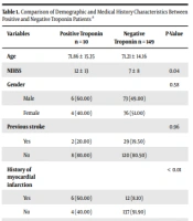
Among 745 patients with ischemic stroke admitted to Imam Reza Hospital, Kermanshah, Iran, from 21 March 2018 to 20 March 2019, just ten patients (1.3 %) (six males and four females; mean age = 71.86) had positive troponin. As a control group, 149 patients (73 males and 76 males; mean age = 71.21) with negative troponin were randomly recruited. The two groups were not significantly different in demographic and medical history characteristics. Only the history of myocardial infarction among the positive troponin group was significantly higher than the negative (Table 1).
| Variables | Positive Troponin, n = 10 | Negative Troponin, n = 149 | P-Value |
|---|---|---|---|
| Age | 71.86 ± 15.35 | 71.21 ± 14.16 | |
| NIHSS | 12 ± 13 | 7 ± 8 | 0.04 |
| Gender | 0.58 | ||
| Male | 6 (60.00) | 73 (49.00) | |
| Female | 4 (40.00) | 76 (51.00) | |
| Previous stroke | 0.96 | ||
| Yes | 2 (20.00) | 29 (19.50) | |
| No | 8 (80.00) | 120 (80.50) | |
| History of myocardial infarction | < 0.01 | ||
| Yes | 6 (60.00) | 12 (8.10) | |
| No | 4 (40.00) | 137 (91.90) | |
| Hypertension | 0.10 | ||
| Yes | 9 (90.00) | 97 (65.10) | |
| No | 1 (10.00) | 52 (34.90) | |
| Diabetes | 0.87 | ||
| Yes | 2 (20.00) | 33 (22.10) | |
| No | 8 (80.00) | 116 (77.90) |
a Values are expressed as mean ± SD or No. (%).
The results for electrocardiogram abnormalities were also compared between the two groups. The highest and lowest frequencies were related to atrial fibrillation and premature ventricular contraction, with 13.2% (n = 21) and 1.9% (n = 3), respectively. A patient with positive troponin and 13.4% (n = 20) of patients with negative troponin had AF in their ECGs (P = 0.67). The frequency of other disorders, including T wave inversion, left branch bundle block, ST-segment elevation, and the presence of pathological Q wave was 6.3% (n = 10), 5.7% (n = 9), 5% (n = 8), and 5% (n = 5), respectively. The results also showed no significant relationship between positive (increase) or negative troponin with electrocardiogram abnormalities (P > 0.05) (Table 2).
| Anomalies | Positive Troponin, n = 10 | Negative Troponin, n = 149 | P-Value |
|---|---|---|---|
| Left branch bundle blocks | 0.99 | ||
| Yes | 1 (10) | 8 (5.4) | |
| No | 9 (90) | 141 (94.6) | |
| Premature ventricular contraction | 0.18 | ||
| Yes | 0 | 3 (2) | |
| No | 10 (100) | 146 (98) | |
| Inversion of the T wave | 0.27 | ||
| Yes | 6 (60) | 4 (2.7) | |
| No | 4 (40) | 145 (97.3) | |
| Increasing T segment | 0.32 | ||
| Yes | 1 (10) | 7 (4.7) | |
| No | 9 (90) | 142 (95.3) | |
| Atrial fibrillation | 0.67 | ||
| Yes | 1 (10) | 20 (13.4) | |
| No | 9 (90) | 129 (86.6) | |
| Pathological Q | 0.24 | ||
| Yes | 1 (10) | 4 (2.7) | |
| No | 9 (90) | 145 (97.3) | |
| Normal ECG | |||
| Yes | 9 (90) | 103 (69.1) | |
| No | 1 (10) | 46 (30.9) |
a Values are expressed as No. (%).
5. Discussion
Cardiac troponins T and I are sensitive and specific markers of myocardial injury (16). Elevated troponin is relatively common in acute ischemic stroke, associated with increased mortality and poor outcome (17, 18). Several mechanisms have been proposed as the possible cause of myocardial damage in stroke patients, including concomitant acute coronary syndrome, ischemic stroke, and “stroke-heart syndrome” (17, 18). Stroke-heart syndrome is related to catecholamine release, increased sympathetic activity, and subsequent myocardial injury (17). This phenomenon is more common within the first three days after stroke and may lead to cardiac dysfunction and arrhythmia (18).
Ischemic stroke and ischemic heart disease have similar risk factors and may coexist in the same patient (17). ECG abnormalities caused by acute stroke are still unknown. However, autonomic dysfunction due to overactive sympathetic activity was suggested. Some consider insular stimulation responsible for abnormal heart function in acute stroke. Inhibiting disorders of the compassionate nervous system increases catecholamine secretion. Previous studies have shown that renal failure, chronic heart disease, hypercholesterolemia, increased stroke severity, and the insular cortex’s involvement are significantly associated with increased troponin levels in patients with ischemic stroke (8).
The present study evaluated troponin’s level and compliance with electrocardiogram findings in patients with acute ischemic stroke. The results showed that the highest rate of electrocardiogram disorders was related to atrial fibrillation, respectively. Ischemic ECG changes are expected and well-known in elderly patients due to the impossibility of examining patients before the stroke and getting an ECG from them. Changes in the ECG can be compared to the previous one (19) because it is clear that it is impossible to do a study to record an ECG a few days before the stroke (9). In the present study, an increase (positive) of troponin was observed in 6.3% of patients. In previous studies, cardiac troponin measurement with standard assays in patients with acute stroke has been reported between 8.7% to 21.4% (10, 20, 21). A 2009 meta-analysis found that 18% of stroke patients had high troponin levels, and ECG changes were more likely in these patients (22).
Depression or elevation of the ST segment is one of the critical ECG changes, which is commonly associated with ischemic heart disease. Pathological Q waves indicated a previous or recent heart attack (15). Simultaneously, depression of ST may indicate an acute coronary heart syndrome (23). ECG changes are even more critical in patients with symptoms of acute coronary syndrome, such as chest pain or shortness of breath (8, 9). Inversion of the T-wave is also significant for neurological reasons (24). In the present study, pathological Q wave, ST-segment elevation, and a T-wave inversion were observed in 10%, 10%, and 60% of patients with increased troponin, respectively. Ahn et al. observed pathological Q wave in 18.2% and ST-segment elevation in 5.8% of these patients (20). Faiz et al. observed pathological Q wave in 5.3% of patients, ST-segment elevation increased, and T wave inversion in 21% (8).
Recently, cardiac troponins have been associated with the prevalence of atrial fibrillation and its diagnosis after stroke in the general population (25). Atrial fibrillation is the most common persistent cardiac arrhythmia, and its presence increases the risk of stroke by five times. There is still uncertainty regarding the pathogenesis of increased troponin in atrial fibrillation (such as tachycardia with rapid ventricular response) or heart failure (26). In the present study, atrial fibrillation prevalence was 10% in patients with elevated troponin levels. Another study showed that 22.3% of these patients have atrial fibrillation (20). The prevalence of atrial fibrillation in the present study was lower than in this study, possibly due to the difference in sample size between the studies.
The left branch bundle block was observed in 10% of patients in the present study. Kral et al. also observed 22% of patients with increased troponin changes in the ST-T segment, a left branch bundle block (27). Another study showed that 5.3% of these patients had left branch bundle block (8). The difference between the results of the present study and the mentioned studies can be due to the difference in the sample size. The primary mechanism of stroke after myocardial infarction is not known. However, factors such as the size of the thrombus, the arteries’ inflammation, and the platelets’ activity are significant. The recurrence of a stroke may also be increased by a history of heart disease and hypertension, which are more prevalent than ischemic stroke. Therefore, preventing and controlling these cases seem necessary (28, 29).
In Denmark, Jensen et al. evaluated 244 patients with acute ischemic stroke regarding cardiac enzymes (troponin and CK MB) and electrocardiogram within five days of admission (30). Elevated troponin T was observed in 10% of patients, and only 3% of patients with elevated cardiac enzymes had electrocardiographic changes (30). Abdi et al. examined 114 Iranian patients with a definite diagnosis of acute ischemic stroke regarding serum troponin level and electrocardiogram (11). Twenty patients (17.6%) had elevated troponin. This subgroup of patients tended to have older age, electrocardiographic change, and more severe stroke (higher NIHSS) than other patients. The electrocardiogram was normal in 6 patients (5%) with elevated troponin (11). The present study showed a much lower frequency of positive test results than these two studies. The patients’ blood samples were obtained very early at the emergency department, and this may suggest that insufficient time has passed for troponin elevation. The differences in the sensitivity of diagnostic laboratory kits may also have a role.
5.1. Conclusions
Based on the results, routine evaluation of troponin levels in all acute ischemic stroke patients is recommended. Troponin elevation may be associated with a cardioembolic source of stroke. Since all acute stroke patients receive routine ECGs, routine troponin assessment is debatable whether it is necessary for all patients or can be limited to those with electrocardiographic abnormalities.
