


1. Background
The thyroid gland is located in front of the trachea and consists of two lobes connected by a narrow tissue (isthmus). The thyroid hormone is very important in regulating metabolism (1). Thyroid volume increases in many diseases of this gland, which is known as thyromegaly or goiter (2). Measurement of the gland size is essential for assessing the need for surgery, calculating the therapeutic dose with radioactive iodine, and examining the treatment response.
Unlike clinical examination, ultrasound is a method of accurately calculating the thyroid volume (1). Determining the thyroid size is a fundamental variable in describing and detecting the disease, and standard ultrasound is mentioned in every report (1).
To evaluate the thyroid size, linear parameters of length, width (W), and thickness (depth) can be used, or its volume can be calculated through the mathematical calculation (length × W × depth × 0.529) (1).
Some authors believe that among the linear measurement parameters, the AP diameter (thickness) of the thyroid lobes is more accurate in detection of thyromegaly and can be used in this regard (1). On the other hand, thyroid volume depends on a person's anthropometric and racial characteristics (1).
Also, all thyroid dimensions don’t increase equally in thyromegaly. There have been very few studies on which of the thyroid dimeters is most affected by thyromegaly. Attempts were made in the present study to use an artificial neural network (ANN) to determine which of the thyroid dimensions was the most accurate measure of its volume and whether measurement of AP diameter, as a thyromegaly measure suggested by some researchers, is true or not in patients studied in the present study.
Artificial neural network systems have acknowledged distinct consideration because of their capability to learn complex issues with the aptitude to preserve accuracy, even in the lack of some data (3-6). Many studies have been performed using an ANN to help physicians diagnose various diseases and classify the types of diseases by using ultrasound (7-9).
However, no study has specifically investigated the relationship between thyroid dimensions and thyromegaly and determining the most important dimension of the thyroid by using ANN. Artificial neural network can determine which ultrasound measurement of the thyroid dimension can be a better estimate for measuring the thyroid volume by knowing which dimensions increase the most in thyromegaly, which accelerate the ultrasound process.
2. Objectives
Considering the need for a decision-making and decision-support systems to help radiologists in order to diagnosing thyromegaly, in this study an ANN-based system was established for automated diagnosing and detecting thyromegaly and find the most important dimension of thyroid size. The results of the proposed ANN-based diagnostic system can be effective in accelerating thyroid diseases diagnosis.
3. Methods
3.1. Participants and Data Collection
The present study is a descriptive-analytical study that began at Imam Reza Hospital in Kermanshah, Iran after being approved by the Ethics Committee of Kermanshah University of Medical Sciences (IR.KUSM.REC.1398.1013). The study population included all patients who referred to the hospital's ultrasound department between 2018 and 2019 at the request of an endocrinologist and underwent diagnostic examinations for thyromegaly. Individuals with normal ultrasound findings were considered as the control group according to the ultrasound standards, and those who were diagnosed with thyromegaly (as widely disseminated thyroid) were considered as the experimental group. Exclusion criteria included history of thyroid surgery, unspecified thyroid margin or any of the dimensions of the thyroid, presence of a huge mass or a very large cyst in a way that it is not possible to measure a clear thyroid volume, or lack of consent for participation in the study. The resident and the radiology professor gave full explanations about the project to eligible patients and they entered the project if they were willing to dos and after completing the informed consent form. At this stage, in the supine position, all patients underwent gray-scale ultrasound evaluation using a surface probe (16 MHz multifunction) of the Samsung Ws80 ultrasound device equipped with 4 active probes, minimum and maximum penetration depth of 2 to 30 cm and automatic imaging capability in B mode and Doppler mode.
Thyroid dimensions including longitudinal (L), anterior-posterior or depth (AP), and W diameter and isthmus thickness were measured and calculated and recorded along with the volume (10). Then, the thyroid volume was calculated using (W × height × depth × 0.52) formula and individuals whose thyroid volume was higher than 11.6 mL in men and 11.1 mL in women based on Nafisi Moghadam et al.'s study, were considered as patient with thyromegaly (11).
The recorded measurements of the two groups, along with the demographic information of the subjects (Table 1), were entered into Excel software based on the main objectives of the research and with the cooperation of all authors of the present article. The sample size formula was calculated by statistics specialist. The data were then analyzed using the MATLAB R2016b neural network toolbox in order to automatically detect the thyromegaly as well as to find the most important the measured ultrasound dimension.
3.2. Neural Network Design
Multi-layer perceptron (MLP) neural network were used in the present study (12). Multi-layer perceptron can differentiate data that is not linearly separable. The mechanism of learning of neural networks used in the present study was based on the concept of decreasing gradient. To teach this type of network, the law of back- propagation is commonly used, which has different algorithms. Levenberg-marquardet and scaled conjugated gradient algorithms were used in the current study (13).
Neurons number in the input layer was equal to the thyroid measured parameters. The number of hidden layers should be as small as possible. This parameter depends on the complexity of the problem and the number of dependent variables. It has been proven that each function can be approximated with a maximum of three hidden layers. There is no universal instruction for choosing the number of these layers. It can be said that most of the networks used have one, two, and three hidden layers, and networks with four layers and above have been rarely used (14). In the present study, the value of this variable is once considered 1 and 2 at another time. Although there are many experimental rules that can be used to select the number of intermediate layer neurons, the trial-and-error method is the best solution for determining the number of neurons in most cases. In order to increase the performance of proposed ANN system in the present study, the neurons number in hidden layers of the ANN system ranged from 1 to 70 and the network performance was considered for the remaining network topology with changed numbers of neurons in the hidden layer. Lastly the current assembly was selected with advanced performance topology (15).
Since there were two outputs (proportional to the presence or absence of thyromegaly), two neurons were considered in the output layer. Concerning neural networks and the implementation of its practical concepts, another parameter is the transfer function.
In this study, "hyperbolic sigmoid tangent" activation function was use because it lead to faster convergence (16).
In order to solve the overfitting problem (17), the solution was to stop the learning process early in the network configuration stage using accreditation data. Early stopping is a method to improve the generality of the network. This method consists of dividing the general data set into three sets of learning or teaching, testing and determining validity or validation instead of using only the first two sets of training and testing. Therefore, a total of 70%, 15%, and 15% of the data were used for training, validation, and testing for proposed ANN system (18).
The proposed ANN system was designed in the MATLAB ver. 2016. To evaluate proposed ANN system with the greatest performance, different networks and different topology were trained and the performance of these networks were obtained. To assess the proposed ANN system, confusion matrix was used and accuracy, sensitivity and specificity were calculated. The processing speed on a Sony VAIO F series laptop was approximately 5 minutes.
4. Results
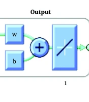
The present research has used a neural network, with a hidden layer and 3 neurons to assess the measured characteristics of a person's thyroid (Figure 1). Initially, measured thyroid characteristics were divided into training and experimental categories and a total of 110 primary data were used for training and 19 final data were also used for testing the neural network.

Neural network, with a hidden layer and 3 neurons
Then, in order to find the significance of each characteristic, regression analysis was performed, the results of which are revealed in Table 2. Finally, the characteristics of the right and isthmus lobes, the right lobe, the left lobe, and the isthmus and the left lobe were considered to be the input of the neural network, which are shown in Table 3.
| Variables | Specifications (R-Value) |
|---|---|
| Width of the right lobe | 0.68 |
| Depth of right lobe | 0.62 |
| Length of the right lobe | 0.61 |
| Volume of the right lobe | 0.59 |
| Width of the left lobe | 0.54 |
| Depth of left lobe | 0.57 |
| Length of the left lobe | 0.56 |
| Volume of the left lobe | 0.49 |
| Total thyroid volume | 0.56 |
| Isthmus | 0.35 |
Importance of Measured Thyroid Characteristics
| Variables | Characteristics of Two Lobes and Isthmus | Characteristics of the Right Lobe | Characteristics of the Right Lobe and Isthmus | Characteristics of the Left Lobe | Characteristics of the Left Lobe and Isthmus |
|---|---|---|---|---|---|
| Percentage of training accuracy | 98.18 | 89.09 | 85.45 | 84.55 | 82.73 |
| Percentage of test accuracy | 100 | 94.74 | 94.74 | 84.21 | 73.68 |
Results of Using Neural Network for Each of the Thyroid Characteristics Measured
As can be observed in Table 2, "width of the right lobe" is the most appropriate characteristic for detecting thyromegaly and "isthmus" is the least important one. Also, in general, the length, W and depth of the right lobe are more suitable characteristics for detecting thyromegaly as compared to those of the left lobe.
As can be observed in the Table 3, the accuracy of the network where all the characteristics are used is better than the rest. However, in general, use of the characteristics of the right lobe produces a relatively good accuracy, and isthmus information does not increase network accuracy very much. Therefore, it seems that there is no need to the isthmus parameter in determining the thyromegaly.
Therefore, the parameters can be ranked as follows based on their quality: (1) Characteristics of right lobe; (2) characteristics of left lobe; (3) characteristic of isthmus.
5. Discussion
The present study was performed on 131 patients referring to Imam Reza Hospital in Kermanshah in 2019. The patients included 87.8% women and 12.2% men in the 14 - 70 years group who referred to the ultrasound section for thyroid examination. The aim of the present study was to investigate the relationship between thyroid volume and the size of its lobes, as well as the isthmus diameter in people with thyromegaly (or goiter) through an artificial network. Based on the results, right lobe is able to detect thyromegaly with an accuracy of 94.74, and the isthmus information does not increase the accuracy of the network very much.
Of the patients enrolled to the project, 52.7% had thyromegaly based on the criteria suggested by Nafisi Moghadam et al.'s study conducted in Yazd. According to this study, which was performed on 314 people during the years 2003 - 2005, the mean thyroid volume was 9.08 ± 2.49 in men and 7.93 ± 3.2 in women (11). The neural network analysis showed that the highest correlation between the measured parameters (dimensions of each lobe and AP diameter of the isthmus) and thyroid volume in people with thyromegaly is related to the W of the right lobe (R = 0.924, P > 0.0001)
Overall, our results revealed that the dimensions of the right lobe are more suitable characteristics for detecting thyromegaly as compared to those of the left lobe, which may be due to the fact that the volume of the right thyroid lobe is relatively larger than the left one.
Tong and Rubenfeld investigated 256 patients with a large thyroid in the United States and concluded that as the thyroid grows, it extends upward, downward, and laterally, and the anterior posterior growth of the gland is relatively less affected (19). To justify their findings, they attributed the anterior posterior growth of the thyroid to the presence of a trachea behind the thyroid and anatomical limitations. However, their study was performed with a nuclear scan.
Rumack and Levine (1) mentioned that among the measured linear parameters of the thyroid, the AP diameter of lobes is the most accurate criterion for diagnosing thyromegaly, which is contradictory with the results of our study. This inconsistency can be because of racial, anthropometric, or regional differences in their studied population and the population of the present study, and further research is needed.
Suzuki et al. (20) showed in a study in Japan that the size of the right lobe was larger than the left lobe, which is consistent with the present study.
Kamran et al. (21) showed in a study in Karachi that there was no significant relationship between isthmus AP diameter and its volume.
The present study also showed the lowest correlation between isthmus volume and AP dimeter.
Although there have been relatively many studies on the relationship between thyroid size and demographic and regional characteristics, as well as height, weight, BMI, and body surface area characteristics, the correlation between thyroid size and volume has not been studied except in Tong and Rubenfeld (19), and Rumack and Levine’s studies (1).
The present study shows the need for further study of the relationship between thyroid dimensions and its volume in different populations. The limitations of the present study include the number of subjects, the hardware required, and the non-uniformity of the subjects. By increasing the number of features required for the neural network and better hardware, the accuracy and speed of detection and processing can be improved.
5.1. Conclusions
The results of this study using ANN system showed that W of the right lobe is the most suitable characteristic for detection of thyromegaly and isthmus is the least important characteristic. Also, in general, the length, W, and AP diameter of the right lobe have more suitable characteristics for diagnosing thyromegaly than those of the left lobe. Network accuracy is better than all others using all the characteristics. However, in general, using the characteristics of the right lobe produces a relatively good accuracy, and isthmus information does not increase network accuracy very much.
