Prevalence of Vancomycin-Resistant Enterococcus spp. Isolates in Southeast Iran, Zahedan

authors:

avatar Zakaria Bameri ORCID 1 , avatar Mojdeh Jahantigh ORCID 2 , * , avatar Abdolrahman Fasihi Nasab ORCID 1 , avatar Amirhossein Vahid ORCID 2 , avatar Mandana Elmi ORCID 3

Infectious Diseases and Tropical Medicine Research Center, Research Institute of Cellular and Molecular Sciences in Infectious Diseases, Zahedan University of Medical Sciences, Zahedan, Iran
Molecular and Cellular Research Center, Cellular and Molecular Research Center, Research Institute of Cellular and Molecular Sciences in Infectious Diseases, Zahedan University of Medical Sciences, Zahedan, Iran
Department of Microbiology, School of Medicine, Zahedan University of Medical Sciences, Zahedan, Iran

how to cite: Bameri Z, Jahantigh M, Fasihi Nasab A, Vahid A, Elmi M. Prevalence of Vancomycin-Resistant Enterococcus spp. Isolates in Southeast Iran, Zahedan. J Kermanshah Univ Med Sci. 2024;28(4):e148366. https://doi.org/10.5812/jkums-148366.

Abstract

Background:

Enterococci are clinically significant, especially in individuals with compromised immune systems, which are recognized as a critical contributor to nosocomial infections.

Objectives:

This study aimed to determine antibiotic susceptibility profiles and vancomycin-resistance Enterococcus (VRE) isolates in Zahedan, Iran.

Methods:

A hospital-based cross-sectional study was conducted at Ali Ibne Abi Talib Hospital from March 2019 to February 2022. Clinical samples included urine, blood, abscesses and other body fluids from patients referred to this hospital during the study period. A total of 3000 patients were included in the study, and the Kirby-Bauer method was used to test isolates identified by morphological and biochemical characteristics for antibiotic susceptibility. Data were analyzed using SPSS software version 20. P-values < 0.05 were considered statistically significant.

Results:

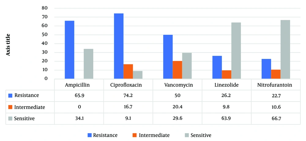
Out of 3000 culture-positive clinical samples comprised of 82 urine, ten blood, one tracheal, two wound, and one pulmonary isolate, were identified as Enterococcus faecalis (69.1%), Enterococcus faecium (7.3%), and Enterococcus spp. (23.6%). Moreover, two-thirds of isolates were multidrug-resistant (MDR). Although 66.7% and 63.9% were sensitive to nitrofurantoin and linezolid, resistance to ciprofloxacin (74.2%) and ampicillin (65.9%) was frequently observed. Nitrofurantoin or linezolid were the only effective antibiotics for UTIs. In addition, 50% of isolates were resistant to vancomycin.

Conclusions:

Based on the results, a combination of vancomycin/or linezolid and nitrofurantoin for urine infections was effective against Enterococci in this clinical center. Nevertheless, continuous and frequent surveillance for resistance patterns is necessary for the reasonable and evidence-based use of antibiotics.

1. Background

Enterococci, the primary opportunistic pathogens implicated in nosocomial infections, are naturally occurring commensal bacteria in the gastrointestinal tracts of humans and animals (1). A significant issue from the past to the present is the deadly opportunistic disease in developing nations, causing a rise in global mortality rates. Diverse microbial agents, including fungi, bacteria, parasites, and viruses, are known to trigger lethal opportunistic infections (2-4). Enterococci usually do not affect healthy individuals, but those with severe underlying health conditions or weak immune systems are at risk. These bacteria are among the most common causes of hospital-acquired infections, leading to bacteremia, endocarditis, and urinary tract infections. Enterococci can enter the environment via sewage systems, and strains resistant to vancomycin are particularly problematic in hospitals, contributing to the prevalence of hospital-acquired infections (5-7). The main reason for their survival in hospitals is their natural resistance to many antibiotics, and they can become resistant to all antibiotics through genetic mutations or acquiring external genetic elements like plasmids and transposons. Vancomycin is often the drug of choice to combat Enterococci, which has developed resistance to multiple drugs. Studying vancomycin-resistant Enterococci (VRE) epidemiology is crucial in environmental contexts. Determining the patterns of antibiotic resistance in VRE strains is essential to prevent their spread (8). The occurrence of VRE differs globally, with significant cases reported in the United States, Britain, Ireland, Saudi Arabia, and Turkey. Conversely, countries like France and Italy report a lower incidence of VRE (9). According to a study by Wada in Nigeria, Enterococcus fecalis was the most, and the pooled prevalence of VRE was estimated at 25.3% (10). In addition, Vancomycin-resistant Enterococci in hospitals on European borders (Netherlands and Germany) were increasingly significant. The biggest difference between the two countries was the studies in Germany showed an increasing trend prevalence in VRE hospitals, and no such trend was observed in studies in the Netherlands (11).

2. Objectives

Previous studies have highlighted the increasing resistance of Enterococcus to vancomycin as a significant concern that calls for epidemiological research. This research analyzes the resistance patterns of vancomycin-resistant Enterococcus strains found in Zahedan.

3. Methods

3.1. Clinical Sampling and Laboratory Identification

In this descriptive-analytical study, among 3000 clinical samples, including Blood culture, Urine, and other fluids, 110 Enterococcus spp. isolates, including E. faecalis and E. faecium, were collected from patients admitted to Ali ibn Abi Talib Hospital in Zahedan, Iran from March 2019 to February 2022. The isolates were cultured in Chocolate and Blood agar Media (Merck, Germany). The causative bacteria were identified based on colony characteristics, Gram stain morphology and biochemical tests (catalase test, growth on bile aesculin agar, tolerance to 6.5% NaCl, arginine dihydrolase test, and fermentation of arabinose, mannitol, raffinose, and sorbitol). The study population included patients of all age groups and both sexes.

3.2. Antibiotic Susceptibility of Enterococcus Species

An antimicrobial susceptibility test using the disc diffusion test (DDT) was carried out for all the confirmed isolates of E. faecalis and E. facume. However, according to the Clinical and Laboratory Standards guidelines, the isolates were categorized as sensitive, resistant, or intermediate to each antibiotic by measuring the respective zone of inhibition. (CLSI-2021) (12). The DDT and antibiotic discs for each pathogen were selected based on CLSI-2021 recommendations as vancomycin (30 μg), ampicillin (10 μg), nitrofurantoin (15 μg) (for urine samples), ciprofloxacin (5 μg), and linezolid (30 μg) (MAST, UK).

3.3. Data Analysis

The data were statistically analyzed using GraphPad Prism version 8 (GraphPad Software, Inc., CA, US) and an appropriate statistical test, i.e., Student’s t-test, Wilcoxon test, chi-square test, or one-way ANOVA.

4. Results

A total of 110 Enterococci were isolated from 3000 culture-positive clinical samples. About 92 urine isolates, 14 blood isolates, one tracheal, two wounds, and one pulmonary isolate were identified as Enterococci (Table 1). All isolates were further speciated as E. faecalis (69.1%), E. faecium (7.3%) and Enterococcus spp. (23.6%). The isolates that were normal flora were not considered.

Table 1.

Incidence and Distribution of Enterococcal Isolates in Different Clinical Samples a

SamplesIsolates of Enterococcus faecalis, N = 76Isolates of Enterococcus faecium, N = 8Isolates of Enterococcus spp., N = 26
Urine70 (92.1)5 (62.5)17 (65.3)
Blood3 (3.9)3 (37.5)8 (30.7)
Tracheal1 (1.3)---
Wound1 (1.3)-1 (3.8)
Pulmonary1 (1.3)--

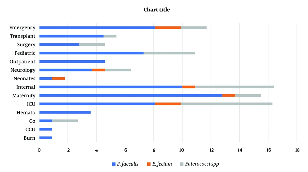
Prevalence of clinical strains in different wards. As shown in Figure 1, isolates from internal (n = 18), ICU (n = 18), and maternity (n = 17) units were the higher isolates (Figure 1).

Prevalence of Enterococci spp. in different wards (%)

4.1. Prevalence of Antibiotic Resistance

The most frequent resistance was observed for ciprofloxacin and ampicillin, with 74.2% and 65.9% of the isolates, respectively, followed by a 50% resistance rate to vancomycin. On the other hand, resistance to nitrofurantoin and linezolid was comparatively less common, detected in 22.7% and 26.2%, respectively (Figure 2).

Antibiotic susceptibility pattern in clinical isolates of Enterococcus

5. Discussion

Given the limited treatment options and increasing prevalence of VRE in the world, VRE remains a severe problem in health care. However, this overall increase in variation has been reported in countries. The patterns of antimicrobial resistance and the prevalence of VRE were examined in Zahedan. This investigation indicated a severe problem of antimicrobial resistance among Enterococci in a hospital in Zahedan. In addition, this pathogen was recognized as a significant nosocomial pathogen causing substantial morbidity and mortality globally. Since the studies have shown the rate of VRE in Germany, the UK, and Italy were 11.2%, 8.5%, 12.5%, and 9%, respectively, in the present study, the 50% rate of VRE prevalence was in agreement with earlier reports of high VRE prevalence in other cities. According to a meta-analysis by Emaneini et al., the estimated prevalence of VRE in Iran was 9.4% (13), whereas it was found to be 50% in the present study. The predominant location of Enterococcal infection in this investigation was the urinary tract (84.7%), frequently observed in patients. Septicemia, at 11.2%, was the subsequent most frequent infection after bacteremia. The dominant species of Enterococcus was identified as E. faecalis, accounting for 82% of cases, aligning with the findings of Chakraborty et al. study in India, which reported a prevalence of 80 - 90% for this bacterium in 2015 (11). The bacteria showed high resistance to gentamicin and cephalexin, while resistance to linezolid was the least observed, and 24% of isolates were resistant to vancomycin (8). However, this study noted a 50% resistance rate to vancomycin. In Razaz Rahmati et al., among 203 Enterococcus strains from Tehran hospitals, resistance was 47.3% to ampicillin, 24.6% to vancomycin, and 9.4% to teicoplanin, with a higher sensitivity to linezolid reported (14). The findings for ampicillin resistance and linezolid sensitivity are consistent with these figures. Goudarzi et al. found no resistance to linezolid among 690 Enterococcal isolates (0%) (15), whereas our study revealed a 63.9% sensitivity rate (Figure 2), indicating a notable level of resistance. Abamecha et al. in Ethiopia found that out of 114 isolated Enterococci, 32.4% were resistant to nitrofurantoin, 49.1% to ciprofloxacin, and 36% to ampicillin, showing an increase in resistance compared to previous results (16). Melese et al. reported that 60.7% of VRE strains were resistant to penicillin and 56.5% to amoxicillin, with a relatively low resistance of 13% to linezolid and daptomycin, which is in contrast to the high sensitivity to linezolid observed in the current study (Figure 2) (17). Various studies, including Devi et al. (18), Harris et al. (19), Karmarkar et al. (20), Sader et al. (21), Pancesso et al. (22), Zhanel et al. (23), Fernandes and Dhanashree (24), and Mihajlović-Ukropina et al. (25), have reported the prevalence of vancomycin resistance in Enterococcal strains to be 10%, 20%, 8.6%, 23.3%, 3.1%, 6%, 23%, 31%, and 31% respectively. According to Figure 1, this bacteria's highest range of contamination was observed in the ICU, maternity and internal ward, which was similar in some research. In Karimzadeh et al. in Shiraz (26), the most contamination by Enterococcus was in the surgical and internal wards, but in Alebouyeh et al. in Tehran, the ICU ward had the highest range of contamination (27). Enterococcal strains are a significant factor in urinary tracheal infection, and they are the third factor of UTI in hospitalized patients, particularly in maternity wards. According to De Francesco et al., in Italy, Enterococci caused 9% of positive cases in outpatients and 12% in inpatients (26).

5.1. Conclusions

Enterococci have become the essential pathogenic bacteria that cause nosocomial infections with multiple-antimicrobial resistance. As mentioned in the results, resistance to ciprofloxacin, ampicillin and vancomycin was widespread among isolates. Therefore, an accurate antibiogram is needed to choose the most effective antibiotics. On the other hand, developing new antibiotics or alternative compounds is necessary.
Acknowledgements

References

  • 1.

    Jia W, Li G, Wang W. Prevalence and antimicrobial resistance of Enterococcus species: a hospital-based study in China. Int J Environ Res Public Health. 2014;11(3):3424-42. [PubMed ID: 24662964]. [PubMed Central ID: PMC3987042]. https://doi.org/10.3390/ijerph110303424.

  • 2.

    Jahantigh M, Larypoor M. Review on the production of effective plant biopolymers in reducing microbial infections. Res Bulletin Med Sci. 2023;27(1). e12.

  • 3.

    Jahantigh M, Tahmasbi H, Bakaeiyan M. [Antibiotic Resistance Pattern of Pseudomonas aeruginosa Strains Isolated from Burn Patients]. Med J Mashhad Univ Med Sci. 2018;60(6):1121-31. FA. https://doi.org/10.22038/mjms.2019.14778.

  • 4.

    Zahedani SS, Tahmasebi H, Jahantigh M. Coexistence of Virulence Factors and Efflux Pump Genes in Clinical Isolates of Pseudomonas aeruginosa: Analysis of Biofilm-Forming Strains from Iran. Int J Microbiol. 2021;2021:5557361. [PubMed ID: 34093708]. [PubMed Central ID: PMC8163533]. https://doi.org/10.1155/2021/5557361.

  • 5.

    Babar N, Usman J, Munir T, Gill MM, Anjum R, Gilani M, et al. Frequency and antibiogram of vancomycin resistant enterococcus in a tertiary care hospital. J Coll Physicians Surg Pak. 2014;24(1):27-9. eng. [PubMed ID: 24411538].

  • 6.

    Bouki C, Venieri D, Diamadopoulos E. Detection and fate of antibiotic resistant bacteria in wastewater treatment plants: a review. Ecotoxicol Environ Saf. 2013;91:1-9. [PubMed ID: 23414720]. https://doi.org/10.1016/j.ecoenv.2013.01.016.

  • 7.

    Breazeal MV, Novak JT, Vikesland PJ, Pruden A. Effect of wastewater colloids on membrane removal of antibiotic resistance genes. Water Res. 2013;47(1):130-40. [PubMed ID: 23089359]. https://doi.org/10.1016/j.watres.2012.09.044.

  • 8.

    Naas T, Fortineau N, Snanoudj R, Spicq C, Durrbach A, Nordmann P. First nosocomial outbreak of vancomycin-resistant Enterococcus faecium expressing a VanD-like phenotype associated with a vanA genotype. J Clin Microbiol. 2005;43(8):3642-9. [PubMed ID: 16081891]. [PubMed Central ID: PMC1233916]. https://doi.org/10.1128/JCM.43.8.3642-3649.2005.

  • 9.

    Woodford N. Glycopeptide-resistant enterococci: a decade of experience. J Med Microbiol. 1998;47(10):849-62. [PubMed ID: 9788808]. https://doi.org/10.1099/00222615-47-10-849.

  • 10.

    Wada Y, Harun AB, Yean CY, Zaidah AR. Vancomycin-Resistant Enterococci (VRE) in Nigeria: The First Systematic Review and Meta-Analysis. Antibiotics (Basel). 2020;9(9). [PubMed ID: 32882963]. [PubMed Central ID: PMC7558171]. https://doi.org/10.3390/antibiotics9090565.

  • 11.

    Chakraborty A, Pal NK, Sarkar S, Gupta MS. Antibiotic resistance pattern of Enterococci isolates from nosocomial infections in a tertiary care hospital in Eastern India. J Nat Sci Biol Med. 2015;6(2):394-7. [PubMed ID: 26283837]. [PubMed Central ID: PMC4518417]. https://doi.org/10.4103/0976-9668.160018.

  • 12.

    Humphries R, Bobenchik AM, Hindler JA, Schuetz AN. Overview of Changes to the Clinical and Laboratory Standards Institute Performance Standards for Antimicrobial Susceptibility Testing, M100, 31st Edition. J Clin Microbiol. 2021;59(12). e0021321. [PubMed ID: 34550809]. [PubMed Central ID: PMC8601225]. https://doi.org/10.1128/JCM.00213-21.

  • 13.

    Emaneini M, Hosseinkhani F, Jabalameli F, Nasiri MJ, Dadashi M, Pouriran R, et al. Prevalence of vancomycin-resistant Enterococcus in Iran: a systematic review and meta-analysis. Eur J Clin Microbiol Infect Dis. 2016;35(9):1387-92. [PubMed ID: 27344575]. https://doi.org/10.1007/s10096-016-2702-0.

  • 14.

    Razaz Rahmati N, Mohabati Mobarez A, Khoram Abadi N, Shokoohizade L. [Antibiotic Susceptibility of Staphylococcus aureus and Enterococcus Spp. Isolated from some Hospitals in Tehran]. Med Lab J. 2015;9(2):78-84. FA.

  • 15.

    Goudarzi M, Mobarez AM, Najar-Peerayeh S, Mirzaee M. Prevalence of biofilm formation and vancomycin-resistant genes among Enterococcus faecium isolated from clinical and environmental specimens in Lorestan hospitals. Iran J Microbiol. 2018;10(2):74-81. [PubMed ID: 29997746]. [PubMed Central ID: PMC6039448].

  • 16.

    Abamecha A, Wondafrash B, Abdissa A. Antimicrobial resistance profile of Enterococcus species isolated from intestinal tracts of hospitalized patients in Jimma, Ethiopia. BMC Res Notes. 2015;8:213. [PubMed ID: 26036911]. [PubMed Central ID: PMC4467607]. https://doi.org/10.1186/s13104-015-1200-2.

  • 17.

    Melese A, Genet C, Andualem T. Prevalence of Vancomycin resistant enterococci (VRE) in Ethiopia: a systematic review and meta-analysis. BMC Infect Dis. 2020;20(1):124. [PubMed ID: 32046668]. [PubMed Central ID: PMC7014939]. https://doi.org/10.1186/s12879-020-4833-2.

  • 18.

    Devi PS, Rao PS, Shivananda PG. Characterization, antibiotic susceptibility pattern and detection of beta-lactamases in Enterococci. Indian J Pathol Microbiol. 2002;45(1):79-82. [PubMed ID: 12593570].

  • 19.

    Harris AD, Nemoy L, Johnson JA, Martin-Carnahan A, Smith DL, Standiford H, et al. Co-carriage rates of vancomycin-resistant Enterococcus and extended-spectrum beta-lactamase-producing bacteria among a cohort of intensive care unit patients: implications for an active surveillance program. Infect Control Hosp Epidemiol. 2004;25(2):105-8. [PubMed ID: 14994933]. https://doi.org/10.1086/502358.

  • 20.

    Karmarkar MG, Gershom ES, Mehta PR. Enterococcal infections with special reference to phenotypic characterization & drug resistance. Indian J Med Res. 2004;119 Suppl:22-5. [PubMed ID: 15232156].

  • 21.

    Sader HS, Moet GJ, Jones RN. Antimicrobial resistance among Gram-positive bacteria isolated in Latin American hospitals. J Chemother. 2009;21(6):611-20. [PubMed ID: 20071283]. https://doi.org/10.1179/joc.2009.21.6.611.

  • 22.

    Panesso D, Reyes J, Rincon S, Diaz L, Galloway-Pena J, Zurita J, et al. Molecular epidemiology of vancomycin-resistant Enterococcus faecium: a prospective, multicenter study in South American hospitals. J Clin Microbiol. 2010;48(5):1562-9. [PubMed ID: 20220167]. [PubMed Central ID: PMC2863891]. https://doi.org/10.1128/JCM.02526-09.

  • 23.

    Zhanel GG, DeCorby M, Adam H, Mulvey MR, McCracken M, Lagace-Wiens P, et al. Prevalence of antimicrobial-resistant pathogens in Canadian hospitals: results of the Canadian Ward Surveillance Study (CANWARD 2008). Antimicrob Agents Chemother. 2010;54(11):4684-93. [PubMed ID: 20805395]. [PubMed Central ID: PMC2976152]. https://doi.org/10.1128/AAC.00469-10.

  • 24.

    Fernandes SC, Dhanashree B. Drug resistance & virulence determinants in clinical isolates of Enterococcus species. Indian J Med Res. 2013;137(5):981-5. [PubMed ID: 23760387]. [PubMed Central ID: PMC3734693].

  • 25.

    Mihajlović-Ukropina M, Medić D, Jelesić Z, Gusman V, Milosavljević B, Radosavljević B. Prevalence of different enterococcal species isolated from blood and their susceptibility to antimicrobial drugs in Vojvodina, Serbia, 2011-2013. African J Microbiol Res. 2014;8(8):819-24. https://doi.org/10.5897/ajmr2013.6517.

  • 26.

    Karimzadeh I, Mirzaee M, Sadeghimanesh N, Sagheb MM. Antimicrobial resistance pattern of Gram-positive bacteria during three consecutive years at the nephrology ward of a tertiary referral hospital in Shiraz, Southwest Iran. J Res Pharm Pract. 2016;5(4):238-47. [PubMed ID: 27843959]. [PubMed Central ID: PMC5084480]. https://doi.org/10.4103/2279-042X.192460.

  • 27.

    Alebouyeh M, Shirvani F, Hassanzadeh R, Azimi T, Ghandchi G, Abdollahi N, et al. Intestinal colonization of vancomycin-resistant Enterococcus in children admitted to Mofid children's hospital intensive care unit at admission and at discharge. Mol Biol Rep. 2023;50(4):3271-81. [PubMed ID: 36710317]. https://doi.org/10.1007/s11033-022-08196-1.