


1. Background
Ectopic pregnancy (EP) is a significant contributor to pregnancy-related morbidity and mortality during the first trimester, occurring in 1 - 2% of all pregnancies (1, 2). Over the years, the management of ectopic pregnancies has evolved substantially, with the preferred approach shifting from surgical intervention to methotrexate (MTX) treatment (3). Methotrexate is the primary drug used for EP treatment and is administered in various forms (2, 4, 5). Multiple studies have validated the safety and efficacy of MTX in managing ectopic pregnancies, leading many physicians to adopt it as the first-line treatment (6).
Methotrexate acts as a folic acid antagonist, inhibiting DNA synthesis by blocking dihydrofolate reductase, thereby effectively targeting rapidly proliferating tissues such as trophoblasts (4). Additionally, it affects other rapidly dividing cells, including malignant cells, bone marrow, intestinal mucosa, and respiratory epithelium (6, 7). While MTX is typically administered via intramuscular injection, it can also be given orally or through intravenous infusion. There are three regimens for MTX administration: Single-dose, double-dose, and multi-dose, with the multi-dose regimen being the most commonly used (4, 7). Despite similar success rates in treatment and future fertility between multiple doses and surgery, medical management avoids the risks associated with surgery and anesthesia, is more cost-effective, requires no specialized skills, and prevents tubal damage, making it the preferred choice for most patients (4, 8-12).
However, despite its advantages, concerns remain regarding the potential negative impact of MTX on ovarian follicle reserve and fertility rates. It is hypothesized that primordial follicles involved in ovulation could be affected by MTX administration (13-15). Methotrexate effectively targets proliferating cells, including malignant, bone marrow, and embryonic cells. While this property makes it effective in treating EP, it may also impair fertility by affecting the division of granulosa cells within the ovary, potentially reducing future ovarian responsiveness and capacity (16).
Anti-müllerian hormone (AMH) is now widely used as a marker of ovarian reserve. It is secreted by granulosa cells of small follicles (4 - 6 mm in size) and remains relatively constant throughout the menstrual cycle, independent of menstruation (17). Women with higher AMH levels, produced by the ovaries, have an almost 2.5 times greater chance of pregnancy success than those with lower levels. Alongside maternal age and ovulation capability, AMH levels serve as a predictor of pregnancy success (18).
Nevertheless, conflicting findings exist in the literature (19). For instance, Shirazi et al. conducted a study comparing AMH serum levels before and after a single dose of MTX for EP treatment. They found no statistically significant difference, indicating that the patient's ovarian reserve remained unaffected (16). Although several studies have validated the safety and efficacy of MTX treatment for ectopic pregnancies and confirmed its negligible impact on ovarian reserve and subsequent fertility (6, 7, 20, 21), the role of MTX in clinical decisions regarding women's fertility remains substantial.
2. Objectives
Given the inconclusive and contradictory findings regarding MTX’s impact on ovarian reserve, this study aims to compare ovarian reserve in patients treated with MTX for EP. The secondary objective is to evaluate the effect of multiple doses of MTX on ovarian reserve.
3. Methods
The present study utilized a cross-sectional analytical design and received approval from the Kermanshah University of Medical Sciences under the ethical code IR.KUMS.REC.1399.790. The research focused on evaluating 40 women diagnosed with tubal EP who sought treatment at Imam Reza Hospital in Kermanshah, Iran, between 2019 and 2023. Sampling was conducted using a census approach, including all women with EP who were candidates for MTX treatment during the study period. Participants who met the inclusion and exclusion criteria were randomly assigned to two groups for investigation.
The inclusion criteria comprised cases of unruptured EP with stable vital signs, a tubal gestational sac diameter of less than 4 cm, serum beta-human chorionic gonadotropin (β-hCG) levels below 10,000 IU/L, a desire to preserve fertility, and the absence of medical contraindications such as severe abdominal pain, liver or kidney dysfunction.
Exclusion criteria included patients younger than 20 or older than 35 years, those with diminished ovarian reserve due to conditions such as endometriosis or previous ovarian surgery, and individuals with hematological, liver, or kidney disorders. Additional exclusions were applied to cases where creatinine levels exceeded 1.5 mg/dL, platelet counts were below 100,000/μL, white blood cell counts were below 2,000/μL, or liver enzymes were elevated by two times or more.
The primary tool used in this research was a checklist designed to capture the study’s key objectives and variables. This checklist was completed for each patient, incorporating demographic details and laboratory results of AMH. Faculty members from the Department of Obstetrics and Gynecology validated the checklist content during the initial project review.
Following the assessment of inclusion and exclusion criteria and obtaining informed consent, a 2 cc blood sample was drawn from each patient and sent to the central laboratory for AMH testing. Patients then underwent treatment and were assigned to either the single-dose MTX (50 mg/m²) group or the multiple-dose MTX (1 mg/kg) group. Both medications were manufactured by Kavosh Gostar Darou Company. In the multiple-dose group, MTX was administered on days 1, 3, 5, and 7. A second 2 cc blood sample was collected and analyzed one week after treatment completion using a Bioactive ELISA kit (Germany) to measure hormone levels. Patients in both groups were matched based on age, Body Mass Index (BMI), and parity.
The collected data were entered into SPSS version 20 for statistical analysis. Quantitative variables were analyzed using t-tests and Mann-Whitney U tests, while qualitative data were assessed using chi-square or Fisher’s exact tests. A significance level of 0.05 was applied to all statistical tests.
4. Results
A total of 40 patients diagnosed with EP were divided into two groups for evaluation: The single-dose MTX group (20 patients) and the multiple-dose MTX group (20 patients). This division was conducted following a thorough assessment of inclusion and exclusion criteria. The AMH levels were measured for all patients before the intervention and one week after treatment.
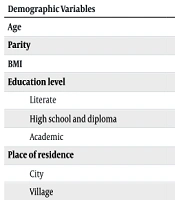
An analysis of the patients' demographic variables revealed that 82.5% resided in urban areas, while 17.5% were from rural villages. The average age of all confirmed EP patients (n = 40) was 30.48 ± 3.66 years. The average parity was 2.10 ± 2, and the mean BMI was 28.28 ± 3.95 kg/m2. The analysis presented in Table 1 showed that the two groups were comparable in terms of age, parity, gravidity, BMI, education level, and place of residence, with no statistically significant differences observed in these variables (P > 0.05) (Table 1).
| Demographic Variables | Single-Dose MTX (n = 20) | Multiple-Dose MTX (n = 20) | Tests | P-Value |
|---|---|---|---|---|
| Age | 29.5 ± 4.34 | 31.45 ± 2.58 | Mann-Whitney U test | 0.242 |
| Parity | 2.15 ± 1.08 | 2.05 ± 1.09 | Mann-Whitney U test | 0.779 |
| BMI | 28.37 ± 3.04 | 28.2 ± 4.77 | Mann-Whitney U test | 0.445 |
| Education level | Chi-square | 0.779 | ||
| Literate | 2 (10) | 2 (10) | ||
| High school and diploma | 13 (65) | 11 (55) | ||
| Academic | 5 (25) | 7 (35) | ||
| Place of residence | Fisher's exact test | 0.447 | ||
| City | 15 (75) | 18 (90) | ||
| Village | 5 (25) | 2 (10) |
Demographic Variables in Single and Multiple-Dose Methotrexate Groups a
The mean AMH level before the intervention was 1.89 ± 1.44 mg/m2. Using the Mann-Whitney U test, no significant difference was found in AMH levels between the single-dose and multiple-dose MTX groups before treatment (P = 0.659), indicating their comparability. The results showed a reduction in AMH levels after treatment in both groups compared to pre-treatment levels. However, this decrease was not statistically significant (P > 0.05) (Table 2).
| Research Groups | Before Intervention | After Intervention | P-Value |
|---|---|---|---|
| Single-dose MTX | 1.99 ± 1.80 | 1.44 ± 1.36 | 0.704 |
| Multiple-dose MTX | 1.79 ± 1.76 | 1.31 ± 1.02 | 0.849 |
| P-value | 0.659 | 0.144 | - |
Mean and Standard Deviation of Anti-Müllerian Hormone Levels Before and After Interventions a
5. Discussion
The importance of treating EP while preserving fertility potential cannot be overstated. However, minimizing the damage and risks associated with EP (22, 23) is crucial for safeguarding the fertility of women of childbearing age. Managing treatment approaches for these patients can be challenging due to potential risks to ovarian reserve (19). The MTX therapy has been identified as a safe and effective alternative to surgical management for asymptomatic EP. As a folic acid antagonist and inhibitor of DNA synthesis, MTX targets actively proliferating cells and, in the case of EP, prevents further growth of fetal cells. However, its effect on other dividing cells, such as oocytes and granulosa cells, remains unclear (21), and conflicting findings have been reported in the literature (19).
For instance, Shirazi et al. conducted a study comparing AMH serum levels before and after administering a single dose of MTX for EP treatment. They found no statistically significant difference, indicating that the patient’s ovarian reserve remained unaffected (16). Similarly, Uyar et al. reported that single-dose MTX treatment for EP did not impact ovarian reserves (22). Boots et al. observed no adverse effects on ovarian reserve or responsiveness after MTX administration for EP treatment, either before or after in vitro fertilization (2). Sekhon et al. also found that ovarian reserve and in vitro fertilization outcomes remained intact following MTX use for EP treatment (24). Orvieto et al. reported no changes in FSH levels, ovarian stimulation characteristics, or the number of retrieved oocytes before and after single-dose MTX treatment for EP in a study involving 14 women undergoing in vitro fertilization (25).
In another study by Oriol et al., AMH levels, cycle length, required gonadotropin doses, maximum E2 levels, the number of retrieved oocytes, and total embryos showed no difference before and after single-dose MTX administration for EP treatment in 14 women undergoing in vitro fertilization or intracytoplasmic sperm injection (ICSI) (20). A retrospective cohort study by Hill et al. found no correlation between the number of MTX doses administered and changes in ovarian reserve, suggesting no dose-dependent effect. Furthermore, MTX did not negatively affect fertility rates (26). In a systematic review and meta-analysis by Ohannessian et al., no statistically significant differences were found in basal plasma FSH levels, total gonadotropin dose for stimulation, stimulation duration, and E2 levels on the day of ovulation before and after MTX treatment for EP. These findings suggested that MTX treatment for EP did not have a detrimental impact on subsequent fertility treatments in infertile patients (27).
Additional studies, which examined different MTX doses and time intervals and compared them with surgical methods for EP treatment, did not identify any significant adverse effects on ovarian reserve and function in subsequent pregnancies. Singer et al. reported equivalent serum AMH levels before and after EP treatment with either MTX or surgery following in vitro fertilization, concluding that single-dose MTX treatment did not reduce AMH levels (28). Sahin Ersoy et al. found no permanent harmful effects on ovarian reserve after EP treatment, regardless of whether single-dose MTX or salpingectomy was used. Furthermore, serum AMH and antral follicle count (AFC) levels remained unchanged in the long term (29). A prospective study by Sahin et al. comparing AMH levels before and after EP treatment using systemic single-dose MTX, unilateral salpingectomy, and salpingectomy after MTX administration indicated that current medical and surgical treatment approaches did not negatively affect ovarian reserve (19). Pereira et al. concluded that treating EP with MTX or salpingectomy may not adversely affect ovarian reserve, ovarian responsiveness, or the outcome of subsequent in vitro fertilization cycles (30).
Although several studies have validated the safety and efficacy of MTX treatment for ectopic pregnancies and confirmed its negligible impact on ovarian reserve or subsequent fertility (6, 7, 20, 21), our study's findings indicate that AMH levels did not exhibit a statistically significant decrease following treatment in either the single-dose or multiple-dose MTX groups compared to their pre-treatment levels. Additionally, when comparing the treatment methods, no significant differences were observed, potentially due to variations in drug dosage and patient geographic locations.
Ovarian reserve markers, including the number of follicles and anti-müllerian hormone levels, have been reported to decrease following single or multiple-dose MTX administration and salpingectomy. These findings align with an animal model study by Ulug and Oner, which evaluated the effects of administering single or multiple-dose MTX and salpingectomy on rat ovarian reserve through AMH level measurements and histological analysis (23). A prospective clinical trial by Askari in a human model produced results similar to our study. They measured AMH levels, a biomarker of subsequent pregnancy, to compare the effects of single-dose MTX and salpingectomy on the ovarian reserve of women with EP. While both methods resulted in decreased AMH levels, the difference was not statistically significant (31). These findings were consistent with our study but differed in their comparison of treatment and surgery, whereas we exclusively compared two treatment methods — single-dose and multiple-dose MTX.
Moreover, Alleyassin et al. compared single and multiple-dose MTX groups concerning drug side effects and treatment outcomes, yielding results consistent with our study, demonstrating no significant difference between the two groups (32). The findings of the present study highlight the need for further, more detailed investigations into the lack of a significant effect of different MTX doses on ovarian reserve. Additionally, the clinical implications of this hypothesis warrant further exploration. Considering the mechanism of action of MTX, this treatment should be recommended with caution and sensitivity, taking into account the individual conditions of each patient to optimize treatment outcomes for women with EP.
The present study had certain limitations that should be considered. It was a cross-sectional analytical study with a limited sample size. Additionally, the cost of AMH measurements constrained the sample size selection. Since hormone levels naturally fluctuate throughout the menstrual cycle, careful timing is required when interpreting results. Moreover, ovarian reserve assessment immediately after treatment may show temporary changes that do not necessarily indicate a permanent decline in ovarian function. Given that the long-term effects of MTX on ovarian function years after treatment remain unknown, further studies with extended follow-up periods are recommended. On the other hand, a notable strength of this study lies in its assessment of AMH during MTX treatment for EP, providing valuable insights into its potential impact on ovarian reserve.
5.1. Conclusions
The findings of our study indicated that AMH levels declined following treatment in both the single-dose and multiple-dose MTX groups compared to their respective pre-treatment levels. However, it is important to note that this observed difference did not reach statistical significance. Additionally, no significant differences were found between the various treatment approaches.
These results suggest that MTX administration in patients with EP may have a potential impact on ovarian reserve. However, further investigations with larger sample sizes, varying MTX dosages, and extended follow-up periods are strongly recommended to comprehensively evaluate the long-term effects of MTX on ovarian reserve. Future research should also explore the clinical implications of these findings and consider potential directions for improving patient management.
Moreover, this study underscores the need for larger-scale research involving diverse populations to enhance the generalizability and impact of its findings.
