Journal of Kermanshah University of Medical Sciences
Peer-reviewed Medical Quarterly
e-ISSN 2588-2570
p-ISSN 2588-2562
Outline
Immunohistochemistry of Inflammatory myofibroblastic tumors
Authors
1Dept. of pathology, Kermanshah University of Medical Science, Kermanshah, Iran
2Dept. of pathology, Molecular Pathology Research Center, Imam Reza Hospital, Kermanshah, Iran
3Dept.of Anatomy & Histology, Kermanshah University of Medical Science, Kermanshah, Iran
*Corresponding Author: Corresponding author: Ahmad Faramarzi, Dept. of pathology, Molecular Pathology Research Center, Imam Reza Hospital, Kermanshah, Iran, Tel: +98 831 4276330-31 Email: [email protected]
Published online:Dec 19, 2009
Article type:Research Article
Received:Apr 09, 2009
Accepted:Sep 08, 2009
How to Cite:Emami Aleagha M, Faramarzi A, Khazaei S, Madani SH, Ghorbani R. Immunohistochemistry of Inflammatory myofibroblastic tumors. J Kermanshah Univ Med Sci. 2009;13(3):e79599. doi:
Abstract
Background: Inflammatory myofibroblastic tumor (IMT) or inflammatory pseudotumor is a tumoral lesion which can be seen in all age groups and in all internal organs. It is in differential diagnosis with some important neoplasms such as Leiomyosarcoma, Rhabdomyosarcoma, and Sarcomatoid carcinoma. Differentiation between these tumors requires a special diagnostic tool such as Immunohistochemistry (IHC). This study aimed to identify Immunohistochemical characteristics of inflammatory myofibroblastic tumors.
Methods: 19 cases included in this cross-sectional study. All cases with diagnosis of :" Inflammatory myofibroblastic tumor" or " Inflammatory pseudotumor" at pathobiology laboratories of Kermanshah university, Tehran Imam Khomaini Hospital and Institute of Cancer were selected and studied by using IHC stains for CK, EMA, SMA, MSA, Desmin, P53, ALK and Vimentin .
Results: Mean age of cases was 40.4 year . 52.6% were male and 47.4% female. Most frequent affected organs were; Stomach (4 cases), Urinary Bladder (3 cases), Small Intestine (3 cases), Lung and Mediastinum (3cases), Omentum (2 cases), Retroperitoneum (1 case), Cervix(1 case), Urethra(1 case) and gluteus Maximus Muscle (1 case), . Vimentin (94.7%) , MSA (57.9%) and SMA (47.4%) were the most frequent expressed IHC biomarkers in diagnosed tumors respectively.
Conclusion: CK is a reliable marker for differentiation between these lesions and Sarcomatoid carcinoma. For differentiation from Leiomyosarcoma, Rhabdomyosarcoma and Postoperative Spindle Cell Nodule, using Desmin is a useful biomarker. It is recommended that "IMT" be used for those lesions that express ALK and/or those that cytogenetic studies reveal a fusion of introducing genes. Other lesions should be classified and reported as "Inflammatory Pseudotumor.
References
- 1.The references of this article is available on PDF.
Copyright
© 2009, Journal of Kermanshah University of Medical Sciences. This open-access article is available under the Creative Commons Attribution-NonCommercial 4.0 (CC BY-NC 4.0) International License (https://creativecommons.org/licenses/by-nc/4.0/), which allows for the copying and redistribution of the material only for noncommercial purposes, provided that the original work is properly cited.
Similar Articles
13
Feb
2023
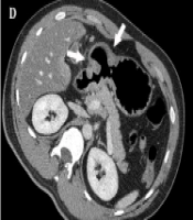
Multidetector Computed Tomography Imaging Features of Inflammatory Myofibroblastic Tumors of the Gastrointestinal Tract in Adults: Radiological, Histopathological, and Immunohistochemical Features
In Young Choi,
Suk Keu Yeom,
Beom Jin Park,
Deuk Jae Sung,
Min Ju Kim,
Na Yeon Han
,et al.Choi IY, Yeom SK, Park BJ, Jae Sung D, Kim MJ, et al. Multidetector Computed Tomography Imaging Features of Inflammatory Myofibroblastic Tumors of the Gastrointestinal Tract in Adults: Radiological, Histopathological, and Immunohistochemical Features. I J Radiol. 2023;20(1):e129661. doi: https://doi.org/10.5812/iranjradiol-129661
19
May
2026
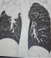
Inflammatory Myofibroblastic Tumor of the Right Upper Lobe in an 11-Year-Old Boy Presenting with Dyspnea: A Case Report
Javad Ghoroubi,
Shahed Faraji,
Leila Azimi
Ghoroubi J, Faraji S, Azimi L. Inflammatory Myofibroblastic Tumor of the Right Upper Lobe in an 11-Year-Old Boy Presenting with Dyspnea: A Case Report. Arch Pediatr Infect Dis. 2026;14(3):e168817. doi: https://doi.org/10.5812/apid-168817
22
Aug
2016
Alteration of Signal Intensity on Follow-Up Magnetic Resonance Imaging in Spinal Epidural Inflammatory Myofibroblastic Tumor: A Case Report and Literature Review
Jinyoung Chang,
Sun-Young Park,
Soo Kee Min,
Sung Hye Koh,
Kwan Seop Lee
Chang J, Park S, Min SK, Koh SH, Lee KS. Alteration of Signal Intensity on Follow-Up Magnetic Resonance Imaging in Spinal Epidural Inflammatory Myofibroblastic Tumor: A Case Report and Literature Review. I J Radiol. 2017;14(1):e13474. doi: https://doi.org/10.5812/iranjradiol.35188
16
Sep
2025
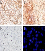
Unexpected Diagnosis of Uterine Inflammatory Myofibroblastic Tumor During Cesarean Section
Jun Tang,
Huaiwen Wang,
Gang Wei,
Dongdong Zhang
Tang J, Wang H, Wei G, Zhang D. Unexpected Diagnosis of Uterine Inflammatory Myofibroblastic Tumor During Cesarean Section. Int J Cancer Manag. 2025;18(1):e163767. doi: https://doi.org/10.5812/ijcm-163767
30
Jan
2016
Follow-Up Imaging of Inflammatory Myofibroblastic Tumor of the Uterus and Its Spontaneous Regression
Biljana Markovic Vasiljkovic,
Vesna Plesinac Karapandzic,
Tomislav Pejcic,
Aleksandra Djuric Stefanovic,
Zorica Milosevic,
Snezana Plesinac
Markovic Vasiljkovic B, Plesinac Karapandzic V, Pejcic T, Djuric Stefanovic A, Milosevic Z, et al. Follow-Up Imaging of Inflammatory Myofibroblastic Tumor of the Uterus and Its Spontaneous Regression. I J Radiol. 2016;13(1):e12991. doi: https://doi.org/10.5812/iranjradiol.12991
Cited by
- Scopus by DOI: 0
Last Update: 1 month ago
- Scopus by Title: 0
Last Update: 1 month ago
- Scopus by Title (Ref): 0
Last Update: 1 month ago
- CrossRef: 0
Last Update: 5 days ago
Comments
Number of Comments:0