1. Introduction
Spinal dermoid/epidermoid cysts are uncommon cystic tumors that may be congenital or acquired. These tumors are normally extramedullary and rarely intramedullary (1, 2). The typical presentation of this tumor in the spinal cord is progressive paraparesis, mmbowel or bladder dysfunction, pain and sensory disturbance (3). Recurrent meningitis is an uncommon presentation of these tumors (4). The case reported is a 2-year-old boy with recurrent meningitis.
2. Case Presentation
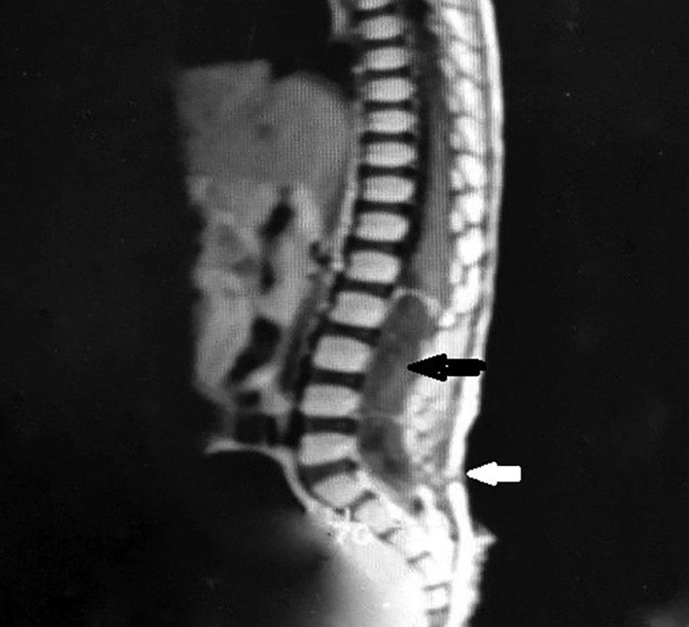
A 2-year-old boy was referred to our clinic with complaints of a headache, fever and neck stiffness. The patient’s medical history revealed that he had been admitted to hospital 4 times because of meningitis. He did not receive any medications and his family history and social history were negative. The physical examination showed that he was conscious although he had malaise and fever. The neurological examination of the patient showed paraparesis with hyporeflexia and hypotonia with bilateral extensor plantar reflex that had started four months before. The MR imaging of the lumbosacral spine revealed an intradural and extramedullary mass located between L2 and s2 levels and also a sinus tract at the level of S1 (Figure 1). The tumor mass was totally resected during surgery and intramedullary dermoid cyst was confirmed through the biopsy and in the follow-up. The patient significantly improved in terms of muscle strength and gait. We obtained the written informed consent from the patient to report the case.
3. Discussion
Dermoid cysts are cystic tumors that originate from different parts of the body, including the scalp, intracranial and intraspinal regions, the skull bone and rarely the spinal cord (5). These tumors are associated with occult dysraphism, split cord malformation, cutaneous lesions and sinus tracts (6), and commonly present in childhood. The usual symptoms include pain, sensory disturbance, muscle weakness and sphincter dysfunction (7). Recurrent meningitis is another presentation of these cysts. Meningitis is caused by the discharge from the cysts into the subarachnoid space or skin flora in cases with a dermal sinus, which may cause abscess formation (8, 9). Yildirim et al. reported a case of dermoid cyst presenting with recurrent aseptic meningitis (10). The dermoid cysts are usually a well-defined mass in imaging, and due to cholesterol components, are hyperintense in T1 and hypo- to hyperintense in T2 (11). A dermoid cyst occasionally ruptures in the subarachnoid space and ventricular system spontaneously or after the trauma. The symptoms of rupture include headache, nausea, vomiting and aseptic meningitis (12, 13). Altay et al. reported a case of spinal dermoid cyst that ruptures into the subarachnoid space, and the particle of fat can be recognized in the central canal, cerebral fissure and basal cisterna (14). Selective treatment is based on the location of the tumor in the extramedullary dermoid cysts. Total excision of the tumor is recommended in intramedullary dermoid cysts due to tumor capsule adhesion to the spinal cord, although it leads to neurological deficits (15). This case was reported to help consider spinal dermoid/epidermoid tumors in the differential diagnosis of patients with recurrent meningitis.

