

1. Background
Changing the level of muscle activity in the muscles around the scapula is one of the important factors in the occurrence of shoulder impingement syndrome (SIS). Some researchers have shown that the pattern of muscle activity changes in people with this syndrome (1, 2). These changes include: (1) Increasing the activity of the upper trapezius, (2) reduced activity of the lower trapezius, (3) decreasing the activity of the middle trapezius, and (4) decreasing the activity of the serratus anterior (1). According to the kinesiopathological model, disturbance in movement patterns and their components is the main cause of pain and pathogenic symptoms. According to this model, it is possible to prevent the occurrence of sports injuries, including SIS, by diagnosing and correcting these inappropriate movement patterns, or in case of an injury, it can be treated more quickly and effectively (3). Research shows that athletes who overuse their hands in overhead movements may have abnormal activity in the muscles of the shoulder girdle and abnormal kinematics in the shoulder joints (1, 3). In case of injury, using the kinesiopathological model, it can be treated more quickly and effectively, and help the athlete return to the sports field. Preventing injuries and treating them more quickly can significantly reduce treatment costs for clubs and athletes. On the other hand, identifying weak muscles and their rehabilitation can play an important role in preventing shoulder pain and movement limitations. By identifying and strengthening weak muscles, it is possible to prevent the occurrence of functional defects and pains related to them. Some studies have reported changes in the activity of different muscles in patients with SIS, but different results have been reported mong studies (4). Despite the significant benefits of the kinesiopathological model in the prevention and treatment of SIS, no comprehensive scientific research has been conducted in this field. Conducting this research can help to better understand this model and its effectiveness in preventing and treating this complication.
2. Objectives
The purpose of this study was to investigate the electrical activity of selected shoulder girdle muscles in athletes with and without SIS at different angles of arm abduction/adduction.
3. Methods
3.1. Subjects
The current research was semi-experimental, applied, and cross-sectional. The statistical population of this research included female overhead athletes from Karaj city of Iran, with a range of 18 - 35 years old and with at least three years of club experience. Thirty athletes (10 volleyball, 10 basketball, and 10 handball players) were selected as a sample by the simple random method using G-Power software. Fifteen of these athletes were suffering from SIS, and fifteen were healthy. It should be noted that there were five athletes from each sport in each group.
Inclusion criteria were: (1) At least three years of club experience in volleyball, basketball, and handball, (2) disabilities of the Arm, Shoulder and Hand (DASH) Questionnaire score of less than 10 for the healthy group, (3) DASH Questionnaire score of more than 25 and confirmed by Neer and Hawkins tests for SIS group, and (4) SIS in the dominant shoulder. Exclusion criteria were: (1) Inability to perform tests, (2) skin sensitivity to surface electrodes (itching of the skin and intolerance of the subject). At first, all participants in the research were asked to read and complete the consent form to participate in the research. This study was approved by the Research Ethics Committees of University of Kurdistan (IR.UOK.REC.1404.008) and all procedures were performed following the Declaration of Helsinki.
3.2. Apparatus and Task
3.2.1. Measuring the Electrical Activity of Muscles
A surface electromyography device (8-channel, Myon Company, Aktos model, made in Italy, with a sampling frequency of 1000 Hz) was used to collect data related to the electrical activity of shoulder girdle muscles. In this method, surface electrodes were placed on the skin at the target muscles and the electrical activity of these muscles was recorded during the arm abduction/adduction movement at different angles.
3.2.2. Initial Preparation
The participants were asked to refrain from vigorous sports activities 15 minutes before the test. Also, they were asked to wear comfortable clothes and remove their jewelry from the area to be measured (shoulder and arm).
3.2.3. Skin Preparation and Electrode Placement
After the skin preparation stage (shaving and cleaning with alcohol), disposable surface electrodes made of silver/silver chloride were placed on the skin at the upper, lower, and middle trapezius, serratus anterior, middle deltoid, and supraspinatus muscles with a center-to-center distance of 2 cm. The exact location of the electrodes was determined based on SENIAM international standards.
3.2.4. Movement Task
Participants were asked to stand with their arms at their sides. They were then asked to abduct their dominant arm and then slowly return it to its original position (adduction) without an external load in saggital plane. This movement was performed at three angles: Forty-five, 90, and 135 degrees. For each 45°, the participant performed the movement for one second and then held it for two seconds. The rhythm of the movement was controlled by a metronome. It should be noted that the adduction movement was performed from an angle higher than 135°, and in the 135 angle was held for two seconds, and from 135 to 90 degrees in one second, and from 135 to 45 degrees in two seconds and held for two seconds. Angles of 45, 90, and 135 degrees were determined for each subject on a metal clamp that could be moved (5). The electrical activity of muscles was recorded at angles of 45, 90, and 135 degrees of arm abduction and adduction.
3.2.5. Maximum Contraction Isometric Contraction
To record the maximum voluntary isometric contraction (MIVC) of the targeted muscles, electrodes are placed on the six muscles including the upper trapezius (between the seventh cervical vertebra and the acromion along the muscle fiber line) (6), middle trapezius (between the third thoracic vertebra and the root of the scapula along the muscle fiber line) (6), lower trapezius (between the seventh thoracic vertebra and the medial border of the scapula along the line of the muscle fiber) (6), serratus anterior (the midline of the surface of the inferior angle of the scapula and the fifth rib just below the axillary area) (7) supraspinatus (distance two cm above the scapular spine in the supraspinatus fossa) and the middle deltoid (along the connecting line from the last appendage to the external epicondyle of the humerus and on the largest part of the muscle) was installed on the dominant side. It should be noted that the MVIC for each muscle was performed for 5 seconds and a one-minute rest was provided to record the MVIC of the next muscle. Muscle activity level was calculated by dividing the average RMS value by the muscle MVIC multiplied by 100.
3.3. Data Analysis
The Shapiro-Wilk test was used to examine the normality of the data distribution. Also, independent sample t-tests were used to compare the means of the variables between the two groups. The significance level for all statistical tests was considered to be 0.05.
4. Results
The characteristics of the research subjects are given in Table 1. According to the results of Table 1, no significant differences were observed between the groups in descriptive and anthropometric indices. Therefore, the research groups are homogeneous in this respect.
| Variable | SIS Group | Health Group | P-Value b |
|---|---|---|---|
| Age (y) | 29.6 ± 4.7 | 27.9 ± 4.9 | 0.14 |
| Height (cm) | 169.5 ± 7.1 | 174.1 ± 8.9 | 0.86 |
| Mass (kg) | 67.5 ± 5.3 | 73.1 ± b7.9 | 0.96 |
| BMI (m/s2) | 23.5 ± 1.5 | 24.1 ± 5.4 | 0.31 |
| Sport experience (y) | 3.5 ± 1.2 | 3.4 ± 1.4 | 0.26 |
Demographic Characteristics of Research Subjects a
Also, the results of the independent sample t-test on the difference between the activity levels of different muscles at different angles of arm abduction between the two groups are presented in Table 2. According to the results, at all three angles of 45, 90, and 135 degrees of abduction, the activity level of the supraspinatus, serratus anterior, middle, and lower trapezius muscles in the healthy group was lower than in the SIS group, but the activity level of the upper trapezius and middle deltoid muscles at all three angles of 45, 90, and 135 degrees of arm abduction in the SIS group was significantly higher than in the control group.
| Muscle | Values (Mean ± SD) | t | P-Value | Effect Size |
|---|---|---|---|---|
| Supraspinatus | ||||
| 45° | 46.2 | 0.001 | 1.25 | |
| Health | 54.9 ± 19.3 | |||
| SIS | 35.3 ± 12.2 | |||
| 90° | 31.07 | 0.001 | 0.71 | |
| Health | 59.3 ± 22.2 | |||
| SIS | 44.8 ± 18.3 | |||
| 135° | 27.5 | 0.001 | 0.69 | |
| Health | 73.5 ± 25.5 | |||
| SIS | 56.5 ± 23.7 | |||
| Upper trapezius | ||||
| 45° | -32.1 | 0.001 | 0.68 | |
| Health | 36.7 ± 19.2 | |||
| SIS | 51.3 ± 23.3 | |||
| 90° | -31.2 | 0.002 | 1.1 | |
| Health | 40.5 ± 16.3 | |||
| SIS | 65.9 ± 29.7 | |||
| 135° | -67.7 | 0.000 | 1.32 | |
| Health | 45.7 ± 23.3 | |||
| SIS | 83.4 ± 33.4 | |||
| Serratus anterior | ||||
| 45° | 41.7 | 0.004 | 0.91 | |
| Health | 47.7 ± 22.2 | |||
| SIS | 30.6 ± 15.3 | |||
| 90° | 38.7 | 0.01 | 0.92 | |
| Health | 56.7 ± 26.2 | |||
| SIS | 36.6 ± 17.4 | |||
| 135° | 53.6 | 0.000 | 1.57 | |
| Health | 89.6 ± 35.5 | |||
| SIS | 44.5 ± 21.6 | |||
| Middle deltoid | ||||
| 45° | -51.1 | 0.000 | 1.16 | |
| Health | 30.6 ± 18.3 | |||
| SIS | 57.3 ± 27.4 | |||
| 90° | -41.1 | 0.000 | 1.13 | |
| Health | 43.7 ± 21.3 | |||
| SIS | 75.9 ± 35.6 | |||
| 135° | -36.1 | 0.01 | 0.92 | |
| Health | 54.2 ± 31.6 | |||
| SIS | 83.8 ± 32.5 | |||
| Lower trapezius | ||||
| 45° | 34.1 | 0.001 | 1.08 | |
| Health | 53.3 ± 26.5 | |||
| SIS | 30.7 ± 15.3 | |||
| 90° | 46.1 | 0.001 | 1.21 | |
| Health | 65.7 ± 31.5 | |||
| SIS | 34.9 ± 19.4 | |||
| 135° | 27.5 | 0.001 | 1.09 | |
| Health | 79.4 ± 35.5 | |||
| SIS | 46.6 ± 24.5 | |||
| Middle trapezius | ||||
| 45° | 37.4 | 0.001 | 1.09 | |
| Health | 53.1 ± 25.9 | |||
| SIS | 31.3 ± 14.1 | |||
| 90° | 62.4 | 0.002 | 1.14 | |
| Health | 69.8 ± 29.5 | |||
| SIS | 40.5 ± 21.8 | |||
| 135° | 66.1 | 0.000 | 0.98 | |
| Health | 80.7 ± 33.2 | |||
| SIS | 51.1 ± 27.1 |
The Comparison of the Activity Level of Muscles in Different Abduction Angles
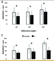
On the other hand, the results of the activity levels of the supraspinatus (Figure 1A), upper trapezius (Figure 1B), serratus anterior (Figure 1C), middle deltoid (Figure 1D), lower trapezius (Figure 1E), and middle trapezius (Figure 1F) muscles at different angles of adduction between the two groups showed that the activity levels of all 6 muscles were higher in the healthy group than in the SIS group (* P < 0.05).
, upper trapezius (B), serratus anterior (C), middle deltoid (D), lower trapezius (E), and middle trapezius (F) muscles at different angles of adduction between the two groups (* P < 0.05).")
The comparison of the activity levels of the supraspinatus (A), upper trapezius (B), serratus anterior (C), middle deltoid (D), lower trapezius (E), and middle trapezius (F) muscles at different angles of adduction between the two groups (* P < 0.05).
5. Discussion
The present study aimed to compare the electrical activity of selected shoulder girdle muscles at different angles of arm abduction/adduction in athletes with and without SIS. The results of the study showed that the electrical activity of the lower and middle trapezius, serratus anterior, and supraspinatus muscles of athletes with SIS was significantly lower at all three angles of 45, 90, and 135 degrees of arm abduction and adduction compared to healthy athletes. Also, the electrical activity of these muscles at lower angles was lower in both groups, with the lowest activity observed at an angle of 45°and the highest activity at an angle of 135°. In contrast, the results of the study showed that the electrical activity of the middle deltoid and upper trapezius muscles was higher in athletes with SIS at all three angles of 45, 90, and 135 degrees of arm abduction, while the activity level of the upper trapezius and middle deltoid muscles was lower in athletes with SIS at all three angles of adduction.
Mehrabian et al. compared electromyographic (EMG) activity of selected shoulder muscles and scapulohumeral rhythm in elite male swimmers with and without SIS. They observed that swimmers with SIS had a later onset of lower scapular upward rotation and higher scapulohumeral rhythm ratio compared to the healthy group. also, swimmers with SIS show abnormal activity and higher onset (delayed activation) and faster offset (early termination of activity); thus, the theory that shoulder impingement may be related to changes in the level of activity and recruitment of the scapulothoracic muscles is confirmed, which overall indicates changes in neuromuscular control (8). In another study by Sabzehparvar et al., which aimed to investigate and compare the EMG activity of selected shoulder girdle muscles in elite swimmers with and without shoulder pain, the results showed that swimmers with shoulder pain had greater activation in the upper trapezius, serratus anterior, and latissimus dorsi muscles compared to swimmers without pain. No significant differences were observed in the activation of the middle and lower trapezius, middle deltoid, and sternocleidomastoid muscles (9). These altered muscle activation patterns may contribute to shoulder pain in elite swimmers. Rehabilitation programs for these swimmers should address potential muscle imbalances. This study emphasizes the importance of proper swimming techniques and training strategies to prevent shoulder pain in elite athletes (10). The reason for the discrepancy in the results of Sabzeh Parvar's study and the present study may be related to the research subjects, as the subjects of the present study were female overhead athletes from volleyball, basketball, and handball sports, while the study by Sabzehparvar et al was elite swimmers, and the nature of the sport may affect the research results.
Diederichsenet al. examined the pattern of shoulder muscle activity in individuals with and without SIS and observed that during external rotation, the activity of the infraspinatus and serratus anterior muscles was significantly lower on the symptomatic side compared to healthy individuals. Also on the asymptomatic side, the groups showed different muscle activity during external rotation. The findings of this study of altered shoulder muscle activity patterns on both symptomatic and asymptomatic sides in patients suggest that different movement patterns may be a pathogenic factor in people with SIS, perhaps due to inappropriate neuromuscular strategies affecting both shoulders (11).
In another study, glenohumeral and scapulothoracic kinematics and scapulothoracic muscle activity associated with SIS were examined at angles (31 - 60°, 61 - 90°, and 91 - 120° degrees) in a group of individuals with SIS compared to a group without symptoms. The results showed that compared to the healthy group, the patient group showed that the EMG activity of the upper and lower trapezius muscles in the patient group increased in the final phase of 91 to 120°, however, the supraspinatus muscle showed less activity in the patient group in all loads and phases (12). The supraspinatus muscle, a vital component of the rotator cuff, plays a vital role in shoulder abduction and stability. The observed decrease in electrical activity in patients with SIS may be attributed to several related factors. Chronic pain, a hallmark of shoulder impingement, can lead to reflex inhibition of muscle activity. This neurophysiological response, known as arthrogenic muscle inhibition, acts as a protective mechanism to prevent further tissue damage, but it can lead to long-term muscle weakness and altered movement patterns. Persistent pain in shoulder impingement may trigger this inhibitory response in the supraspinatus region, leading to decreased electrical activity (13).
The serratus anterior muscle is critical for scapular stability and upward rotation during arm elevation. The decreased electrical activity observed in this muscle in patients with SIS may be explained by the following mechanisms. It has been shown that shoulder impingement often involves altered scapulohumeral rhythm and scapular dyskinesia. Limited glenohumeral motion may lead to compensatory changes in scapular kinematics, potentially leading to reduced serratus anterior activation. This adaptation may be an attempt to minimize pain or compensate for the limited range of motion in the glenohumeral joint (12, 14, 15). The deltoid muscle, which consists of anterior, middle, and posterior portions, is essential for shoulder abduction and flexion. The decreased electrical activity observed in the deltoid of patients with SIS may be attributed to several factors. In healthy shoulders, the deltoid works with the rotator cuff muscles to produce smooth, coordinated movements. Pathological changes in SIS may disrupt this synergistic relationship and lead to altered deltoid activation patterns. Decreased supraspinatus activity, as observed in our study, may require compensatory changes in deltoid function, potentially leading to an overall decrease in activation (16). Similar to the supraspinatus muscle, Norte et al. 2021 reported that the deltoid may experience arthrogenic muscle inhibition due to chronic pain associated with SIS (17). This inhibition can lead to a decrease in motor unit recruitment and firing rates, which manifests as decreased electrical activity (17, 18).
5.1. Conclusions
This study's reduction in electrical activity in the supraspinatus, serratus anterior, and deltoid muscles provides new insight into the complex neuromuscular adaptations associated with shoulder impingement. These findings have significant implications for understanding, diagnosing, and managing this challenging condition. By understanding mechanisms of altered muscle function, this research provides effective treatment strategies that potentially improve outcomes for patients with SIS.
5.2. Limitations
While our study provides valuable insights into the neuromuscular aspects of SIS, some limitations can be noted. The cross-sectional nature of this study prevents us from determining whether the observed muscle activity changes are a cause or consequence of shoulder impingement. We did not assess the deep rotator cuff muscles, which may play an important role in the pathomechanics of this condition. Also, careful screening of subjects for shoulder, neck, and spine abnormalities and the lack of recording of kinematic data during the arm abduction and adduction tasks were other limitations of the present study. We suggest that future researchers investigate these issues.
