Journal of Skin and Stem Cell

Image Credit:Journal of Skin and Stem Cell

Cutaneous Plasmacytoma: A Case Report

Author(s):
Kalaprapa AsavisanuKalaprapa Asavisanu1,*, Christos Kasparis Christos Kasparis 1, Ye Lin HockYe Lin Hock2
1Department of Dermatology, Walsall Manor Hospital, Walsall, England
2Department of Histopathology, Walsall Manor Hospital, Walsall, England

Journal of Skin and Stem Cell:Vol. 6, issue 2; e100545
Published online:Jan 27, 2020
Article type:Case Report
Received:Dec 27, 2019
Accepted:Jan 20, 2020
How to Cite:Asavisanu K, Kasparis C, Hock YL. Cutaneous Plasmacytoma: A Case Report. J Skin Stem Cell. 2019;6(2):e100545. doi: https://doi.org/10.5812/jssc.100545

Abstract

Introduction:

Multiple myeloma is a rare haematological malignancy of plasma cells commonly presenting in the fifth and sixth decade. Uncommonly, multiple myeloma can present with cutaneous manifestations. Usually these lesions occur as a result of an infiltrative extension of osteolytic bone lesions.

Case Presentation:

A 72-year-old gentleman with a large malodorous fungating lesion on his right thigh. A skin biopsy demonstrated that the lesions are deposits of known kappa light chain plasma cell myeloma.

Conclusions:

Cutaneous plasmacytoma typically presents as erythematous or violaceous hard and tender nodules or plaques. Treatment involves chemotherapy, steroids, local radiotherapy and surgery. The discovery of cutaneous metastatic plasmacytomas can be the first sign of progression of the disease or signal a deteriorating clinical course in multiple myeloma therefore prompt investigation is essential.

1. Introduction

Multiple myeloma (MM) is a rare haematological malignancy of plasma cells commonly presenting in the fifth and sixth decade. It involves malignant monoclonal proliferation of plasma cells. This proliferation is usually confined to the bones and the bone marrow.
Uncommonly, multiple myeloma can present with cutaneous manifestations and these occur in 1% - 5% of patients (1, 2). Usually these lesions occur as a result of an infiltrative extension of osteolytic bone lesions (3, 4).

2. Case Presentation

A 72-year-old Caucasian gentleman presented with a 6-month history of an expanding, malodorous and painful fungating plaque on his right medial thigh.
He had a history of Kappa light chain multiple myeloma that was previously treated with complete remission.
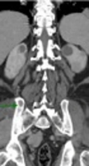
X-ray of the right femur: Rounded lucent areas are seen within the cortex just distal to the prosthesis posteriorly and proximal to the knee in keeping with myelomatous deposits.
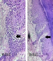
Histopathology and immunohistochemistry demonstrated dermal and subcutaneous deposits of monoclonal neoplastic plasma cells showing kappa light chain restriction (clonal population demonstrated) (Figure 1). Positivity for CD138 (Figure 2) and aberrant cyclin D1 positivity supports that these are neoplastic plasma cells.
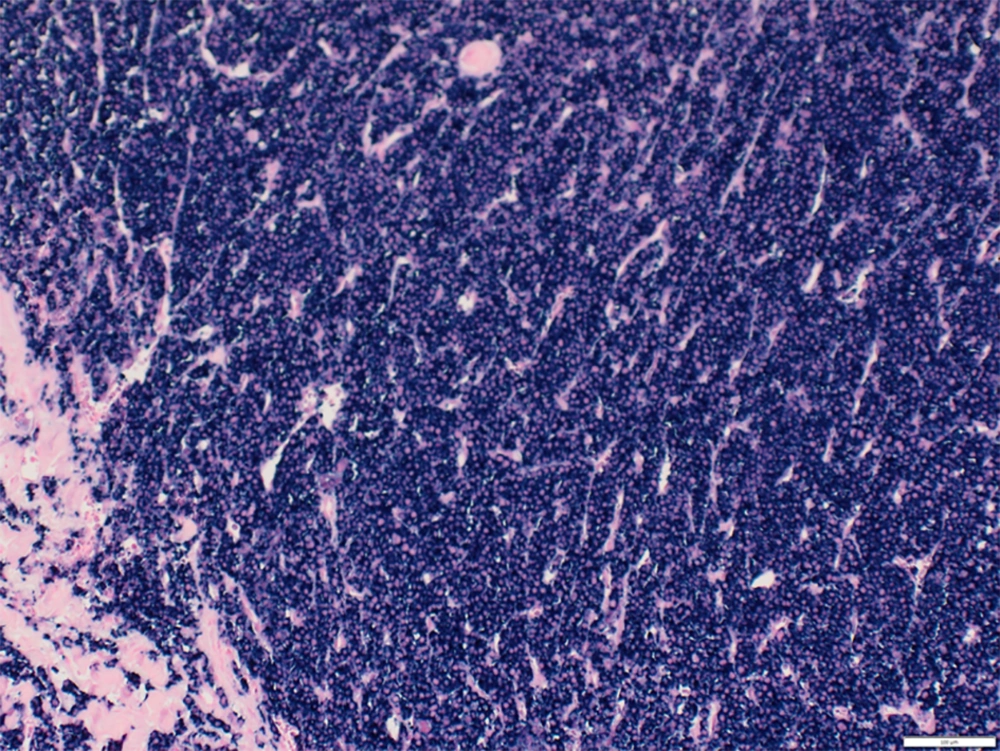
Immunohistochemistry showing kappa light chain restriction in the neoplastic cells
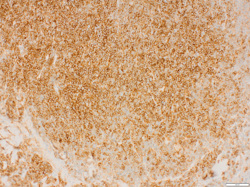
Figure 1.

Immunohistochemistry showing kappa light chain restriction in the neoplastic cells

Immunohistochemistry showing CD138 positivity in neoplastic cells
Figure 2.

Immunohistochemistry showing CD138 positivity in neoplastic cells

These same neoplastic cells were found to the be in the bone marrow.
Therefore, the skin and bone marrow findings are entirely consistent with the skin lesions being deposits of known kappa light chain plasma cell myeloma (multiple myeloma).

3. Discussion

Typically, plasmacytomas present as erythematous or violaceous hard and tender nodules or plaques with a smooth surface. They may be dermal or subcutaneous or both and can be solitary or multiple. These lesions can occur anywhere in the skin but are most commonly seen in the chest and abdomen (44%) (5).
Histopathological findings are characterised by monomorphic dermal and subcutaneous infiltrates of plasma cells. Immunohistochemically we see monoclonality of the plasma cells which have strong immunoexpression for CD138 (6).
Cutaneous plasmacytomas tend to occur with MM relapses and in highly aggressive and progressive forms of MM. In both cases the prognosis is poor with an average life expectancy of 12 months post diagnosis with less than 20% of patients found to be progression free at 5 years (6-9).
Treatment for cutaneous plasmacytoma in multiple myeloma involves chemotherapy, steroids and local radiotherapy. Surgical excision has a role in lesions resistant to radiotherapy or where lesions are very large and symptomatic.
Due to the large size, pain and odour of the lesion in our patient a surgical excision with a wide margin and skin graft repair was elected. He subsequently had chemotherapy with Revlimid and dexamethasone. The lesion resolved completely with no recurrence. Unfortunately despite treatment of the skin lesion, his MM progressed and he passed away 4 years later. We believe that our case represents cutaneous plasma cell neoplasm from direct extension of osteolytic lesions (from his right femur) associated with a relapse of his multiple myeloma.
In conclusion, MM can present with cutaneous findings in both primary and recurrent disease. The discovery of cutaneous metastatic plasmacytomas can be the first sign of progression of the disease or signal a deteriorating clinical course in multiple myeloma. Therefore prompt investigations including a skin biopsy are essential in guiding further treatment.
Footnotes

References

  • 1.
    James W, Berger T, Elston D. Andrews' diseases of the skin clinical dermatology. 11th ed. Canada: Elsevier Saunders Publication; 2011.
  • 2.
    Souza DAFD, Freitas THPD, Paes RAP, Muller H, Hungria VT. Mieloma multiplo com plasmocitomas cutaneos. An Bras Dermatol. 2004;79(5):581-5. https://doi.org/10.1590/s0365-05962004000500009.
  • 3.
    Requena L, Kutzner H, Palmedo G, Calonje E, Requena C, Perez G, et al. Cutaneous involvement in multiple myeloma: A clinicopathologic, immunohistochemical, and cytogenetic study of 8 cases. Arch Dermatol. 2003;139(4):475-86. [PubMed ID: 12707095]. https://doi.org/10.1001/archderm.139.4.475.
  • 4.
    Patterson JW, Parsons JM, White RM, Fitzpatrick JE, Kohout-Dutz E. Cutaneous involvement of multiple myeloma and extramedullary plasmacytoma. J Am Acad Dermatol. 1988;19(5):879-90. https://doi.org/10.1016/s0190-9622(88)70249-2.
  • 5.
    Jurczyszyn A, Olszewska-Szopa M, Hungria V, Crusoe E, Pika T, Delforge M, et al. Cutaneous involvement in multiple myeloma: A multi-institutional retrospective study of 53 patients. Leuk Lymphoma. 2016;57(9):2071-6. [PubMed ID: 26726867]. https://doi.org/10.3109/10428194.2015.1128542.
  • 6.
    Santos G, Sousa L, Fernandes T, Joao A. Case for diagnosis. Cutaneous involvement associated to multiple myeloma. An Bras Dermatol. 2014;89(1):173-4. [PubMed ID: 24626671]. [PubMed Central ID: PMC3938377]. https://doi.org/10.1590/abd1806-4841.20142431.
  • 7.
    Yoo J, Jo M, Kim MS, Jue MS, Park HJ, Choi KH. Cutaneous plasmacytoma: Metastasis of multiple myeloma at the fracture site. Ann Dermatol. 2017;29(4):483-6. [PubMed ID: 28761299]. [PubMed Central ID: PMC5500716]. https://doi.org/10.5021/ad.2017.29.4.483.
  • 8.
    Usmani SZ, Heuck C, Mitchell A, Szymonifka J, Nair B, Hoering A, et al. Extramedullary disease portends poor prognosis in multiple myeloma and is over-represented in high-risk disease even in the era of novel agents. Haematologica. 2012;97(11):1761-7. [PubMed ID: 22689675]. [PubMed Central ID: PMC3487453]. https://doi.org/10.3324/haematol.2012.065698.
  • 9.
    Woo YR, Kim JS, Lim JH, Hwang S, Kim M, Bae JM, et al. Prevalence and clinicopathologic characteristics of multiple myeloma with cutaneous involvement: A case series from Korea. J Am Acad Dermatol. 2018;78(3):471-478 e4. [PubMed ID: 29107338]. https://doi.org/10.1016/j.jaad.2017.08.054.

Similar Articles

28
Jul
2019
International Journal of Cancer Management

Multifocal Extramedullary and Multiple Solitary Bone Plasmacytoma: A Case Report and Review of the Literature

Mozaffar Aznab,
Mansour Khazaei

Aznab M, Khazaei M. Multifocal Extramedullary and Multiple Solitary Bone Plasmacytoma: A Case Report and Review of the Literature. Int J Cancer Manag. 2019;12(7):e91498. doi: https://doi.org/10.5812/ijcm.91498

22
Jul
2015

Non Secretory Multiple Myeloma With Extensive Extramedullary Plasmacytoma: A Diagnostic Dilemma

Soo Fin Low,
Nor Hanani Mohd Tap,
Thean Yean Kew,
Chai Soon Ngiu,
Radhika Sridharan

Low SF, Mohd Tap NH, Kew TY, Ngiu CS, Sridharan R. Non Secretory Multiple Myeloma With Extensive Extramedullary Plasmacytoma: A Diagnostic Dilemma. I J Radiol. 2015;12(3):e11760. doi: https://doi.org/10.5812/iranjradiol.11760v2

30
Mar
2020
Journal of Skin and Stem Cell

Acral Lentiginous Melanoma on the Palm: Unusual Site with a Fatal Outcome

Ravindranath Brahmadeo Chavan,
Vasudha Abhijit Belgaumkar,
Nitika Sanjay Deshmukh,
Nupur Vasudeo Warke

Chavan RB, Belgaumkar VA, Deshmukh NS, Warke NV. Acral Lentiginous Melanoma on the Palm: Unusual Site with a Fatal Outcome. J Skin Stem Cell. 2019;6(3):e101299. doi: https://doi.org/10.5812/jssc.101299

20
Jun
2020
International Journal of Cancer Management

Successful Treatment for Solitary Bone Plasmacytoma of the Maxilla: A Rare Case Report

Arghavan Etebarian,
Mohamadreza Moravej

Etebarian A, Moravej M. Successful Treatment for Solitary Bone Plasmacytoma of the Maxilla: A Rare Case Report. Int J Cancer Manag. 2020;13(6):e91352. doi: https://doi.org/10.5812/ijcm.91352

31
Jan
2008

Lepromatous leprosy presenting with lichenoid papules

Mohammad Omidian,
Mohammad Ali Mapar,
Department of Dermatology, School of Medicine, Ahvaz Jundishapur University of Medical Sciences, Ahvaz, Iran,
Zahra Omidian

Omidian M, Mapar MA, DODSOMAJUOMSAI, Omidian Z. Lepromatous leprosy presenting with lichenoid papules. Jundishapur J Microbiol. 2008;1(1):. doi:


Crossmark
Crossmark
Checking
Share on
Cited by
Metrics

Ordering Reprints

Articles are published under the Creative Commons license stated on each article. No permission or royalty fee is required for uses permitted by that license. CCC handles optional bulk and customized reprint orders. Any quotation covers production and delivery services only, not copyright permission. > Request Reprints from CCC 

Search Relations

Author(s):

Related Articles