1. Introduction
A transmissible venereal tumor (TVT) is a transplanted tumor with an uncertain origin, which affects only canine species and has no sexual or racial predilection. Although it is a neoplasm that has been reported worldwide, it is more prevalent in areas with limited dog population control programs (1).
Its transmission is associated with coitus or social interactions, such as licking. Therefore, stray or free-roaming dogs are more likely to develop tumors (2).
TVT can be classified according to the affected anatomical region, and the most common primary sites of involvement are the external genitalia (3). Initial lesions are superficial, pink to red, and the neoplasm may be single or multiple, hemorrhagic, friable, and in some cases, the tumor mass has a cauliflower-like shape (4).
Diagnosis includes a careful history taking, evaluation of clinical signs, physical examination, and other supplementary tests. Cytological analysis, obtained by imprinting, swabs, or fine-needle aspiration (FNA), enables the definitive diagnosis of the disease (4, 5).
Antineoplastic chemotherapy is the most commonly used therapy for these cases, especially with vincristine sulfate, as it is the most efficient strategy and is associated with fewer refractory cases (4).
Patients with no resistance to chemotherapy achieve a complete remission and generally have a good to excellent prognosis (5).
This report aimed at describing an atypical and extremely aggressive presentation of cutaneous TVT, since this kind of manifestation and the presence of metastasis is very uncommon in clinical routine.
2. Case Presentation
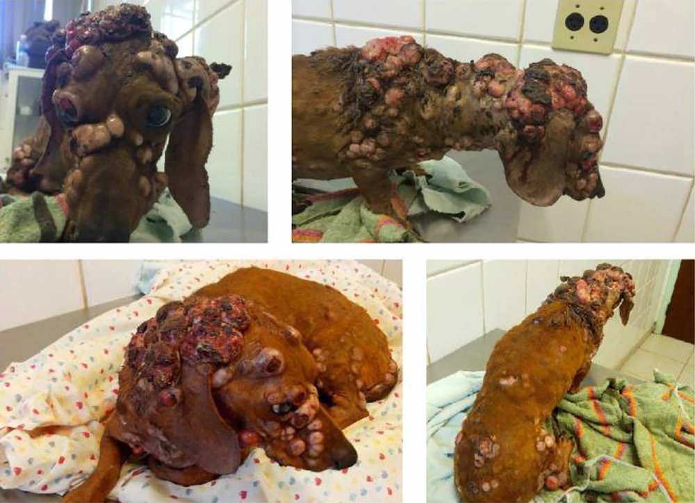
On May 14, 2015, an approximately nine-year-old male Teckel dog was admitted at the oncology service at the Veterinary Hospital of the Universidade Estadual Paulista (UNESP), Jaboticabal - São Paulo, Brazil, with several nodules throughout the body (Figure 1). The case had been rescued 15 months ago (February 2014) with cutaneous nodulation in the cervical region and was taken to a veterinary clinic. At that clinic, cytological analysis of the nodule and near lymph nodes was performed, and based on the results, the cutaneous TVT with lymphatic involvement had been confirmed. The case was subjected to weekly chemotherapy sessions for four weeks with vincristine sulfate (0.75 mg/m2 intravenous). These sessions induced the clinical remission of the disease; though, no additional cytological analysis was performed to evaluate a possible microscopic disease.
After five months, the dog was taken again to the clinic due to submandibular lymphadenomegaly. A new FNA had been conducted, and the cytological diagnosis was compatible with TVT. Chemotherapy with vincristine sulfate had restarted, but after two sessions, there was submandibular lymph nodes enlargement with an emergence of a cutaneous nodule with a diameter of 1 cm at the left side of the neck. The dog was subjected to incisional biopsies with subsequent histopathological and immunohistochemistry analyzes. The results were compatible with undifferentiated round cell tumors. Immunohistochemistry analysis was performed, by which TVT was confirmed. However, before the definitive diagnosis using immunohistochemistry, treatment with doxorubicin (30 mg/m2 intravenous) had already been instituted every 21 days for five sessions. During therapy, the owner reported a slow progression of the dog’s lesions; therefore, it was taken to the UNESP’s oncology service.
The dog was referred to the Veterinary Hospital, and the first physical examination showed multiple crusted, ulcerated, coalescent nodules 0.6 to 9 cm in diameter (Figure 1), peripheral lymphadenomegaly (submandibular, superficial cervical, inguinal, and popliteal lymph nodes) and hyperthermia. The disease was staged using exams, such as fine-needle aspiration of peripheral lymph nodes, abdominal ultrasound, thorax radiographs, complete blood count, biochemical tests, and urinalysis.
Hematological tests indicated severe anemia, thrombocytosis, and leukocytosis by neutrophilia with no left shift. Biochemical analysis revealed hypoalbuminemia, increased alanine aminotransferase (ALT), and alkaline phosphatase (AF). Urinalysis indicated proteinuria (urinary protein/creatinine ratio of 2.6) and bilirubinuria. No alteration on blood pressure was evidenced.
The abdominal ultrasound indicated increased echogenicity and nodules in the liver parenchyma, with the largest nodule approximately 6 cm in diameter. Splenic parenchyma was irregular, and iliac and mesenteric lymph nodes were enlarged. It was suggested to perform the FNA of the liver structures and lymph nodes; however, the owner refused it.
Supportive therapy was conducted with blood transfusion and the following medicines: Amoxicillin/Potassium Clavulanate (22 mg/kg PO q12h) and Ranitidine (2 mg/kg PO q12h) for 10 days; Meloxicam (0.1 mg/kg PO q24h) for four days; S-Adenosyl L-Methionine (SAME) (22 mg/kg PO q12h), and Silymarin (15 mg/kg PO q12h) until further advice.
After seven days of therapy, no clinical improvement of the lesions was observed, but the case was more responsive. New hematological tests and biochemical analysis were taken, and complete blood count showed thrombocytosis, moderate anemia, and mild leukocytosis by neutrophilia, whereas biochemical analysis showed the persistence of hypoalbuminemia and an increase in ALT and AF rates.
As the improvement was not satisfactory, it was decided to try administering chemotherapy medicines that had not been previously prescribed; therefore, lomustine in combination with bleomycin was suggested. The recommended protocol was bleomycin (0.5 IU/kg, subcutaneous) in the first and the second weeks, lomustine (70 mg/m2 PO) in the third week, and reusing the protocol from the fourth week. On the same day, bleomycin was administered.
One week later, the dog underwent reevaluation and a second chemotherapy session with bleomycin. Although there was still no clinical improvement or progression of the lesions, hematological analyzes showed a slight improvement compared with the previous week.
In the third week, the owner reported deterioration of the patient’s condition, hyporexia, and prostration. Accordingly, nasoesophageal tube was inserted. Due to the case’s condition, the third chemotherapy session was postponed. Two days later, the owner made telephone contact and reported that the dog had died, completing 16 months of survival time.
3. Discussion
TVT is a naturally occurring neoplasm that mainly affects sexually active male and female dogs with access to infected animals (4). In this case report, a stray dog was presented with possible interaction with unhealthy dogs, which led to contamination and subsequent development of the disease.
Non-aggressive behavior is commonly associated with TVT, but metastases can occur from 0 to 17% of the cases affecting the skin, lymph nodes, nervous system and viscera, and oral, nasal, and ocular mucous membranes, especially in immunosuppressed dogs (6). Due to the case’s unknown history, little is known regarding its immune status. However, even after an improvement in the dog’s immune condition, an unusual and extremely aggressive presentation of TVT had developed, since there was no response to commonly instituted therapies, and despite the treatment, disease progression was observed.
The extragenital presentation may or may not coexist with genital involvement (6). According to a study conducted by Valençoela et al (7), 32% of the patients presented with extragenital TVT, whereas in 26.1% of the cases, skin or subcutaneous tissue were involved. In this report, it was not possible to clarify whether the implantation site in the skin was primary or secondary to a previous genital tumor. Meantime, atypically, there were multiple ulcerated nodules throughout the patient’s body, located in the trunk, abdomen, thoracic and pelvic limbs, head, and cervical and sacrococcygeal regions (7).
According to de Araujo Santos et al. (8), cutaneous TVT lesions are usually circumscribed and are 2 - 5 cm in diameter. In contrast, lesions in our case were 0.6 - 10 cm and were so anomalous that they lost their circumscribed shape, becoming coalescent to each other.
Hematological analysis revealed anemia and leukocytosis by neutrophilia. Costa and Castro reported that these blood disorders might be present in patients with TVT due to the anatomical sites of the tumor, which may favor bacterial contamination, trauma, or chronic blood loss. In this case report, blood loss occurred due to ulcerated nodules, which could also explain the occurrence of leukocytosis, as they became inflammatory and infectious (5). However, according to Mangieri (9), such disorders can possibly be paraneoplastic syndromes (PNSs).
Since the considered therapy could not control the progression of the disease, the occurrence of such disorders cannot be attributed to TVT-associated PNS, and the bone marrow involvement could not be ruled out because its puncture was not performed.
The laboratory findings (thrombocytosis and increased AF, ALT, and bilirubinuria) and liver abnormalities observed on ultrasound were consistent with those reported in patients with hepatocellular carcinoma (10). It was not possible to perform microscopic analysis of the liver nodules for the case because it was refused by the owner. Therefore, finding an association between these abnormalities and TVT or even another primary liver neoplasia was not possible.
Hypoalbuminemia could be associated with decreased liver function, as well as glomerular loss since proteinuria was evidenced in this case. Glomerulopathies are disorders that affect the glomerular membrane and eventually result in urinary protein loss. Several diseases can culminate with glomerular lesions, including neoplastic disorders (11).
Diagnosis of TVT is based on a defined history, clinical signs, and supplementary tests, such as cytological, histopathological, and immunohistochemistry analyzes, as well as polymerase chain reaction (PCR). The last two have been used in dedifferentiated tumors (3). Usually, cytology is preferred to histopathology, since cytology causes less cellular distortion to microscopic analysis and is a simple, minimally invasive, painless, and low-cost technique (7). Also, because of the morphological characteristics of the tumor cells, the diagnosis is often based only on cytological findings (5). Due to cellular dedifferentiation observed in this case, immunohistochemistry analysis was necessary for definitive diagnosis.
Several treatment options have been described for TVT, including chemotherapy, radiotherapy, surgery, electrochemotherapy, and immunotherapy. The first one is considered as the most effective and practical method, and vincristine sulfate is the drug of choice, which can induce remission in up to 95% of the cases (4). However, refractory cases have been reported. Accordingly, other chemotherapy medicines, such as doxorubicin, cyclophosphamide, vinblastine, methotrexate, and bleomycin sulphate have been used (4, 6). The dog in this report presented apparent clinical remission after the first chemotherapy protocol based on vincristine; nonetheless, after relapse, even using different protocols, it was not possible to obtain clinical remission again.
Early discontinuation of chemotherapy and/or maintenance of microscopic disease may be associated with tumor recurrence, and chemotherapy resistance observed in this case. When clinical remission was reached, the microscopic analysis was not remade; therefore, little can be inferred about the maintenance of the disease microscopically.
Chemotherapy resistance can be due to several factors, including naturally resistant tumor cells, resistance acquired by p-glycoprotein overexpression, defects in the regulation of genes controlling apoptosis, increased intracellular detoxification mechanisms, or mutation in DNA repair systems (2). Plasmocytic subtype appears to cause greater resistance to chemotherapy, as well as a greater predisposition to metastasis (7). However, evaluation of the cytological subtype was not performed in this report.
In most cases, TVT has a favorable prognosis that is based on its good response to chemotherapy and low chemotherapy resistance (5), which was not observed in this report. Because of an aggressive microscopic feature and clinical behavior of the tumor, the patient passed away 16 months after diagnosis.
Based on the results, it can be concluded that TVT can be more aggressive, unlike the behavior usually expected for this tumor. Thus, the importance of an appropriate diagnosis and treatment was emphasized to avoid recurrences or even chemotherapy resistance. This case also showed the importance of cytology after the treatment to check the lack of microscopic disease, allowing deciding to stop the treatment at the correct moment.

