History of Dermal and Subdermal Injectable Fillers Before Collagen: The Early Years

authors:

avatar Omeed Memar ORCID 1 , *

Academic Dermatology and Skin Cancer Institute, Chicago, USA

how to cite: Memar O. History of Dermal and Subdermal Injectable Fillers Before Collagen: The Early Years. J Skin Stem Cell. 2020;7(1):e104559. https://doi.org/10.5812/jssc.104559.

Abstract

Dermal fillers are a mainstay of aesthetic medicine. Currently, the most common ingredient in fillers is hyaluronic acid (HA). However, there was an evolution of products that brought us to where we are today. We review the fillers from fat to synthetic fillers in this review paper. We present notable figures in history who used such fillers and key legislation on the use of such fillers. The early evolution of the use of volumization has been reviewed. This is an important milestone in the history of fillers that is rather brief in the present literature.

1. Context

Dermal fillers are a mainstay of aesthetic medicine. Currently, the most common ingredient in fillers is hyaluronic acid (HA). However, there was an evolution of products that brought us to where we are today. This review aims to cover the data on soft tissue volumizers before the advent of collagen.

2. Discussion

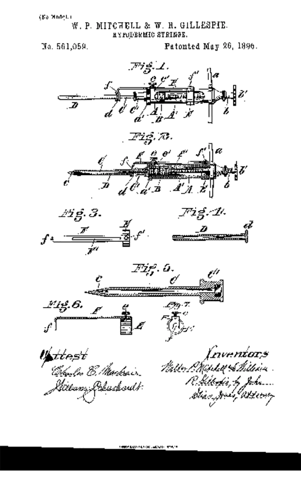
Soft tissue augmentation dates back to the 19th century, when Neuber (1) excised fat lobules from the arm for facial scar, and concluded that larger grafts were less predictable than smaller ones. Czerny (2) used fat from a lipoma to introduce for breast reconstruction in 1895. Lexer (3) studied fat survivability, and concluded that excised fat grafts should be handled with care in both excision and implantation in order to achieve good results. By 1890s, the hypodermic syringe was being developed in glass and silver (Figure 1) (4, 5).

Hypodermic needle: patent drawing of the device designed by Mitchell and Gillespie, 1896.

Lexer (6) presented successful volumization of the face and breasts. By 1911, the hypodermic needle/syringe was use to inject particles of excised fat for post-rhinoplasty irregularities, with excellent results in the short term, and significant subsequent resorption (7). In 1912, the first photographic evidence of fat transplantation using a hypodermic needle/syringe for facial lipoatrophy was published (8). In 1923, the first histologic evidence of transplanted fat that had engrafted was published, and therefore, fat was not just a mass that would be converted to scar tissue, but a true live tissue transplantation (9). In 1926, the term cannula was introduced in the realm of fat transplantation, when Miller (10) presented his technique of face and neck scar correction with injection of fat through a cannula. In 1931, facial lipoatrophy was treated successfully with fat grafting (11). Next, breast reconstruction with fat grafting was reported in 1941, with one breast receiving a large fat graft and another receiving a combination fat/fascia graft, with the fat/fascia graft having better volume retention (12). In 1950, Peer predicted that his technique yielded a 50% survivability of fat grafts (13). He examined histologic samples of transplanted fat to develop the “cell survival theory,” concluding that fat viability was correlated to graft volume (14).

In 1951, Gray et al. (15) performed dermis/fat grafts, and reported complications, including cyst formation. In 1969, Sawhney et al. (16) reported animal studies of dermis/fat grafts done on pigs. Fat/dermis grafts measuring 1.5 × 1.5 cm were transplanted with dermis side down. At one week, the grafts had a reduction of volume by 6.7%; 2 weeks, a 9% reduction of volume; 3 weeks a 20% reduction of volume; 4 weeks, a 33.3% reduction of volume vis-a-vie the original transplant. All the fat was replaced with fibrous tissue, yet the volume was maintained in the 4-week study. Ben-Hur and Neuman (17) determined that epithelial cells were causing cysts and hence the cyst formations reported by Gray et al. were most likely due to inadequate de-epithelialization (18). The game changes came in 1975, when a father/son gynecologist team changed the way fat was harvested, and set in motion a technique that is ever more popular today (17). The Fischer (18) used a blunt metallic suction cannula and invented modern-day liposuction. This allowed procurement of abundant viable fat cells (19).

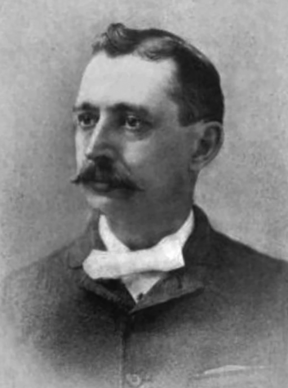
Concurrently, petroleum was discovered, from which came mineral oil. Refined mineral oil produced liquid paraffin. Gersuny (20) published the first medical use of paraffin with a hypodermic needle/syringe in 1900 for creation of a testicular prosthesis in a man who had a bilateral orchiectomy due to tuberculosis. From 1899 to 1914, paraffin was used for breast augmentation. The Derma Featural Company, incorporated in England, was focused on cosmetic surgery, and a court case in 1908 describes the use of hot paraffin injected into the nasal skin and molded to the desired shape. Derma Featural’s largest account was a dermatologist named John Humphrey Woodbury (Figure 2) in New York, NY, who had established cosmetic surgery centers in New York, Boston, Philadelphia, Chicago, St. Louis, and Washington, DC. However, he got tied up with many law suits and committed suicide in 1909 (21). The most notable victim of paraffin injection was the beautiful Gladys Marie Spencer-Churchill, Duchess of Marlborough (Figure 3). She tried to even a small nasal tip asymmetry with paraffin and had some injected into her jaw. The procedure caused major deformities to the point that she became a recluse. She died at the age of 96 in 1977 (22). By 1912, complications of paraffin injection were reported, including, draining persistent fistulas, pulmonary embolism, ulceration, necrosis, breast amputation, and death (23, 24).

John Humphrey Woodbury
Portrait of Gladys Marie Spencer Churchill, Duchess of Marlborough; the most notable victim of paraffinoma syndrome

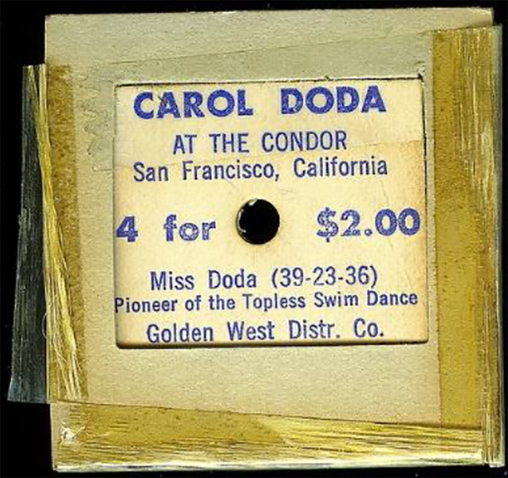
Paraffin complications opened the door to a new comer, “Cleopatra’s needle” (25), where liquid silicone was used for breast augmentation. Liquid silicone was developed and used during world war II as insulation for electrical transformers (26). This began in Japan, where stolen Army stocks of industrial silicone were being injected into the breasts of local prostitutes, creating a more Western appearing contour. Dr. James Brown applied silicone to soft tissue supplementation in 1947 (27). He further studied the safety and complications, concluding that silicone is biocompatible and a safe tissue enhancer (28). Dow Corning introduced a liquid silicone called MDX4-4011, which was FDA approved for coating syringes, but started being used in an unregulated manner for soft tissue augmentation. In 1964, Carol Doda became the face of silicone injections, as she flaunted her breasts in topless burlesques. (Figure 4). Some even mixed different oils, including olive oil and paraffin to the silicone oil to develop their individualized mixtures (29). However, many complications began being reported (30), including granulomas, product migration (31), granulomatous hepatitis, and death (32). In 1975, Nevada became the first state to outlaw the use of injectable silicones. Currently, only two liquid forms of silicone are FDA approved, and only for intra-ocular injection to treat retinal detachment (33). Many have used medical grade silicone off-label for soft-tissue augmentation and by using the droplet-technique. This technique induced fewer complications. However, the illegal use of non-medical grade liquid silicones in the hands of non-medical or ill trained individuals has skyrocketed and resulted in multiple deaths (34).

Ticket to a show by Carol Doda

3. Conclusion

The hunt for the ideal filler was now hotter than ever, since a market had been developed, which demanded easy and affordable soft-tissue volumization. Collagen and a slew of other fillers followed, but the ideal filler is still quite elusive.

References

  • 1.

    Neuber FF. Chir kongr verhandl dsch gesellch chir. 22. 1893.

  • 2.

    Czerny M. Reconstruction of the breast with a lipoma. Chir Kongr Verh. 1895;2:216.

  • 3.

    Lexer E. Zwanzig jahre transplantatiosforshung in der chirurgie. Arch Klein Chir. 1925;138:294.

  • 4.

    Kotwal A. Innovation, diffusion and safety of a medical technology: A review of the literature on injection practices. Soc Sci Med. 2005;60(5):1133-47. [PubMed ID: 15589680]. https://doi.org/10.1016/j.socscimed.2004.06.044.

  • 5.

    Mitchell W, Gillespie WR. Hypodermic syringe. 1896. Report No.: USPTO US561059A.

  • 6.

    Lexer E. Freie fett transplantation. Dtsch Med Wochenschr. 1910;36:640.

  • 7.

    Brunning P. Contribution a letude des greffes adipeuses. Bull Mem Acad R Med Bel. 1919;28:440.

  • 8.

    Hollander E. Die kosmetische chirurgie. In: Joseph M, editor. Handbuch der kosmetik. Leipzig: Verlag van Veit; 1912. p. 690-1.

  • 9.

    Neuhof H, Hirshfeld SD. The transplantation of tissues. New York: Appleton; 1923. p. 1-297.

  • 10.

    Miller C. Cannula implants and review of implantation techniques in esthetic surgery. Chicago: The Oak Press; 1926.

  • 11.

    Eitner E. Fettplastik bei gesichtsatrophie. Med Klin. 1931;27:624.

  • 12.

    Billings EJ, May JJ. Historical review and present status of free fat graft autotransplantation in plastic and reconstructive surgery. Plast Reconstr Surg. 1989;83(2):368-81. [PubMed ID: 2643129]. https://doi.org/10.1097/00006534-198902000-00033.

  • 13.

    Peer LA. Loss of weight and volume in human fat grafts. Plast Reconstr Surg. 1950;5(3):217-30. https://doi.org/10.1097/00006534-195003000-00002.

  • 14.

    Peer LA. Cell survival theory versus replacement theory. Plast Reconstr Surg (1946). 1955;16(3):161-8. [PubMed ID: 13266544]. https://doi.org/10.1097/00006534-195509000-00001.

  • 15.

    Gray DB, Mansberger AJ, Yeager GH. The fate of buried full-thickness skin; an experimental study. Ann Surg. 1951;134(2):205-9. [PubMed ID: 14847491]. [PubMed Central ID: PMC1802772]. https://doi.org/10.1097/00000658-195108000-00006.

  • 16.

    Sawhney CP, Banerjee TN, Chakravarti RN. Behaviour of dermal fat transplants. Br J Plast Surg. 1969;22(2):169-76. [PubMed ID: 4891594]. https://doi.org/10.1016/s0007-1226(69)80061-5.

  • 17.

    Ben-Hur N, Neuman Z. The fate of implanted living epithelial cells into the subcutis. Experimental study in mice. Plast Reconstr Surg. 1964;34:37-49. [PubMed ID: 14200518]. https://doi.org/10.1097/00006534-196407000-00006.

  • 18.

    Fischer A, Fischer G. First surgical treatment for molding body’s cellulite with three 5 mm incisions. Bull Int Acad Cosmet Surg. 1976;3:35.

  • 19.

    Boschert MT, Beckert BW, Puckett CL, Concannon MJ. Analysis of lipocyte viability after liposuction. Plast Reconstr Surg. 2002;109(2):761-5. discussion 766-7. [PubMed ID: 11818867]. https://doi.org/10.1097/00006534-200202000-00054.

  • 20.

    Gersuny R. Ueber eine subcutyane Prothese. Z Heilk. 1900;30:1-5.

  • 21.

    Denkler KA, Hudson RF. The 19th century origins of facial cosmetic surgery and John H. Woodbury. Aesthet Surg J. 2015;35(7):878-89. [PubMed ID: 26069152]. https://doi.org/10.1093/asj/sjv051.

  • 22.

    Fielding D. The face on the sphinx: A portrait of gladys deacon, duchess of marlborough. H. Hamilton; 1978.

  • 23.

    Hollander E. Abstract from berliner gesellschaft fur chirurgie. Munch Med Wochenschr. 1912;59:2842.

  • 24.

    Kolle FS. Plastic and cosmetic surgery. Ann Surg. 1911;54(5). https://doi.org/10.1097/00000658-191111000-00021.

  • 25.

    Webb MS. Cleopatra's needle: the history and legacy of silicone injections. 1997. Available from: https://dash.harvard.edu/bitstream/handle/1/8889460/mwebb.pdf?sequence=1.

  • 26.

    Foreman.m J. Women and silicone: A history of risk. Sunday, City Edition: Boston Globe; 1992.

  • 27.

    Brown JB, Fryer MP, Ohlwiler DA. Study and use of synthetic materials, such as silicones and teflon, as subcutaneous prostheses. Plast Reconstr Surg. 1960;26(3):264-79. https://doi.org/10.1097/00006534-196009000-00002.

  • 28.

    Brown JB, Fryer MP, Randall P, Lu M. Silicones in plastic surgery; laboratory and clinical investigations, a preliminary report. Plast Reconstr Surg (1946). 1953;12(5):374-6. [PubMed ID: 13111916]. https://doi.org/10.1097/00006534-195311000-00007.

  • 29.

    Ortiz-Monasterio F, Trigos I. Management of patients with complications from injections of foreign materials into the breasts. Plast Reconstr Surg. 1972;50(1):42-7. [PubMed ID: 5032325]. https://doi.org/10.1097/00006534-197207000-00007.

  • 30.

    Winer LH, Sternberg TH, Lehman R, Ashley FL. Tissue reactions to injected silicone liquids. A report of three cases. Arch Dermatol. 1964;90:588-93. [PubMed ID: 14206865]. https://doi.org/10.1001/archderm.1964.01600060054010.

  • 31.

    Delage C, Shane JJ, Johnson FB. Mammary silicone granuloma. Migration of silicone fluid to abdominal wall and inguinal region. Arch Dermatol. 1973;108(1):105-7. [PubMed ID: 4268578]. https://doi.org/10.1001/archderm.108.1.105.

  • 32.

    Ellenbogen R, Rubin L. Injectable fluid silicone therapy. Human morbidity and mortality. JAMA. 1975;234(3):308-9. [PubMed ID: 1174244].

  • 33.

    Ellis LZ, Cohen JL, High W. Granulomatous reaction to silicone injection. J Clin Aesthet Dermatol. 2012;5(7):44-7. [PubMed ID: 22798975]. [PubMed Central ID: PMC3396457].

  • 34.

    Graziani C, Hsieh J, Smith D, Neveu W, Demoss B, Grodzin C. Acute cor pulmonale and cardiovascular collapse following illicit cosmetic silicone injections. B58. WHO'S BAD: Case reports in pulmonary vascular medicine I. USA: American Thoracic Society; 2018. A3724 p.