1. Introduction
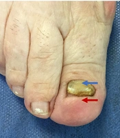
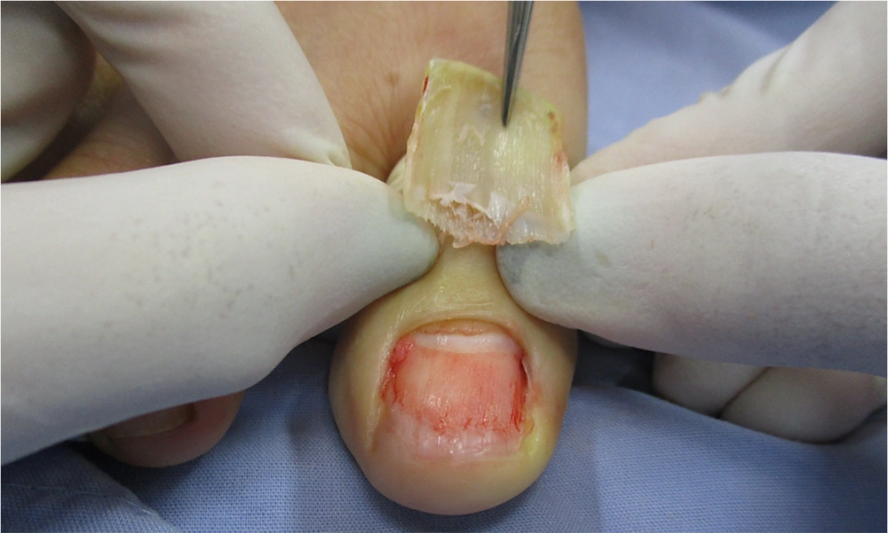
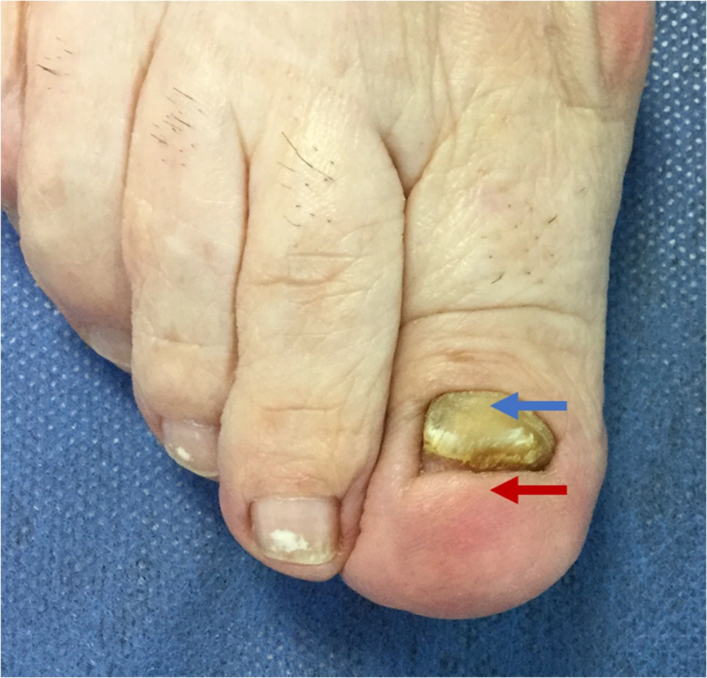
The nail bed is a specialized epithelium attached to the ventral portion of the nail plate. Its appearance is a sequence of ridges and grooves that perfectly correspond to the nail plate (1) (Figure 1). It is not yet clear whether the migration of the nail bed is due to the movement of the cells that compose it or to the growth of the nail plate that slides longitudinally over it, "stretching" it (2). What is clear, in functional terms, is that if the nail bed changes, the shape of the nail also changes and that a large part of the nail dystrophies corresponds to an underlying alteration of the nail bed. Thus: (1) if the nail bed presents a shortening, keratinization, or what is known as "disappearing nail bed" (3), the nail is necessarily forced to thicken since the mitotic activity of the matrix is the same, but the space to deposit its product is less in length (Figure 2); (2) if the nail bed is thin or depressed, the nail increases its thickness and fills the space. If the nail bed is thick, the nail in this portion will be thin (Figure 3).
This behavior allows us to conclude that if we have a flat nail bed with normal morphology and extension, the shape and adhesion of the nail will also be adequate. To illustrate this theory, we use the example of two patients seen in our nail unit service.
We describe through clinical observations the functioning of the nail bed and its response to mechanical dermabrasion.
2. Case Presentation
2.1. Case 1
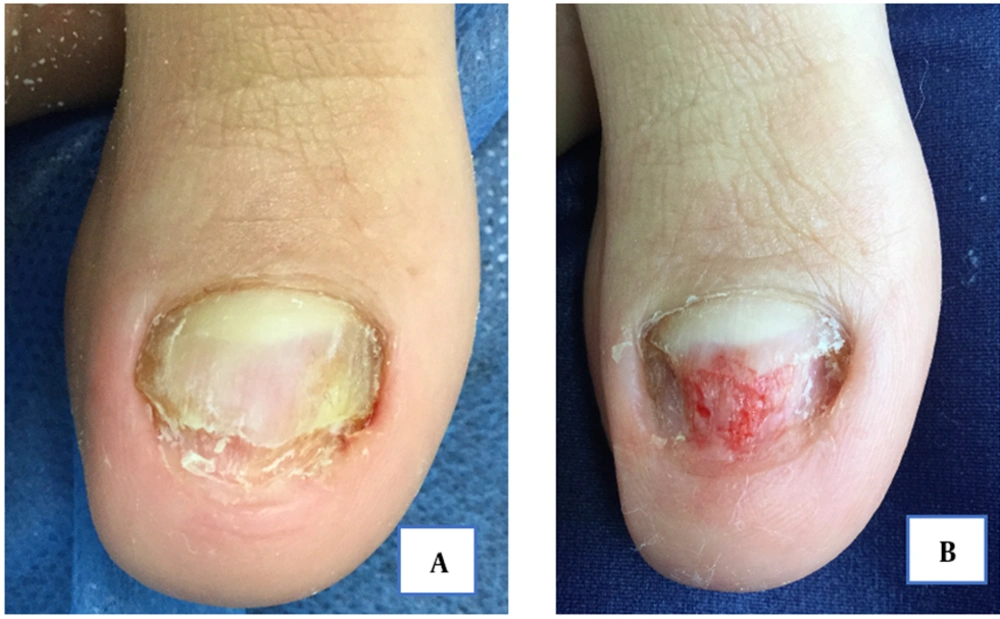
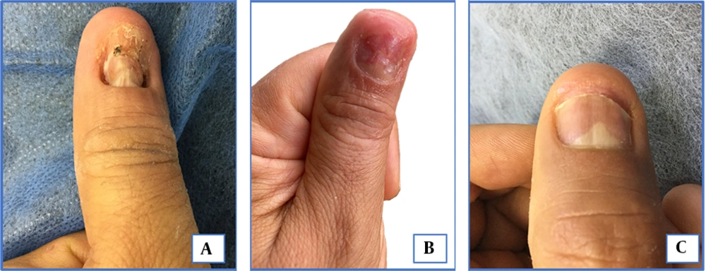
A patient consulted after 11 months of blunt trauma to the left thumb with complete loss of the nail plate and injury to the nail bed (Figure 4A). Mechanical dermabrasion on the nail bed was performed at medium speeds with diamond tips, seeking to sweep away the hyperkeratosis and recover the flat morphology of the nail unit. Improvement was seen since the third postoperative day (Figure 4B), and almost complete recovery was obtained after six months (Figure 4C). These changes favor the observations of the dynamic and migratory nature of the nail bed cells.
A, Hyperkeratotic and disappeared nail bed in the distal portion, adhered dystrophic nail plate in the central region, distorted lunula, and rounded fingertip; B, Day 3 after the procedure. Flattening of the nail bed and change in the morphology of the fingertip; C, Sixth months after the procedure. Considerable improvement in the shape of the nail and almost complete recovery of the nail bed and the shape of the fingertip. Triangular lunula can be observed.
2.2. Case 2
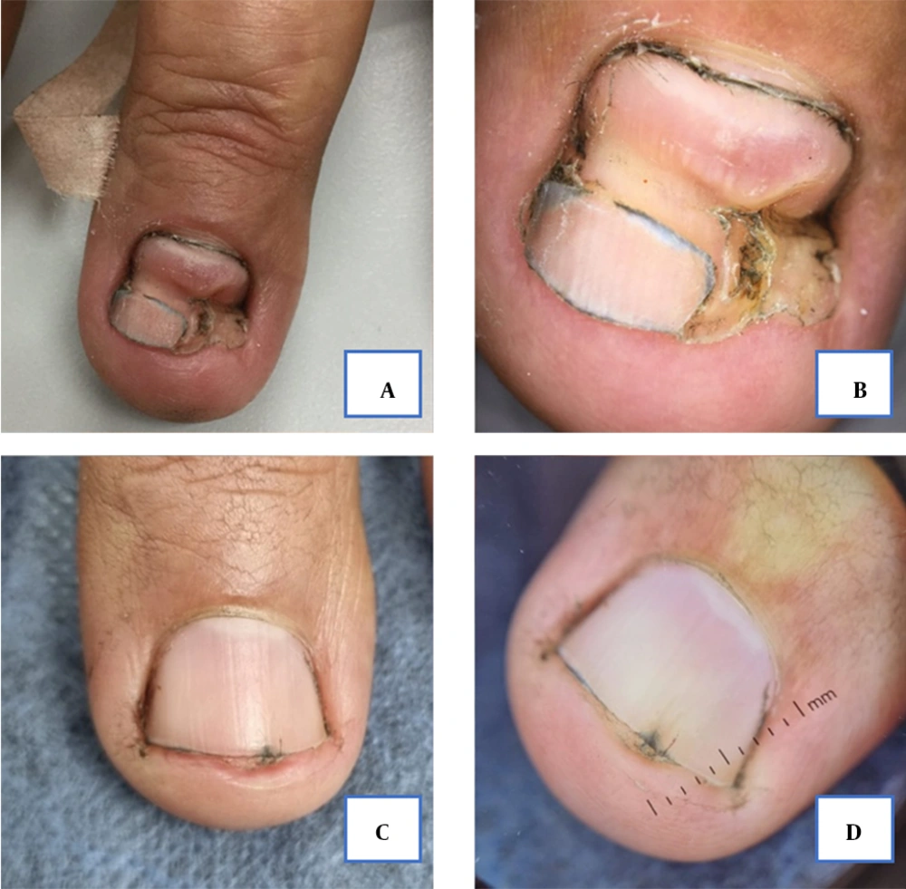
A patient consulted five months after trauma with a hydraulic jack in the right thumb, he presented destruction of the nail bed and alteration of the morphology of the nail plate (Figure 5A and B).
A and B, Initial clinical and dermatoscopic view. A distally small portion of preserved nail plate; in the proximal and medial aspect depressed and folded nail bed with areas of hyperkeratosis. Rounded fingertip. C and D, Clinical and dermatoscopic view nine months postoperatively. Recovery of the nail plate, normal shape of the fingertip, and healthy visible portion of the lunula.
Mechanical dermabrasion was performed, with the technique described in the previous case, to flatten and “stretch” the nail bed and allow better adherence to the nail plate. After nine months of treatment and follow-up, almost complete improvement of the nail was achieved, leaving only a minimal portion of nail bed hypertrophy (Figure 5B and C).
3. Discussion
Historically for the repair of nail bed defects, particularly large ones, surgical methods such as suturing the nail bed, the use of acrylate-based glues (4), or even skin grafts (5) have been the first-line treatments, especially in acute events. The cases described in this article allow us to broaden our perspective and consider other therapeutic options in the approach to nail dystrophies.
Taking into account the premises proposed during the introduction, these results allow a deeper understanding of the functioning of the nail unit, which is, without a doubt, a dynamic structure that responds to forces applied to it. We further hypothesize that mechanical dermabrasion is useful not only for its effect on nail bed hyperkeratosis, but that there must also be an additional effect of the heat and vibration produced during the procedure, which can stimulate the regeneration of the underlying onychocytes. More research is required to understand further the complex response of the nail bed to this therapy, and it would be interesting to carry out a subsequent controlled study to evaluate the efficacy of dermabrasion for the management of this difficult-to-treat entity.
With the previous findings, we present a less invasive technique for recovering the lost nail bed based on its dynamic response to external stimuli and forces.
