1. Introduction
The aesthetic balance of the face is strongly related to the auricles. Shape, angle, and size determine the architecture of the external ear. Ear piercing has been a practice to improve the cosmesis of the ear since ancient times. Skin defects and deformities are associated with a significant negative impact on the quality of life. Other studies have demonstrated the positive effect of reconstructive surgery on patients of all ages (1). Skin cancer of the auricle is a common neoplasm, especially in older people. Melanoma accounts for a small minority of cases, while basal cell carcinoma (BCC) and squamous cell carcinoma (SCC) are responsible for most skin cancers (2).
Age, sun exposure, contour irregularities, and large shape are all contributing risk factors for malignant transformation (3). Early interventions are imperative to avoid tumor unresectability or extensive reconstructive methods with high failure rates. In addition, the oncologic outcome is strongly correlated with the cancer stage, with poor results in SCC stage IV classification cases (4). Biopsies are mandatory before any intervention. However, histologic confirmation often delays the cure, and a two-stage operation is unavoidable (3). Our study aimed to present the benefits of frozen sections on ear skin tumors with high clinical suspicion to avoid a second surgery and the necessity to restore the auricle with an acceptable cosmetic outcome even in people of the third age.
2. Case Presentation
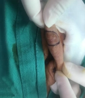
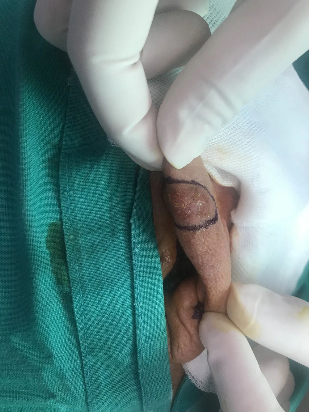
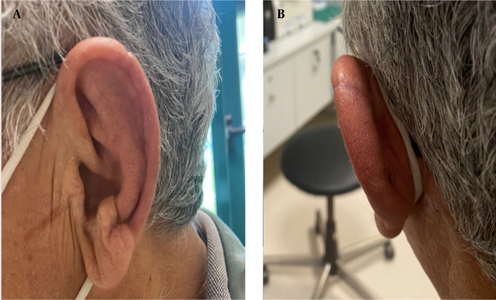
A 76-year-old male presented to our Ear, Nose, and Throat Department with the chief complaint of progressive exophytic growth on his left auricle (Figure 1). He reported no pain, weight loss, or bleeding. The patient was a former smoker with a referred history of 30 cigarettes per day for 42 years. Alcohol consumption was also a social habit of his past. His medical history included diabetes mellitus, arterial hypertension, dyslipidemia, cardiac insufficiency, lupus erythematosus, and a family history of BCC. Specifically, the patient’s father had a known BCC of the tip of the nose, and the patient’s mother had been diagnosed with a BCC of the forehead. Acetylsalicylic acid was the only anticoagulant medication he consumed. He had worked as a woodworker for 45 years. The patient was referred from another hospital due to the inability of the other department to perform an extensive reconstructive restoration.
On physical examination, a skin growth was detected on the helix of his left auricle. The mass was exophytic, not friable, and located in the superior part of the helix with slight posterior extension. The remainder of his facial skin, scalp, external auditory canals, and oral cavity were lesions-free. Neck palpation was normal, with no enlarged lymph nodes. Imaging modalities were not prescribed, and the patient was informed of the probable skin cancer diagnosis. However, he was informed that before any reconstructive intervention, a biopsy is mandatory. After receiving patient consent, a frozen section biopsy was determined. In case of positive results, the operation was planned to continue with full tumor excision and reconstruction.
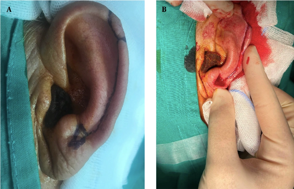
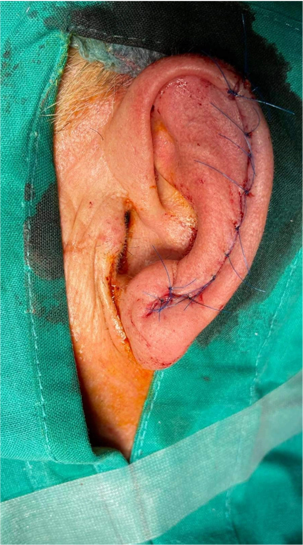
The other day, the patient was transferred to the operating theatre and underwent a biopsy under local anesthesia. A part of the sample was sent for a frozen section. The frozen section results confirmed the presence of a pigmented BCC. Therefore, the surgery proceeded to reach the preoperative goals. Under local anesthesia, total lesion excision was performed with a 4-mm safety margin. A helical rim advancement flap was recruited as the best option for an excellent cosmetic result (Figure 2). After total mobilization of the skin and especially the posterior part, a tension-free closure was achieved (Figure 3). Hemostasis was normal, and the patient was discharged after 2 h. In a one-month follow-up, the cosmetic result was excellent (Figure 4), and the patient was in an excellent emotional condition. Histology was indicative of a pigmented BCC excised on wide negative surgical margins. The patient was discharged with no suggested adjuvant therapy.
3. Discussion
Patients with skin cancer have the advantage of early diagnosis due to its presentation in sun-exposed areas (5). A simple excision usually suffices to reach oncologic clearance, but often a primary closure does not secure a satisfactory cosmetic result. Therefore, versatile local flaps offer tissue-matching native structures and help successful healing. The design of the flap is crucial because it relies on random blood supply and a suitable geometry is mandatory to avoid necrosis (3). Surgeon who selects to recruit a helical rim advancement flap should be aware of tricks and tips to perform a flawless reconstruction. The helical rim should be incised perpendicular to the sulcus fashion, and the defect might be needed to be extended to reach the sulcus (at least its base). Blunt dissection should be aimed at undermining the auricle skin. The posterior part is more vulnerable to mobilization and should be recruited greater than the anterior part. Finally, sutures should not be dense to allow a better vascular supply of the skin (2, 3).
The helical rim advancement flap is mainly used for defects of up to 2 cm on the upper part of the auricle (6). Advantages include the similarity of the color and thickness of the flap with the native tissue. The posterior auricular artery is the workhorse of blood supply, and a broad pedicle is needed to prevent wide necrosis (7). Advancement flaps seem ideal for medium size defects, but it is better to avoid superficial or minor defects. According to the literature, superficial defects are better treated with full-thickness free grafts from the postauricular skin. On the other hand, minor defects could be easily dealt with side-to-side repairs (8). Defects > 2 cm are more difficult to be repaired and usually need a staged transposition flap from the postauricular area because they lack adequate tissue when a helical rim flap is used. When the tumor includes the scaphoid fossa, a reduction in pinna size should be considered an option to achieve a tension-free closure (9).
The helical rim advancement flap has been associated with some minor complications. The most known well-described is a slight decrease in the width and height of the lobule. Consequently, an asymmetry with the contralateral auricle might cause a minor cosmetic deformity (6). Our case shows that using advancement flaps in the reconstruction practice of ear skin cancers should be routine. A good cosmetic result should be offered independent of the patient’s age. In addition, frozen sections are extremely useful to avoid a second operation. However, the result is better tested in a broader sample and not only on a single case. Our case is important because it emphasizes the unique advantages of a specific flap reconstruction technique. Helical rim advancement should be the backbone of reconstruction for the skin cancer of the ear because it secures oncologic clearance and gives a great cosmesis. Physicians should be aware of this technique and employ it independently of the patient’s age. Both indications and surgical techniques are described and discussed meticulously.
