1. Introduction
PG is a rare neutrophilic dermatosis (ND) characterized by rapidly evolving painful ulcers with undermined borders and peripheral erythema. Clinically, there are five distinct clinical variants, including classic, bullous, vegetative, pustular, and peristomal PG. Legs are the most commonly affected site in classic PG (1). Based on the literature, PG incidence ranged from 0.4 - 2.6% among IBD patients. This is significantly higher than its prevalence among the general population (3 - 58 people per million) (2). The etiology of PG is not fully understood, but trauma has been introduced as the culprit of PG. Trauma induces inflammatory cytokines like IL-8 and IL-36 from keratinocytes as well as the expression of autoantigens. Pathogenic variants of genes related to the inflammasome pathway are accompanied by PG (3). Oral corticosteroids and biological agents such as infliximab and adalimumab are the most common therapies for IBD-associated PG (4). Here, we report an atypical case of PG in a patient with UC, successfully treated by infliximab.
2. Case Presentation
A 34-year-old man with a history of UC was referred for an IBD flare-up. There were abdominal cramps, bloody diarrhea (15 - 20 times/day), significant weight loss, and laboratory findings that indicated a severe disease state (Mayoclinical score = 3). The patient’s hemodynamic status was stable, and the abdominal examination was unremarkable. His father had a history of UC. The patient’s UC was in clinical remission for years with mesalamine and azathioprine. His rectosigmoidoscopy revealed multiple segmental and diffuse pseudopolyps in the sigmoid and descending colon, generalized ulcers, loss of vascular pattern, marked friability, and spontaneous bleeding compatible with Mayoendoscopic score = 3. Laboratory tests showed CRP: 48 mg/L, Albumin: 2.8 g/dL, mild leukocytosis, and severe anemia. The stool test was inflammatory with a calprotectin level of more than 1100 µg/g feces.
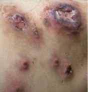
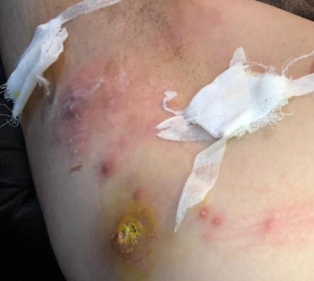
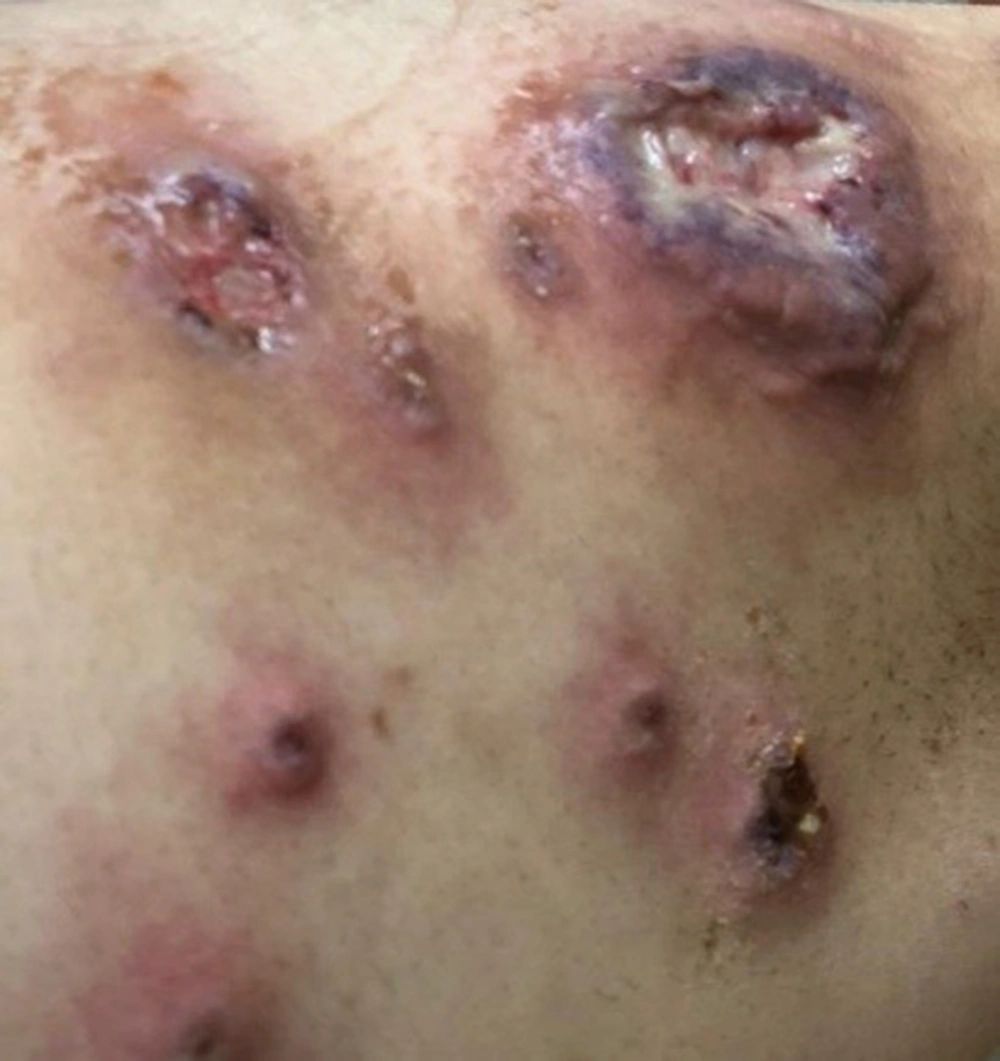
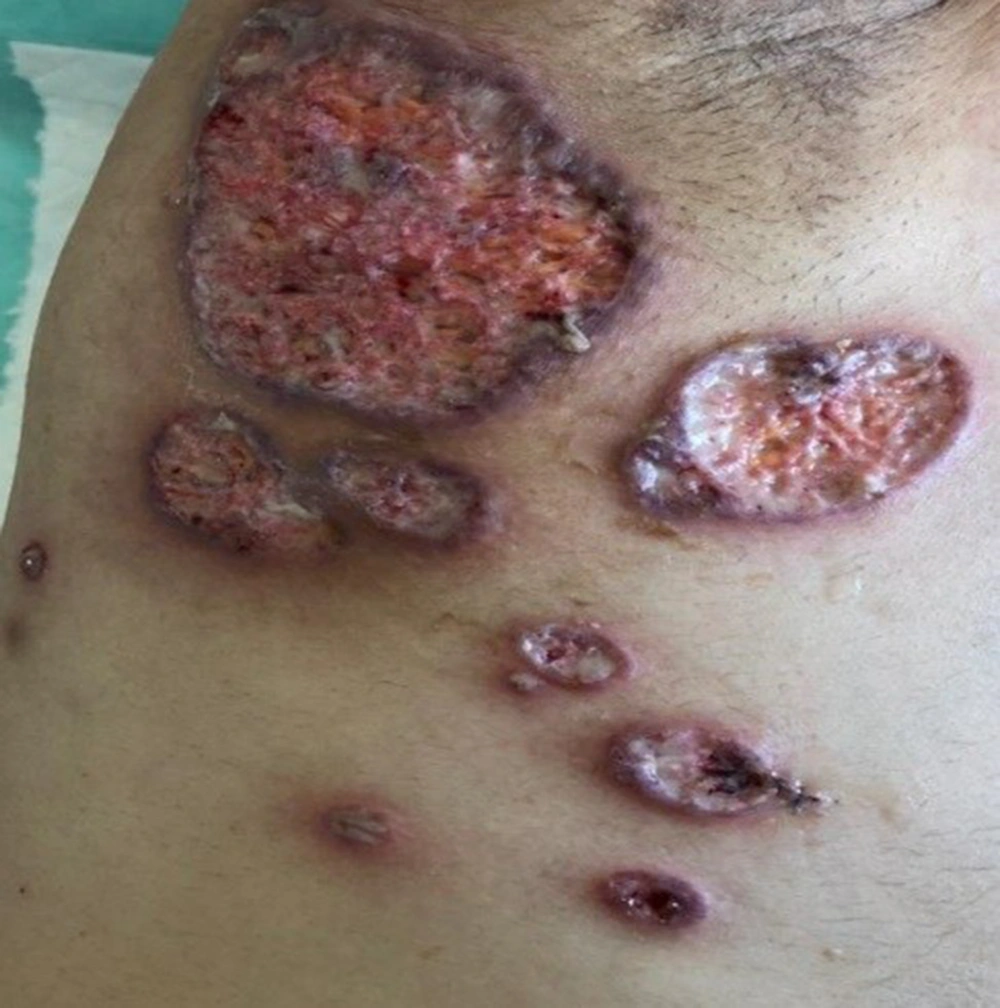
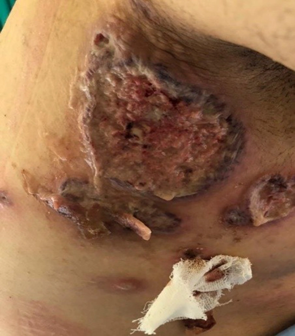
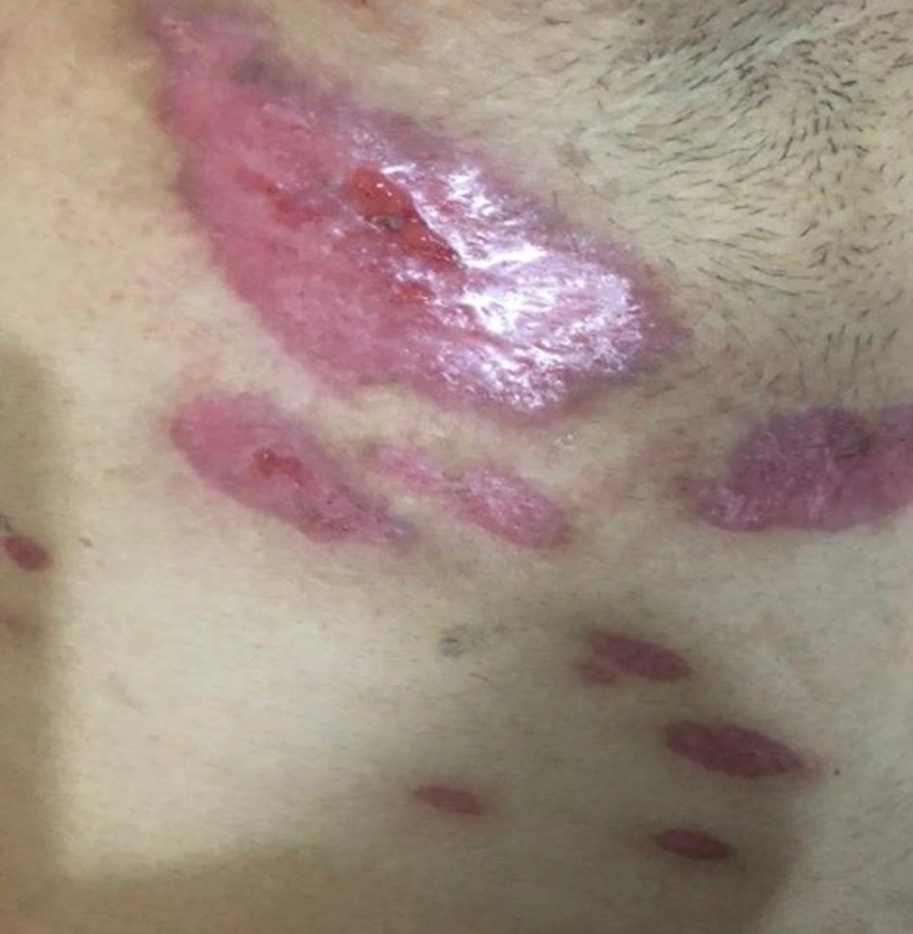
Skin examination revealed follicular lesions (Figure 1), initially seen in the axillary area. It rapidly evolved into a giant ulcer with a yellow discharge surrounded by purple edges in a few days (Figure 2). The ulcer eventually became a large necrotic ulcer (Figure 3). PG was suspected, and infective processes were also in the differential diagnoses. A definite diagnosis of PG was made by pathologic evaluation of the lesion, which was consistent with a sterile abscess. The CT-enterography revealed normal small bowel loops and moderate to severe thickness of the distal colon wall accompanied by prominent pericolic lymphadenopathies, fat infiltration, and mucosal enhancement. The patient was started on intravenous steroids as the first step in the treatment of an episode of IBD flare. Empiric wide spectrum antibiotics and topical tacrolimus were also started for skin lesions. Response to therapy was not impressive until the initiation of high-dose infliximab (10 mg/kg), which helped reduce the size and severity of the ulcer (Figure 4). The response to treatment started within a week. Complete healing of the ulcer was seen two months after the initiation of infliximab (Figure 5).
3. Discussion
PG was first described in the first decade of the 20th century as an erythematous, invasive, and painful skin lesion with purulent cavities and histopathology of neutrophil infiltration. The word pyoderma gangrenosum was coined by Brunsting in 1930. PG typically appears as a painful nodule or pustule (5), which can be found everywhere in the body, although they almost exclusively arise in the legs, arms, and adjoining stoma. PG is an autoimmune inflammation associated with inflammatory processes and is seen in 1 - 5% of IBD patients. The rapid spread of an atrophic and cribriform painful lesion surrounded by purple or red edges with a positive pathergy skin test when other causes of such ulcerations have been excluded would be diagnostic, especially in the presence of a concurrent autoimmune disorder (6). The presented case fulfilled the criteria for diagnosis of PG, but a simultaneous flare of IBD was not expected. Involvement of the axillary area was also atypical for PG. A brief review shows that interruption of the equilibrium of the host immune system and immune tolerance might be responsible for PG (7). The relationship between UC and PG is not entirely clear, and the interfering role of some other pathogenic factors, like interleukins, cannot be ignored. Shwartzman phenomenon associated with colonic endotoxins is one of the proposed hypotheses. Studies have reported no direct relationship between PG and UC (8), in contrast to the presented case with synchronous presentation of IBD flare and PG. Therapy aims to control inflammation, reduce pain, and heal wounds. Topical and systemic treatments with corticosteroids and cyclosporine have historically been known as the mainstay of therapy, but recent studies have shown that anti-TNF agents are more successful. Infliximab is a chimeric monoclonal antibody that blocks TNFα (4). Patients' quality of life was also introduced as an important factor in the management of PG, which can be improved by administering infliximab to patients with the debilitating disease of PG (9).
