1. Introduction
Seborrheic melanosis (SM) is a definition with hyperpigmentation in the sebaceous areas of the face, such as the alar grooves, nasolabial folds, angles of the mouth, and labiomental crease. The term “seborrheic melanosis” is denoted by Indian dermatologists. Dark-skinned Asian, Hispanic, and African populations with Fitzpatrick skin types 4 and above are more likely to have SM (1). Generally, the characteristics and locations of skin lesions are distinct from other types of facial hyperpigmentation. Because hyperpigmentation presents itself with sebum production and scaly flakes on the seborrheic parts of the face, SM presents distinct features in terms of facial hyperpigmentation.
In SM, hyperpigmentation can vary in severity and location, but its distinctive characteristics make it easily identifiable. Also, treatment options for SM are different from other types of facial melanoses (2). Therefore, to select the most appropriate treatment method for SM, clinicians need to distinguish it from other facial melanoses.
2. Case Presentation
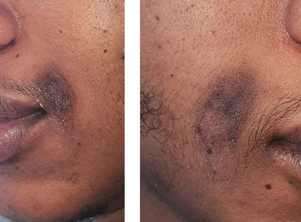
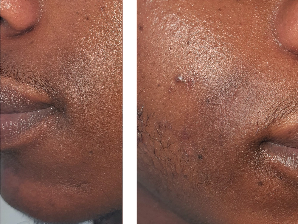
A 19-year-old male patient presented with dark brown spots on both sides of his nasolabial folds. He applied an over-the-counter cream that contained hydroquinone to the hyperpigmented areas for bleaching for one month before he consulted our dermatology department, but instead, he had irritation with deep hyperpigmentation. While there was a mild itching problem, he did not scratch or traumatize the lesions. He declared that there was no family or genetic history of the findings he had. Additionally, neither he nor his family had a history of atopy. His skin type was Fitzpatrick 6. According to the skin examination, there were nummular oval macules with hyperpigmentation and mild desquamation at the junction of the nasolabial fold and the supralabial area bilaterally (Figure 1). The pigmentation was homogeneous, brown-black, and showed no contrast under Wood’s light examination. Additionally, there were hyperpigmented macules on a seborrheic basement at the alar-supralabial junction bilaterally, and he had brownish hyperpigmentation with a greasy texture on the nasal alar folds. The pigmented areas included mild squama on the surface. There were no lesions on other parts of the face and body, including the forehead, retroauricular area, or trunk. Although there was a seborrheic appearance on the scalp, he did not present with pityriasis capitis. Based on the anamnesis and clinical findings, the patient was diagnosed with SM. Skin biopsy was not considered because of cosmetic concerns. Compatible with therapy for SM (1), tacrolimus ointment 0.1% once every night and isoconazole nitrate cream once daily were prescribed for a period of one month. A water-based moisturizer and sunscreen were also added to the treatment. Over the course of one month, the patient experienced a significant improvement in hyperpigmentation and desquamation, proving the efficacy of the prescribed regimen. He stopped utilizing the main treatment after one month; however, he continued to apply moisturizers and sunscreen. Further, he used a facial peeling combination of alpha-hydroxy acid 10% and beta-hydroxy acid 2% two times per week for hyperpigmented areas. Hyperpigmentation was barely discernible on his face five months after the beginning of the therapy (Figure 2).
3. Discussion
Clinical presentations of SM include specific findings on the skin as well as the patient’s origin. Particularly Fitzpatrick skin types 4 and 6 are prone to hyperpigmentation in the seborrheic areas. Specifically, Asians, Africans, and Hispanics with darkening of their seborrheic areas are more likely to suffer from SM. These seborrheic areas include the alar groove, angles of the mouth, and labiomental groove (1). Additionally, Verma et al. described early SM lesions as localized scaly erythema of alar grooves with nasolabial dyssebacia that progresses to develop hyperpigmentation. Furthermore, SM is defined to include pigmentation of the alar crease, pigmentation of the labiomental crease, involvement of the angles of the mouth, and involvement of the nasolabial fold (1). Herein, the clinical pattern and the localization of hyperpigmentation in the patient led us to diagnose the case as SM. Since darker-skinned populations are more likely to have higher levels of melanin, they are more likely to suffer from this type of hyperpigmentation. Additionally, the characteristics of the lesions and the locations they appear in are unique to SM, making them easily identifiable.
Seborrheic melanosis has also been discussed as "postinflammatory hyperpigmentation", which may result from any papulosquamous dermatitis affecting the face, including seborrheic dermatitis, acne, atopic dermatitis, perioral dermatitis, psoriasis, allergic contact dermatitis, and angular cheilitis (3). Furthermore, frictional melanosis, photomelanosis, and Riehl’s melanosis are the other causes of facial melanoses. These conditions should be treated with topical bleaching creams or rubbing; however, these treatment methods could be hazardous for SM (2). Riehl melanosis is a pigmented contact dermatitis developed in reaction to cosmetic allergens. It is brown-black pigmentation that is seen mostly on the forehead and temples of dark-skinned patients. If topical bleaching creams and rubbing fail to solve the issue, laser or light therapy may reduce the melanin production associated with Riehl’s melanosis (4). The other common facial hyperpigmentation condition is frictional dermal melanosis. This condition is usually associated with repeated damage to the skin and is primarily seen on superficial bony prominences. Treatment methods for frictional dermal melanosis are similar to those for Riehl’s melanosis (5). Although bleaching methods for the skin could be used for other facial melanoses, as mentioned above, they can cause controversial effects such as irritation and postinflammatory hyperpigmentation in the exacerbation period of SM patients. On the other hand, topical bleaching agents can also be chosen appropriately after SM exacerbations (2).
Because of the fact that hyperpigmentation on the face poses cosmetic concerns, defining the characteristics of the SM poses a significant challenge. Having a skin biopsy from the face can exacerbate aesthetic concerns in patients with darker skin tones, while histopathology of SM may not provide a certain diagnostic result (2). Even though dermatoscopic findings may lead to the diagnosis of SM, they still may remain ambiguous and may not be conclusive for a decisive diagnosis without clinicopathologic correlation (3).
Limitations of this case report are dermatoscopic evaluation and lack of information regarding the clinicopathologic correlation of a skin biopsy. On the other hand, we defined hyperpigmentation with desquamation and seborrhea, which allowed us to determine the right course of treatment. Furthermore, this case report provided a detailed discussion of the results of the treatment, demonstrating the efficacy of the chosen method.
Despite the lack of adequate diagnostic tools, the hallmarks of clinical presentation and associated findings can be used to differentiate SM from other forms of facial melanoses. By understanding the patient’s medical history and performing a meticulous physical examination, it is possible to identify any underlying causes contributing to the condition. This helps determine the most appropriate course of treatment and ensures that it is tailored to the individual’s needs. However, more studies are still needed to develop diagnostic techniques to define SM with concrete evidence.

