A Clutching Claw: Unexpected Coexistence of Pediculosis Capitis and Tinea Capitis

authors:

avatar Dalia Ibrahim Halwag ORCID 1 , *

Department of Dermatology, Venereology and Andrology, Faculty of Medicine, Alexandria University, Alexandria, Egypt

How To Cite Halwag D I. A Clutching Claw: Unexpected Coexistence of Pediculosis Capitis and Tinea Capitis. J Skin Stem Cell. 2023;10(4):e143263. https://doi.org/10.5812/jssc-143263.

Abstract

Introduction:

Pediculosis capitis and tinea capitis are very common superficial infections/infestations, primarily affecting school-aged children. The overall pediculosis capitis infestation rate exceeds 25% in some studies, and the occurrence rate for tinea capitis may be as high as 45%. They are more prevalent, but not exclusively, in crowded conditions and environments with low socioeconomic status. Scaly scalp is a common presentation of tinea that may be, at first glance, confused with pediculosis nits.

Case Presentation:

Here, we describe a case report showing the coexistence of both tinea capitis and pediculosis capitis in a young girl, supplemented with an interesting photograph of a nymph of Pediculus humanus capitis attached by its claws to a hair, with dystrophic endothrix hairs in the background. We also describe further fungal culture performed, with the result and the treatment the patient received.

Conclusions:

Tinea and pediculosis should always be excluded first in case of a scaly scalp in children. Whether their coexistence is attributed to a causal relation and not simply a poor environment is still to be investigated. This is especially important because body louse is a proven vector for a number of pathogens; however, this is still a matter of debate for head louse.

1. Introduction

Pediculosis capitis and tinea capitis primarily affect school-aged children. The causative dermatophyte species of tinea capitis varies across geographical regions (1). In contrast, Pediculus humanus capitis is responsible for pediculosis capitis worldwide. The coexistence of both infections in the same patient is rare (2).

The overall pediculosis capitis infestation rate amongst school-age children may reach more than 25% in some studies (3). Children with lice infestations may be completely asymptomatic; however, there is usually some pruritus. The itching can be severe, leading to excoriations, secondary impetiginization, and cervical lymphadenopathy. Identification of crawling adult lice and/or the nits, mainly in the occipital and retroauricular scalp, confirms the diagnosis (4).

Tinea capitis also mainly affects children worldwide, with a higher frequency in tropical and subtropical regions and an estimated occurrence rate of up to 45% in some studies (5). Tinea capitis can present with a multitude of clinical manifestations, including alopecic patches, scaly scalp that may be confused with nits, inflammatory changes in kerion with purulent discharge, and the possibility of cicatricial changes.

2. Case Presentation

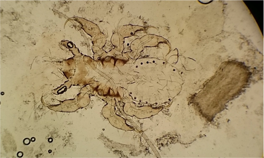
Herein, we present a 9-year-old girl presenting to our Mycology Clinic for an extensively scaly scalp with multiple focal areas of alopecia. The alopecic patches showed broken hairs, consistent with the clinical diagnosis of black dot tinea capitis. Hence, to confirm the diagnosis of tinea capitis, scrapings from the hair stumps were collected for potassium hydroxide (KOH) smears and fungal culture. Scrapings of hair stumps from the alopecic patches were examined with 10% KOH under light microscopy. At 200X magnification, to our surprise, a nymph of P. humanus capitis was seen attached by its claws to a healthy hair, with dystrophic hairs seen in the background (Figure 1). Clinically, pediculosis might have been obscured by the heavy scalp scaling. The dystrophic hairs on higher (400X) magnification showed endothrix fungal spores. The culture of the scrapings on Sabouraud dextrose agar (SDA) revealed, after 2 weeks, the growth of Trichophyton violaceum, which is endemic to our geographical region (1). The child was prescribed both systemic griseofulvin at a dose of 10 mg/kg for tinea capitis and topical 5% permethrin lotion for pediculosis.

Six-legged nymph (according to appearance, the third nymph) of Pediculus humanus capitis attached by its claws to a hair. Next to the nymph, there is a dystrophic hair parasitized by fungal spores (endothrix). The sections are visualized using 10% potassium hydroxide under light microscopy at 200X magnification

3. Discussion

The simultaneous acquisition of pediculosis capitis and tinea capitis is rarely reported. Also, the visualization of pediculi in scrapings is unusual and, hence, rather an interesting site.

The definitive diagnosis of pediculosis capitis can be made upon identifying crawling lice on the scalp hair. Nits alone are not diagnostic of active infestation unless they are very close to the scalp, are dark-colored, and contain larvae (6). In our case, the nymph was clearly demonstrated on a wet mount of scraped hairs.

Clinical diagnosis of tinea capitis is usually confirmed by KOH-prepared wet mount of plucked hairs or hair stumps collected by scraping or brushing. The location of the fungal spores in relation to the hair shaft (endothrix or ectothrix) can give a clue about the causative dermatophyte species, which should be confirmed by fungal cultures as SDA. Sabouraud dextrose agar contains dextrose, peptone, agar, and chloramphenicol to inhibit bacterial growth. Fungal growth is expected within 2 to 4 weeks. The gross appearance of the culture and stained culture mounts can accurately specify the causative dermatophyte subtype (7). The presence of endothrix suggests that the etiological agent belongs to the Trichophyton fungal genus. Waxy, glabrous, wrinkled, deep purple colonies (like the ones that were isolated from our case) are characteristic of T. violaceum (8).

Permethrin, a synthetic pyrethroid, interferes with sodium transport in the parasite, subsequently causing neural depolarization and, hence, respiratory paralysis. Permethrins are considered safe with low toxicity, and retreatment in 7 to 10 days is advised for the recently hatched nymphs (9). Although the 5% concentration is mainly used for scabies, it is currently used for pediculosis to overcome the resistance to the 1% preparations. Other lines of treatment for pediculosis include mechanical lice/nit removal using wet combing or other chemical preparations, such as lindane 1% and malathion 0.5% (6). Griseofulvin is FDA (Food and Drug Administration)-approved for treating tinea capitis. It is considered the drug of choice, especially in children, with high cost-effectiveness, cure rate, and easy accessibility. Griseofulvin is a microtubule assembly inhibitor that interferes with the formation of the mitotic spindle; hence, it inhibits mitosis in dermatophytes (10).

Body louse is a known vector for Bartonella organisms (B. quintana, B. recurrentis) (11), as well as for louse-borne relapsing fever, trench fever, and epidemic typhus, by transmitting different Rickettsia species (R. prowazekii, R. rickettsii) (12). However, whether head lice also serve as a vector is still inconclusive (13).

Studies found that head lice can pass infective Rickettsiae in their feces (14). Other studies showed that head lice could harbor a number of bacterial pathogens, such as the DNA of B. quintana (15), Coxiella burnetii (16), Borrelia recurrentis (17), and even Acinetobacter (16). However, the mere presence of a bacterial organism in an arthropod does not necessitate the ability to transmit it to humans and cause a disease in the infested individual (13).

The question remains whether the coexistence of tinea and pediculosis capitis is mainly attributable to overcrowding, poverty, and poor hygiene or whether the head lice might have a role in harboring and transmitting dermatophytes (2).

References

  • 1.

    Nouh AH, Rageh MA. A rare case of coexisting pediculosis capitis and tinea capitis in a healthy adult female. Skin Appendage Disord. 2021;7(6):486-8. [PubMed ID: 34901182]. [PubMed Central ID: PMC8613637]. https://doi.org/10.1159/000517198.

  • 2.

    El-Khalawany M, Shaaban D, Hassan H, Abdalsalam F, Eassa B, Abdel Kader A, et al. A multicenter clinicomycological study evaluating the spectrum of adult tinea capitis in Egypt. Acta Dermatovenerol Alp Pannonica Adriat. 2013;22(4):77-82. [PubMed ID: 24336943].

  • 3.

    Kassiri H, Mehraghaei M. Assessment of the prevalence of pediculosis capitis and related effective features among primary schoolchildren in Ahvaz County, Southwest of Iran. Environ Sci Pollut Res Int. 2021;28(18):22577-87. [PubMed ID: 33420692]. https://doi.org/10.1007/s11356-020-12284-9.

  • 4.

    Bloomfield D. Head lice. Pediatr Rev. 2002;23(1):34-5. discussion 34-5. [PubMed ID: 11773591]. https://doi.org/10.1542/pir.23-1-34.

  • 5.

    Dogo J, Afegbua SL, Dung EC. Prevalence of tinea capitis among school children in nok community of kaduna state, Nigeria. J Pathog. 2016;2016:9601717. [PubMed ID: 27471603]. [PubMed Central ID: PMC4947659]. https://doi.org/10.1155/2016/9601717.

  • 6.

    Ko CJ, Elston DM. Pediculosis. J Am Acad Dermatol. 2004;50(1):1-12. quiz 13-4. [PubMed ID: 14699358]. https://doi.org/10.1016/s0190-9622(03)02729-4.

  • 7.

    Ely JW, Rosenfeld S, Stone MS. Diagnosis and management of tinea infections. American family physician. 2014;90(10):702-11.

  • 8.

    Magill SS, Manfredi L, Swiderski A, Cohen B, Merz WG. Isolation of Trichophyton violaceum and Trichophyton soudanense in Baltimore, Maryland. J Clin Microbiol. 2007;45(2):461-5. [PubMed ID: 17151204]. [PubMed Central ID: PMC1829009]. https://doi.org/10.1128/JCM.02033-06.

  • 9.

    Rasmussen J. Antiparasitic agents. In: Wolverton SE, editor. Comprehensive dermatologic drug therapy. Philadelphia: WB Saunders Co; 2001. p. 537-46.

  • 10.

    Gupta AK, Mays RR, Versteeg SG, Piraccini BM, Shear NH, Piguet V, et al. Tinea capitis in children: A systematic review of management. J Eur Acad Dermatol Venereol. 2018;32(12):2264-74. [PubMed ID: 29797669]. https://doi.org/10.1111/jdv.15088.

  • 11.

    Drancourt M, Mainardi JL, Brouqui P, Vandenesch F, Carta A, Lehnert F, et al. Bartonella (Rochalimaea) quintana endocarditis in three homeless men. N Engl J Med. 1995;332(7):419-23. [PubMed ID: 7529894]. https://doi.org/10.1056/NEJM199502163320702.

  • 12.

    Sundnes KO, Haimanot AT. Epidemic of louse-borne relapsing fever in Ethiopia. Lancet. 1993;342(8881):1213-5. [PubMed ID: 7901534]. https://doi.org/10.1016/0140-6736(93)92190-5.

  • 13.

    Feldmeier H. Head lice as vectors of pathogenic microorganisms. Trop Med Health. 2023;51(1):53. [PubMed ID: 37730694]. [PubMed Central ID: PMC10510260]. https://doi.org/10.1186/s41182-023-00545-5.

  • 14.

    Robinson D, Leo N, Prociv P, Barker SC. Potential role of head lice, Pediculus humanus capitis, as vectors of Rickettsia prowazekii. Parasitol Res. 2003;90(3):209-11. [PubMed ID: 12783309]. https://doi.org/10.1007/s00436-003-0842-5.

  • 15.

    Bonilla DL, Kabeya H, Henn J, Kramer VL, Kosoy MY. Bartonella quintana in body lice and head lice from homeless persons, San Francisco, California, USA. Emerg Infect Dis. 2009;15(6):912-5. [PubMed ID: 19523290]. [PubMed Central ID: PMC2727331]. https://doi.org/10.3201/eid1506.090054.

  • 16.

    Louni M, Amanzougaghene N, Mana N, Fenollar F, Raoult D, Bitam I, et al. Detection of bacterial pathogens in clade E head lice collected from Niger's refugees in Algeria. Parasit Vectors. 2018;11(1):348. [PubMed ID: 29907122]. [PubMed Central ID: PMC6003154]. https://doi.org/10.1186/s13071-018-2930-5.

  • 17.

    Amanzougaghene N, Akiana J, Mongo Ndombe G, Davoust B, Nsana NS, Parra HJ, et al. Head lice of pygmies reveal the presence of relapsing fever borreliae in the republic of congo. PLoS Negl Trop Dis. 2016;10(12). e0005142. [PubMed ID: 27911894]. [PubMed Central ID: PMC5135033]. https://doi.org/10.1371/journal.pntd.0005142.