1. Introduction
Dermoid cysts (DC), also known as mature teratomas, can be classified as either primitive or secondary. In the primitive form, they arise from a graft of ectodermal cells during embryogenesis. In the secondary form, intra-peritoneal epithelial implantation may occur post-traumatically or as a result of surgery. We report the incidental discovery of a DC in a young patient, which required surgical removal.
2. Case Presentation
A 36-year-old man, who is asthmatic, presented with a right thoracoabdominal contusion resulting in a fracture of the anterior arch of the 8th right rib. An emergency body scan performed as part of the injury assessment incidentally revealed a right mesocolic cystic formation. It is worth noting that a few months prior to his accident, the patient had complained of right lumbar pain suggestive of renal colic, which had not responded to minor painkillers taken via self-medication. Clinical examination revealed tenderness in the right iliac fossa.
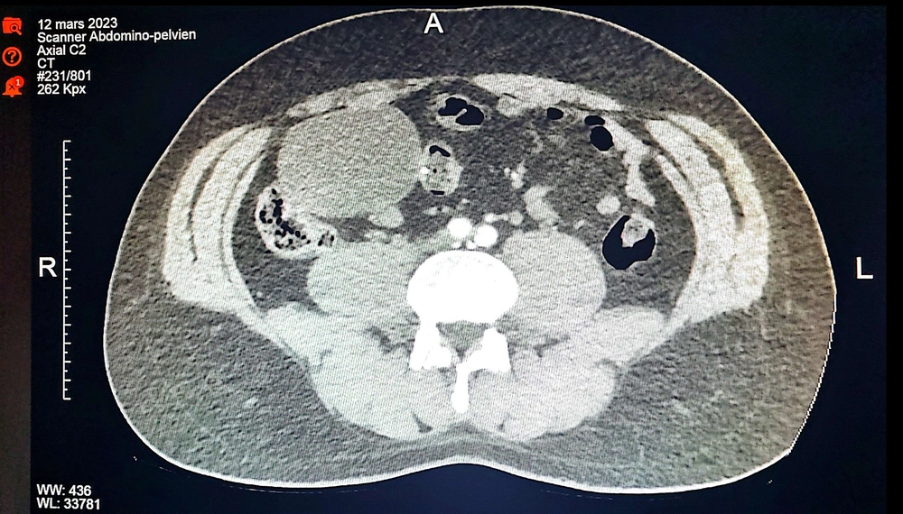
Imaging revealed an intraperitoneal cystic mass of fluid density (08 HU), measuring 71 × 56 mm in long axis and extending over 93 mm. The mass was located opposite the cecum and was cleaved from it. It had a thin wall with no partitions or calcifications, suggesting a cystic lymphangioma or caecal duplicity. The internal latero-caecal appendix was of normal size (Figure 1). Ileocolonoscopy showed no abnormalities.
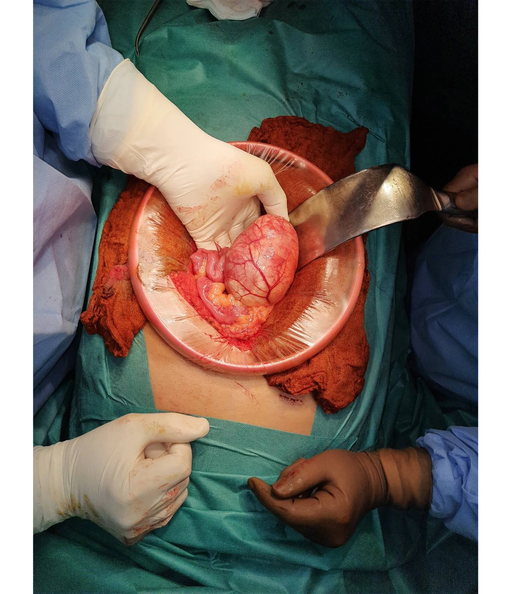
Given the discomfort caused by the compression on the digestive and urinary structures, an operative decision was made to remove the cystic mass. A median coeliotomy revealed a well-limited liquid mass measuring 10 cm, emanating from the right mesocolon and intimately attached to the cecum (Figure 2). Complete removal of the mass was performed in one piece. The patient was discharged on the 4th day post-operatively with no complications.
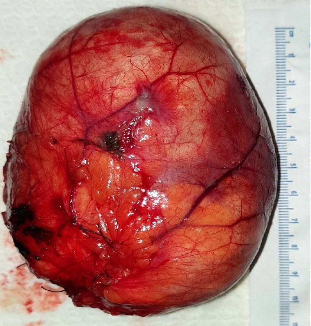
Macroscopic examination revealed a cystic sac measuring 10 × 8 cm with a soft consistency and smooth surface (Figure 3). The opening revealed a pasty material and a smooth unilocular cavity. The wall was thin and flexible.
Microscopic examination showed a cystic wall lined by regular atrophic squamous epithelium, delimiting a cavity filled with lamellar eosinophilic material. In conclusion, this was a mesenteric dermoid cyst with no evidence of malignancy.
3. Discussion
Mesenteric cysts represent a rare group of diseases that can be classified into six subtypes based on their tissue origin:
(1) Lymphatic cyst (lymphangioma)
(2) Mesothelial cyst (benign cystic mesothelioma, malignant cystic mesothelioma)
(3) Enteric cyst (duplication cyst)
(4) Urogenital cyst
(5) Mature cystic teratoma (dermoid cyst)
(6) Pseudocysts (infectious and post-traumatic cysts) (1).
Abdominal location of DC is rarer than pelvic location. The volume of the cyst may compress neighboring structures, causing abdominal pain, nausea/vomiting, and a palpable mass. Acute symptoms may arise, such as volvulus (2), peritonitis due to intraperitoneal rupture of the cystic contents (3), or infection of the cystic contents. Rarely, anemia may occur due to an autoimmune reaction caused by the cyst and directed against the subject's own erythrocytes (4). Imaging plays a crucial role in diagnosing these cystic lesions. In our case, the cyst was discovered incidentally during imaging following a thoracoabdominal contusion. The patient had not previously consulted a physician, despite a history of right lumbar pain. Histological studies confirmed the diagnosis and ruled out malignancy.
The DC should not be confused with a cyst that contains epithelial content but lacks dermal cells, or a benign cystic teratoma. In our case, the radiologist who interpreted the imaging suggested cystic lymphangioma and probable caecal duplicity as possible diagnoses for this cystic mass due to its proximity to the cecum. However, because there were no suggestive calcifications, the diagnosis of DC was not considered by the radiologist.
In children, the sacrococcygeal site is most common, while in adults, the gonadal site predominates (5, 6). Abdominal location is rare. Dermoid cysts result from abnormal implantation of primordial germ cells (7). This is a poorly understood, multifactorial process that depends on complex migratory factors (8). The most likely explanation for this implantation in our case appears to be the migration of these cells from the dorsal mesogastrium through the mesentery to establish themselves in the caecal region.
Dermoid cysts are treated surgically. Marsupialization of cysts was attempted in the past but led to the formation of fistulae, and as such, it is no longer recommended. A case of enucleation of a caecal dermoid cyst has been described with good results. Complete surgical excision, with or without bowel resection, remains the preferred treatment option. Although most cysts are removed through laparotomy, cases of successful laparoscopic excision have been reported (9, 10). However, the laparoscopic approach carries a higher risk of DC rupture.
When surgery is contraindicated or the patient refuses it, non-steroidal anti-inflammatory drugs (NSAIDs) can be used to reduce the size of the DC or slow its growth.
The parietal thickening of the DC is explained by the histological structure of its epithelium. This epithelium is keratinized and made up of scales. It continuously secretes sebum, which leads to the growth of the cyst, sometimes reaching considerable volumes, resulting in extrinsic compression of neighboring organs. The sebaceous content of the cyst can be recognized by trained imagers and may even be pathognomonic for DC. In addition to sebum, some DCs may contain hairs or calcifications.
The prognosis remains generally good, although clinical and ultrasound monitoring of patients over time is advisable, given the risk of recurrence.
3.1. Conclusions
Abdominal DC are rare, unlike gonadal, cervical, and nerve-related ones. The reported cases help us better understand their pathogenesis and work towards a potential classification. When a patient presents with a cystic mass near the cecum, the diagnosis of DC should be considered. All abdominal DCs must be carefully resected due to the risk of rupture. In some very selected cases, laparoscopic excision may be performed by highly trained teams, although this approach carries an increased risk of rupture. Malignant degeneration is possible but remains exceptional.


