1. Introduction
Kaposi Sarcoma (KS) is an uncommon type of malignant vascular tumor, which has become more frequent in the recent years due to Human Immunodeficiency Virus (HIV) infection and post-organ transplant immunosuppression (1). This lesion consists of proliferating spindle cells, which originate from vascular endothelial cells. Classic KS usually presents purple patches, plaques, and nodules on the lower limbs in elderly males (2). Rarely KS presents an exophytic vascular lesion with an epidermal collaret named Pyogenic Granuloma-like KS (PG-like KS) (1-3). Human Herpesvirus 8 (HHV8) detection and immunohistopathological clues differentiate PG-like KS from PG (2-4).
We could not find two PG-like KS lesions in an individual patient in prior reports, so we present this patient.
2. Case Presentation
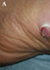
A 57-year-old male was presented to our clinic with chief complaint of an asymptomatic protuberant lesion on the left foot for 3 months with no history of previous trauma or bleeding. He was diabetic and smoker. He had left-sided nephrectomy due to complicated renal stone 30 years ago. He was on metformin, acarbose, and aspirin. On the physical examination, a peduculated nodule surrounded by an epidermal collaret on the left foot, an exophytic papule associated with a collaret, and a deeply seated nodule on the left palmar surface were noted (Figure 1). All lesions were excised and immunohistopathologic evaluation confirmed PG-like KS in exophytic lesions and KS in the nodule (Figure 2). Polymerase chain reaction (PCR) and immunostaining were positive for HHV8. All laboratory data were within normal limits except blood sugar. HIV antibody were negative. Ultrasound showed compensated hypertrophy of the right kidney and bilateral varicocele.
At low magnification, the foot lesion shows ulcerated epidermis with well-circumscribed lobular proliferation of well-formed capillaries, solid areas of spindle cells, slit-like spaces filled by red blood cells, and edema (D, magnification × 10)) (H&E stain). High magnification shows fascicular arrangement of the spindle cells with mild atypia and lymphoplasmacytic infiltration (E, F, magnification × 40) (H&E stain). Positive immunostaining for CD34 in the well-formed capillaries and in the areas of spindle cells (G, magnification × 10). Positive immunostaining for HHV-8 LNA-1 is seen in the spindle cells (H, magnification × 40).
3. Discussion
The PG-like KS is a non-classic form of KS, which could be an imitator of PG clinically and histopathologically. Furthermore, KS may initially grow as a PG-like lesion or rapid enlargement of the tumor may result in secondary ulceration and granulation tissue formation (5).
The PG-like KS has been reported as a solitary lesion without bleeding tendency on the lower limbs and rarely on the upper limbs (6). In histopathology, it was shown that solid areas of spindle cell proliferation was associated with lymphoplasmacytic infiltration and slit-like spaces. On the contrary, PG usually presents as a trauma-induced solitary lesion with bleeding tendency mainly on the upper limbs, which consists of lobulated mature capillary proliferation with fibrous septa formation (1-6).
Immunohistochemistry helps with making a diagnosis, as PG has dual cell groups; pericytes are highlighted with Smooth Muscle Actin (SMA) while mature endothelial cells are highlighted with Factor VIII, CD34, and CD31. On the contrary, spindle cells seen in KS are positive for CD34 and CD31, and negative for SMA and Factor VIII (6).
The HHV8 has been identified in the spindle cells of KS, using immunostaining, which detects latent nuclear antigen-1 (LNA-1). Latent viruses within endothelial cells cause up-regulation of lymphatic-associated genes, which makes these cells look like lymphatic endothelium (1).
Polymerase chain reaction (PCR) is a highly sensitive method for detecting HHV8 (1) but is time consuming with some false-positive results due to infected-by stander cells other than spindle cells or blood-borne viremia in endemic regions for HHV8 (3).
The HHV8 has been detected almost in all KS lesions yet has never been reported in PG lesions, which helps differentiate these lesions. The HHV8 has rarely been present in other vascular neoplasms, such as angiosarcoma, reactive angioendotheliomatosis, and in the lesions of patients, who harbor HHV8, so clinicopathologic correlation is mandatory (7). It has been reported that smoking may interfere in classic presentation of KS, similar to the current patient (8). Nephrectomy in this case was due to abscess formation after complicated renal stone 30 years ago, thus it seems it has not had any association with development of the lesions.
In the current case, all lesions were small-sized, thus, primarily all of them were totally excised with 3-mm safe margin and in histopathologic evaluation all margins were tumor-free. During the 6-month follow-up, there was no recurrence yet he was recommended to return in 6 months for a follow-up visit. No treatment other than excision was used.
This study found about 20 cases of PG-like KS in the English literature up to year 2016, yet it seems that this lesion has been under-reported or misdiagnosed as PG, especially on unusual locations, thus it is more common than we think and immunostaining should be considered in ambiguous cases.
3.1. Conclusions
Researchers should consider immunostaining methods in pyogenic granuloma-like lesions with unusual presentations.


