1. Background
Since 5000 years ago, honey has been used for the treatment of burns, cough, and sore to shorten the length of treatment (1, 2). The emergence of serious bacterial strains with different patterns of multiple resistances reduced the efficiency of current therapies. Thus, there is a need for effective and faster treatment. Given that honey-impregnated dressings with anti-bacterial effects are easily accessible, scientists were forced to reevaluate traditional treatments (2, 3). Honey is a saturated mixture of fructose and glucose monosaccharides and provides little water to grow pathogenic microorganisms. On the other hand, honey has anti-bacterial properties because of a pH range of 3.2 to 4.5. Antimicrobial properties of honey are partly due to their ability to produce hydrogen peroxide (1). Hydrogen peroxide concentration in honey is 1 mmol/L. When diluted, hydrogen peroxide is activated and released slowly without any damage to tissues (4).
On the other hand, honey has an insulin-like effect on cells involved in wound healing and stimulates the growth of cells responsible for replacing damaged tissue. It also stimulates development of new vessels and activates protein-digesting enzymes in the involved tissues. Honey's ability to quell inflammation could be related to its ability to quench free radicals (5). Antioxidants prevent free radical formation and are responsible for the anti-inflammatory effect of honey. They also provide a risk-free moist wound healing environment for the growth of bacteria and reduce edema, eczema, and wound odor because of their anti-inflammatory properties (1).
Honey contains several vitamins, including B6 and C, thiamine, niacin, riboflavin, pantoic acid as well as minerals, such as calcium, iron, magnesium, manganese, phosphorus, potassium, sodium, zinc, and amino acids. Antioxidant compounds in honey include chrysin, pinobaksin, pinocembin, vitamin C, and catalase (1). Vitamin C content in honey is 3 times that in plasma and acts as a nutrient in tissue reconstruction. Honey shows mitogenic properties on B and T lymphocytes (1, 6). Burn repair is done with minimal scarring and no harmful effects. It rarely has allergic and irritating effects and it is very easy to insert and remove dressings impregnated with honey (6).
Albumin is a water-soluble protein, essential for the health of many living organisms. Different types of proteins have been found in nature. Most plants and animals contain albumin or secrete it. Physiologically, albumin maintains plasma oncotic pressure in the vascular system and the liquid state plasma flow in the body by keeping water in the circulatory system. In addition, albumin helps transfer of essential fatty acids from adipose tissue to muscle and transport of small molecules, such as calcium, unbound bilirubin, hormones, like cortisol and thyroxine, and drugs in the blood serum, such as warfarin, phenybutazone, chlorpheniramine, and toxins. Dehydration results in high concentrations of albumin in plasma. In other words, dehydration is the only sign of increased concentration of albumin in plasma.
An increase in albumin level in plasma leads to an artificial increase in other analytes, such as hemoglobin, lipids, and calcium. Decreased albumin levels result in a decrease of oncotic pressure and thereby entrance of blood liquid to tissues and pores leading to edema and ascites. In addition, edema delays entrance of nutrients to the body (7). Given the important role of albumin in the body, it is essential to maintain the normal structure of this protein under different conditions.
Among trace elements in the body, zinc is the most abundant element after Iron (8). Zinc is found in all tissues and body fluids at relatively high concentrations. The average concentration of zinc in adults is 1.4 to 2.3 g. The zinc content needed to maintain health and proper growth is dependent on diet, climate, and stress caused by trauma or infection. However, the daily average of 20 to 15 mg of zinc is required (9). Zinc plays 3 important biological roles, including catalytic, structural, and regulatory roles. Regarding the catalytic role, zinc is needed for the function of over 300 enzymes and acts as a direct co-catalyst for enzymes. Zinc is involved in controlling many cellular processes, such as DNA synthesis, normal growth, brain development, behavioral responses, reproduction, membrane stability, bone formation, and wound healing (8). Ultra-small particles of Zinc Oxide (ZnO) are highly transparent and thus are used in sunscreen creams, paints, polished oil, plastics, and cosmetics, especially to prevent the passage of ultraviolet light. Zinc oxide nanoparticles have been recently used in the manufacture of sunscreen glasses. By downsizing ZnO particles to nanoscale, their behavior will change when exposed to light and electromagnetic waves. Chemical, physical, and micro-structural properties of ZnO powders are dependent on the synthesis method. Due to the extensive application of ZnO in advanced technologies in the recent years, most researchers have focused on the synthesis of ZnO nanoparticles with desired characteristics (10). Zinc plays a key role in the structure of zinc finger proteins, which are involved in regulating gene expression and prevention of cancer.
Increased use of metal oxides in the industry increases human exposure to nanoparticles at the workplace and the periphery environment. However, the effects of nanoparticles on the nervous system and health have been thoroughly investigated. Therefore, the physiological and pharmacological studies of nanoparticles are of special importance.
2. Methods
In this experimental study, 16 adult male albino mice with a weight of 25 to 30 g were used. To ensure the absence of any infection in the body, animals were examined. Mice were kept in the animal room in appropriate conditions at 22 ± 2°C, relative humidity of 40 to 60 percent and a 12-h photoperiodic cycle. The mice were fed with pellets for laboratory animals and drinking water. All applicable international, national, and institutional guidelines for the care and use of animals were followed.
For years, different compounds were used to control infection in burn wounds. In this study, water-soluble tetracycline was used orally. Due to the use of antibiotics as adjunctive therapy in the removal of specific microorganisms, it seems logical to use tetracycline because of its positive effects on gram-negative bacteria and anaerobes. In very destructive cases that do not respond to conventional therapies, tetracycline is used more than other antibiotics. It is worth mentioning that in addition to antimicrobial effects, the synthetic and natural forms of tetracycline are able to inhibit the activity of lytic collagen enzymes (11). Herein, antibiotic powder was poured in water (12). Like other experimental groups, mice in the control group received the same antibiotic.
For the preparation of test solutions, the mother solution was separately prepared for each ingredient using Neutral Lubricant Gel. After preparing the mother solutions, certain amounts of solutions were mixed and kept in separate containers. To avoid light, the containers were wrapped in foils and placed in a refrigerator. The mother solution was used to prepare the following mixtures (Table 1):
| Groups | Honey | Nano albumin | Nano zinc |
|---|---|---|---|
| Group 1 | + | - | + |
| Group 2 | + | + | - |
| Group 3 | + | + | + |
| Group 4 | - | - | - |
The Prepared Treatment Mixture for Experimental Groups
Applied volumes were as fallow: Honey 8 mL, Nano-Albumin 8 mL, and Nano-Zinc 5 mL.
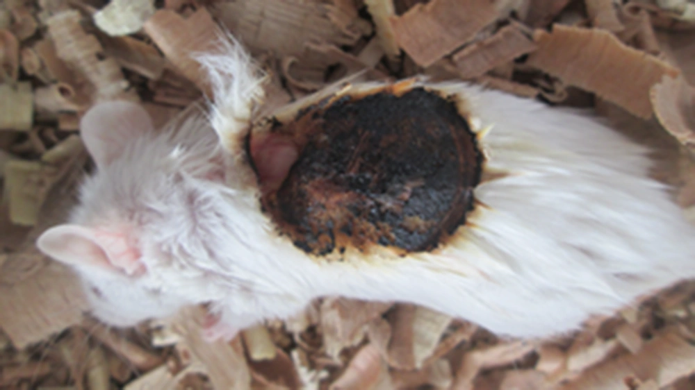
Mice were randomly divided to 4 groups (n = 4). First, the mice were anesthetized by ether. For this purpose, animals were placed inside a closed transparent chamber containing a high dose of inhaled ether. After anesthesia, the abdomen was placed on the pan and the hair on the back was removed by a blade. The shaved area was disinfected with cotton soaked in ethanol. The skin between the shoulders and the neck was burned for 8 seconds by a 2-cm coin, which was heated for 3 minutes with an alcohol burner. In this way, similar severe thermal burns were generated (Figure 1).
Overall, 0.9% sodium chloride solution was not applied to burned areas to evaluate disinfection properties of treatments. The experimental groups included: (a) experimental group 1 including injured mice treated by honey and Nano Zinc (b) experimental group 2 including injured mice treated by honey and Nano Albumin (c) experimental group 3 including injured mice treated by honey, Nano Albumin, and Nano Zinc, (d) control group that remained without any treatment after severe deep burns. Topical treatments were done once a day at a certain time. At the end of each week, a mouse from each experimental group was randomly selected and scarified after anesthesia. The hair of the treated area was shaved with a shaver. After taking images of the burn wounds, the skin was separated using a scalpel blade for microscopic investigation of the rate, quality of healing, and wound shrinkage.
After dehydration and preparation of paraffin templates, 6-µm thin sections were prepared and stained with hematoxylin-eosin. The thin sections were prepared to examine the thickness of skin layers and count the number and diameter of hair follicles and hair shafts.
3. Results
3.1. Macroscopic Findings
Macroscopic images of treated wounds confirmed shrinkage and complete wound closure in the experimental group 1 that were treated with honey and Nano Zinc as compared to other groups. Wound healing and shrinkage in the first group was better than the other 2 experimental groups and control. The group that received topical treatment of the combination of honey and Nano Zinc (group 1) showed less scar than other experimental groups (Figure 2).
3.2. Microscopic Findings
The results showed the significant effect of the mixture of honey and Nano Zinc on third-degree burns so that the healing process in this group was faster with less scarring than the control group and those received other mixtures (Tables 2 - 5).
| Group 1 | Honey + Nano Zinc |
|---|---|
| 1st week | Deep ulcer (severe inflammation) |
| 2nd week | Ulcer (severe inflammation) |
| 3rd week | Skin ulcer |
| 4th week | Normal skin |
| 5th week | Normal skin |
| 6th week | Normal skin |
Microscopic Results on Healing of the Burn Wounds Within 6 Weeks (Group 1)
| Group 2 | Honey + Nano Albumin |
|---|---|
| 1st week | Deep ulcer (severe inflammation) |
| 2nd week | Deep ulcer (severe inflammation) |
| 3rd week | Deep ulcer (severe inflammation) |
| 4th week | Deep ulcer (severe inflammation) |
| 5th week | Ulcer (severe inflammation) |
| 6th week | Normal skin (less hair follicles) |
Microscopic Results on Healing of Burn Wounds Within 6 Weeks (Group 2)
| Group 3 | Honey + Nano Zinc + Nano Albumin |
|---|---|
| 1st week | Deep ulcer (severe inflammation) |
| 2nd week | Deep ulcer (severe inflammation) |
| 3rd week | Deep ulcer (severe inflammation) |
| 4th week | Skin with acanthosis and mild acute to chronic inflammation |
| 5th week | Focal ulcer (mild inflammation) |
| 6th week | Superficial ulcer (moderate inflammation) |
Microscopic Results on Healing of Burn Wounds Within 6 Weeks (Group 3)
| Group 4 | Control |
|---|---|
| 1st week | Deep ulcer (severe inflammation) |
| 2nd week | Deep ulcer (severe inflammation) |
| 3rd week | Deep ulcer (severe inflammation) |
| 4th week | Ulcer (mod inflammation) |
| 5th week | Skin ulcer |
| 6th week | Normal skin (less hair follicle) |
Microscopic Results on Healing of Burn Wounds Within 6 Weeks (Control for Group 4)
In the first group, edema and inflammation were eliminated at the end of the fourth week and no mononuclear, multi nuclear cells or red blood cells were observed on the wound surface. In addition, epidermis and stratum corneum were formed. A significant difference was found between the first and other groups in terms of wound healing speed and number of dermal fibroblasts (Figure 3).
A, experimental group 1, agglomeration of fibroblasts in the dermis, formation of a regular epidermis and dermis; B) experimental group 2, severe secretion of inflammatory cells; C, experimental group 3, formation of too much vascularized tissue and thickened dermis; D, control, abnormal formation of epidermis and vascularization in the tissue.
A significant increase in the overall thickness of the skin, keratinocyte layer, and hypodermis was observed in the first experimental group compared to the control and the 2 other groups. Also a significant increase in the thickness of the dermis indicated the positive effects of honey and Nano Zinc on accelerating the shrinkage of deep burn wounds, and repair and differentiation of the skin layers and appendages (P < 0.05). The significant difference between the control group and other experimental groups in the dermal thickness was due to the delay in initiation of skin cell differentiation, which appeared as a grainy dermal tissue in the control group.
The dermal thickness in the third experimental group showed a significant increase in comparison to the control group, which may be due to scar formation (P < 0.05) (Figure 3C).
4. Discussion
According to the literature, factors that increase blood flow, disinfection, and reduce inflammation have a positive effect on the process of burn wound healing (13). It has been proven that skin elasticity is mainly dependent on the content and contact of collagen fibers. Previous studies on hexoses (fructose, sucrose, galactose, and glucose) in cell culture showed that these compounds with positive metabolic effects stimulate synthesis of protein, collagen, extracellular matrix, and growth factors (14). Astragalus honey contains hexoses and different combinations and is able to increase collagen synthesis by increasing fibroblasts and the biosynthesis of nucleic acids and proteins. In addition, Astragalus honey has a positive effect on maturity and correct orientation of collagen fibers and increased skin elasticity.
According to Huang et al.’s (2002) study, TGF-β antagonists have an accelerating role on reducing scars caused by burn wounds and thus wound healing (15). Some studies showed that these metal ions react with proteins by binding to sulfhydryl groups (-SH) in enzymes to inactivate proteins. If these metals are present in very small sizes, they will show improved antimicrobial properties because of increased surface to volume ratio (16). Atmaka et al. (1997) examined the effect of zinc on the growth of microorganisms, such as staphylococcus aureus and staphylococcus epidermidis, and found that zinc is able to inhibit the growth of these microorganisms (17).
Steven et al. (2005) used zinc oxide photocatalyst to inhibit fungi and bacteria and found that Candida albicans disappeared after 120 minutes (18). Several factors are associated with antimicrobial properties of nanoparticles, including concentration, size, and surface area. Smaller ZnO particles have more antimicrobial properties (19). Hsiao et al. (2006) examined antimicrobial activity of copper particles against some bacteria (20). However, nanoparticles may be harmful to health for 2 major reasons; first, they diffuse quickly from biological membranes, and second, their toxicity is still not fully understood because of novelty. On the other hand, toxicity of nanoparticles depends on their concentration, shape, and diameter (21-24). Accordingly, more research should be conducted on nanoparticles and their effects on organs and blood factors. According to microscopic findings of the present study and other reports, it could be concluded that natural honey by increasing biosynthesis of nucleic acids and protein, collagen synthesis, and Nano ZnO, with analgesic and anti-microbial properties, could significantly increase the speed of healing burn wounds and skin strength.
In fact, a nanocomposite was designed for burn wound healing in male mice. After damage, especially severe damage to tissues, free radicals increase in the body. These free radicals cause further damage to the injured tissue, such as the surrounding skin. According to Gutteridge and Hallivell’s study (1990), various types of trauma, such as burns, are associated with imbalance in the production of free radicals and the function of antioxidant system (25). Angiogenesis is an important factor in wound healing, during which the wound is saturated with blood vessels. Angiogenesis is necessary to feed the wound so that the wound will not be healed in the absence of angiogenesis.
The factors, which could stimulate angiogenesis, could promote a normal healing process. An important part of wound healing process is angiogenesis so that in the absence of this phenomenon, invasion of macrophages, and fibroblasts into the wound will not be useful due to lack of oxygen and nutrients (1). The number of blood vessels in the first experimental group 1 (honey + Nano Zn) increases until the 14th day, which is consistent with the proliferative phase. Then, it gradually decreases, which is consistent with the restructuring phase. It seems that oxygen peroxide in honey is responsible for therapeutic effects observed in the treatment of wounds. Oxygen peroxide stimulates the growth of cells that must be replaced and has an insulin-like effect on wound healing.
On the other hand, wound bacteria consume glucose and produce lactic acid, which plays a key role in angiogenesis. According to the study of Molan et al. honey accelerates angiogenesis and granulated tissue formation in the wound area (26). Fibroblast cells play an important role in the wound healing process and reach the wound on the third day of treatment. During proliferation, the synthesis of collagen increases continuously for 3 weeks to establish a balance. The samples of the second week showed more fibroblast cells in the first experimental group than the control and other groups. On the third week, concentration of collagen was higher in the first experimental group when compared with the control and other experimental groups. It seems that honey and Nano Zinc accelerates the synthesis of collagen so that wound collagen reaches its maximum level faster and thus the restructuring phase arrives earlier. Honey provides fibroblasts with nutrients and oxygen by increasing angiogenesis and accelerating the release of oxygen from hemoglobin by increasing acidity.
According to other work by Massague et al. (1990), on skin damage or burns, platelet-derived TGF-β1 molecules appear and accelerate wound healing (27). The TGF-β1 molecules increase the mitosis effect on human dermal fibroblasts (28). At the wound site, TGF-β stimulates angiogenesis, proliferation of fibroblasts, differentiation of myofibroblasts, and the formation of extracellular matrix. The first goal in healing is fast wound closure. Thus, drugs that modulate the inflammatory response seem to be an appropriate treatment. Unsaturated fatty acids are a progenitor of many lipoic compounds involved in inflammatory reactions. N3 and N6 fatty acids are abundant in fish and soybean oils. In addition to being involved in the formation of lipoic compounds, N9 is involved in the synthesis of membrane phospholipids. The N9 fatty acids accelerate the wound healing process (29).
In addition, less edema with a thinner fibrin clot are observed. These fatty acids induce gene expression of collagen type III and reduce the expression of cyclooxygenase II. Collagen type III is the main collagen produced during wound healing (30). Collagens are first organized in a typical grid network and then the skin starts to show signs of fibrosis path rather than reconstruction. Accordingly, it could be concluded that topical use of honey and Nano Zinc on the wound bed has significant effects on accelerating wound shrinkage, healing, repairing skin layers, appendages, and reducing scars after healing wounds of severe burns.


