Fabrication and Characterization of Fibrin/Bioactive Glass Nanoparticles Composite for Bone Tissue Engineering

authors:

avatar Alireza Noori 1 , * , avatar Seyed Jamal Ashrafi 2 , avatar zahra mohammadi 3 , avatar Javad Mohammadnejad Arough 3 , avatar Abdoreza Sheykhmehdi Mesgar 3 , **

Department of Tissue Engineering and Applied Cell Sciences, School of Medicine, Semnan University of Medical Sciences, Semnan, Iran
Department of Nanotechnology, School of Medicine, Shahrud University of Medical Sciences, Semnan, Iran
Division of Biomedical Engineering, Department of Life Science Engineering, Faculty of New Sciences and Technologies, University of Tehran, Tehran, Iran
Corresponding Authors:

How To Cite Noori A, Ashrafi S J, mohammadi Z, Mohammadnejad Arough J, Sheykhmehdi Mesgar A. Fabrication and Characterization of Fibrin/Bioactive Glass Nanoparticles Composite for Bone Tissue Engineering. koomesh. 2024;26(2):e149784. https://doi.org/10.69107/koomesh-149784.

Abstract

Introduction:
Bone tissue engineering is one of the emerging strategies that has been developed to restore the bone of the damaged area without provoking an adverse immune reaction. In this context, the tissue engineering scaffold must be as similar as possible to the natural bone tissue. Bone is a nanocomposite material composed of hydroxyapatite and collagen; hence, the development of nanocomposite scaffolds has been viewed as an appropriate choice for bone tissue restoration. In many situations, these composite materials combine a bioactive mineral phase with a biodegradable polymer phase. The current study aimed to create and analyze a new composite scaffold for bone tissue engineering applications employing bioactive glass nanoparticles (nBG) and fibrin.
Methods:
The nBG used in this study was based on the 70:30: SiO2-CaO system, which was synthesized using the sol-gel method. Scanning electron microscopy (SEM), X-ray crystallography (XRD), and Fourier transform infrared (FTIR) spectroscopy were used to characterize the fabricated nanoparticles. On the other hand, the whole blood was centrifuged twice at 3000×g to separate the plasma from the blood, and during the next steps, fibrinogen and thrombin were separated from the platelet-free plasma. These components were then mixed with nBG to create an injectable composite scaffold. The composites were subjected to physicochemical characterization, such as degradability and clot formation rate, while human osteoblast-like cells (G-292 cell line) were used to assess the scaffold's biocompatibility as well as cell proliferation and differentiation using the MTT and alkaline phosphatase activity tests, respectively.
Results:
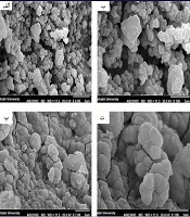
In the case of bioactive glass nanoparticles, SEM analysis verified the formation of spherical nanoparticles with a diameter of 50 to 110 nm. XRD analysis showed its non-crystalline nature, and the FTIR spectrum demonstrated the presence of Si-O-Si and O-H functional groups. Investigations on the composite of fibrin and bioactive glass nanoparticles (nBG) revealed that incorporating nBG into the fibrin hydrogel enhances its stability and reduces the degradation rate of the scaffold by approximately 40%. In vitro investigations on G-292 cells revealed that including nBG in the fibrin hydrogel improves cell viability, cell proliferation by approximately 150%, and alkaline phosphatase activity by around 45%.
Conclusion:
Fibrin gel is widely used in bone tissue engineering applications. However, our studies show that when combined with bioactive glass nanoparticles, it is more effective at repairing damaged bones.

Fulltext

The full-text is available in the PDF file.