1. Introduction
Cardiac Hydatidosis is a rare and ominous complication of hydatid disease (1). Cardiac echinococcosis may be asymptomatic for several years but could be discovered after the development of lethal complications (2). With this, we report a rare case of ruptured hydatid cyst resulted in the pericardial effusion and cardiac tamponade.
2. Case Presentation
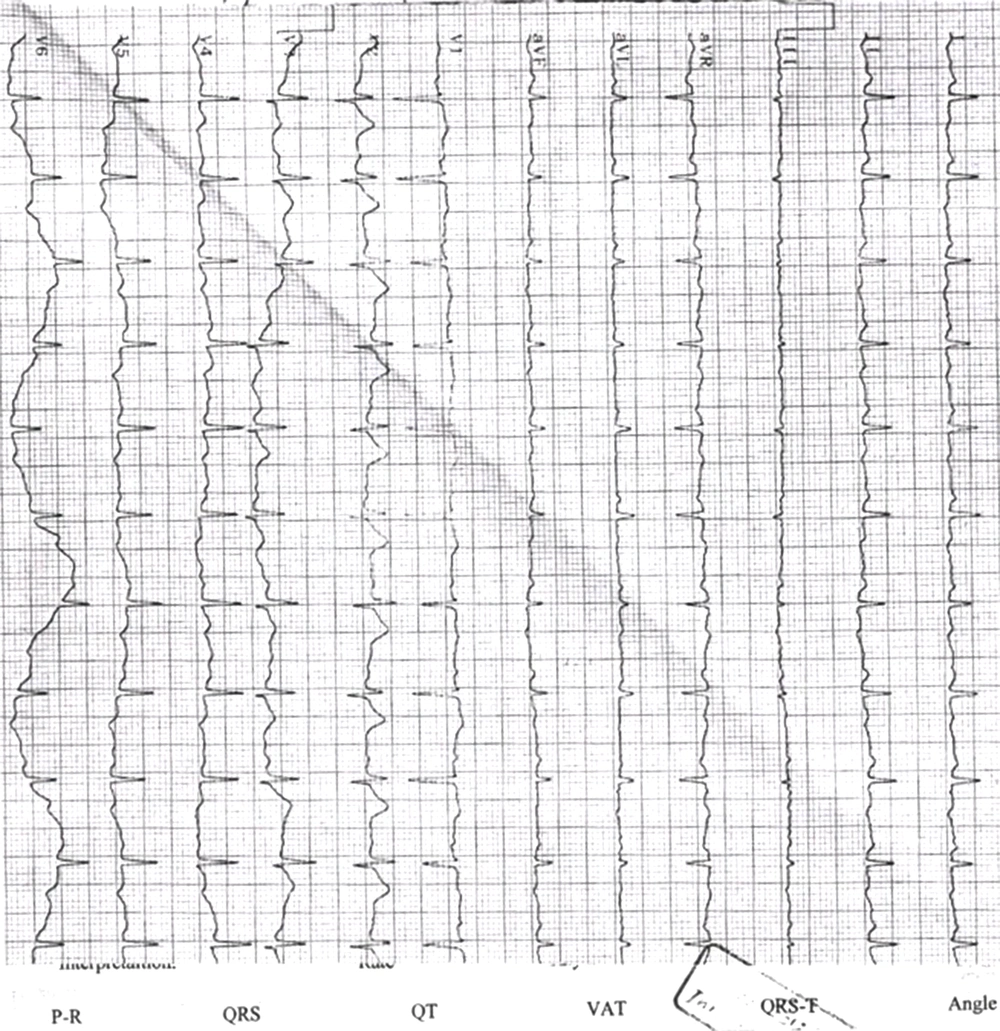
A 31 years-old-male referred with a chief complaint of pleuritic chest pain with a possible diagnosis of acute pericarditis. electrocardiography (ECG) showed new T inversion in V5-V6 that was absent in the available ECG from last year. The initial trans-thoracic echocardiography (TTE) showed no pericardial effusion. The chest pain was diminished using non- steroid anti-inflammatory drugs. After four days of hospitalization, he suffered from repetitive chest pain episodes that were resistant to nitrate and analgesic treatment. One fever episode about 40OC was also noticed. ECG at this time showed low voltage in all leads (Figure 1).
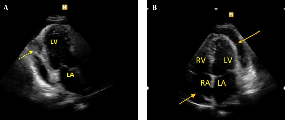
Laboratory data showed leukocytosis, normal erythrocyte sedimentation rate (ESR) level, elevated pro-calcitonin level and liver enzymes. Abdominal and pelvic spiral CT scan showed no hepatic mass, but showed focal and heterogeneous increased thickness of lateral LV wall with protrusion into LV and bulging into pericardial space without central enhancement after contrast injection. His condition deteriorated suddenly, and TTE showed moderate size pericardial effusion resulted in cardiac tamponade with a round cystic lesion suspected to hydatid cyst (Figure 2).
Moderate size pericardial effusion resulted in cardiac tamponade with a round cystic lesion suspected to hydatid cyst . A, Arrow indicates to the cystic lesion posterior to LV (left ventricle). B, Arrows indicate to circumferential pericardial effusion. LV: left ventricle, RV: right ventricle, LA: left atrium, RA: right atrium.
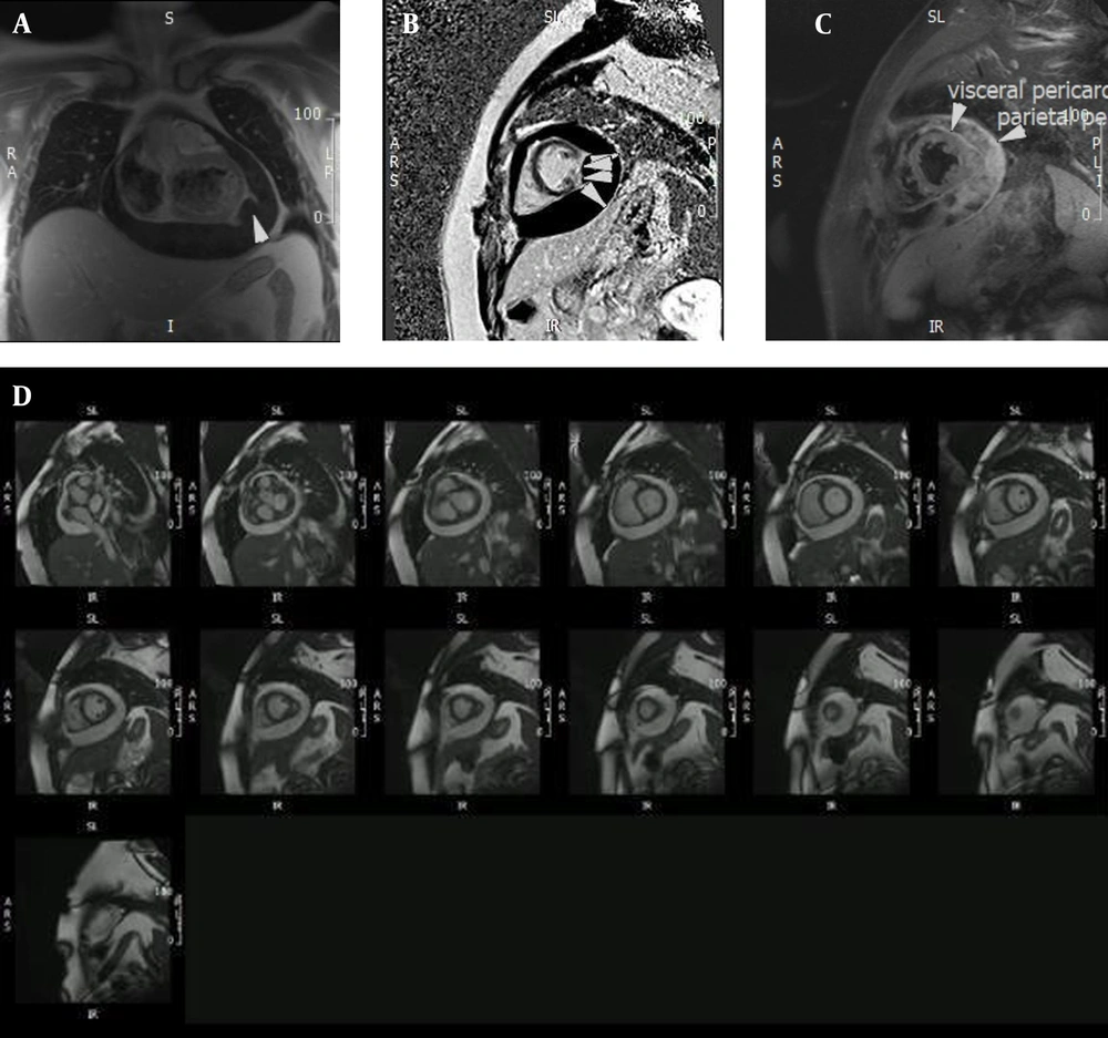
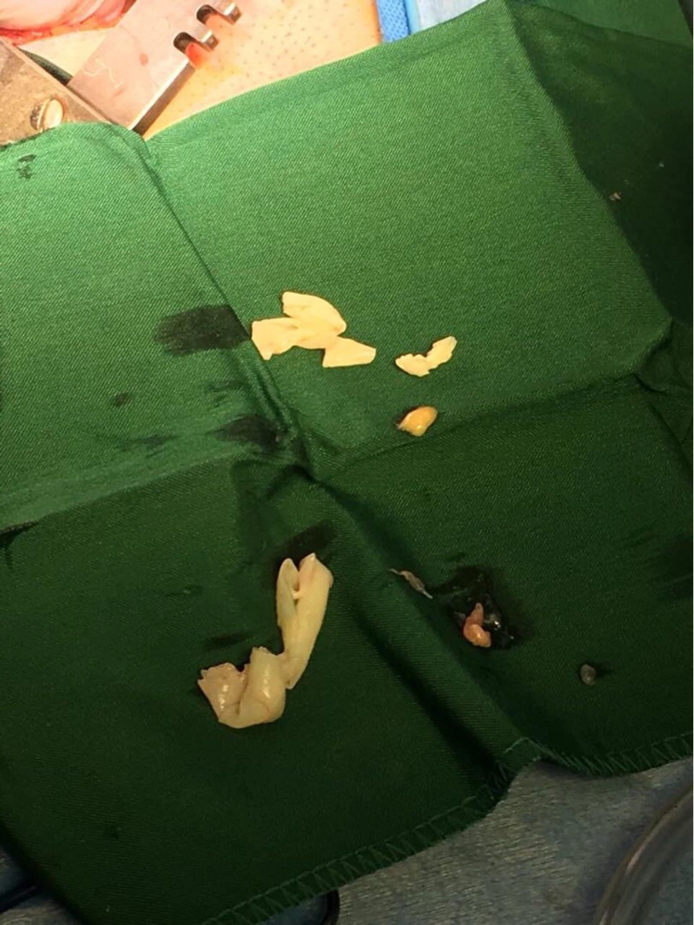
Cardiac MRI showed extensive and severe circumferential pericardial effusion with CMR evidence of cardiac tamponade. Some round and small particles within effusion suggestive of possible scolex around LV were also detected. There was round, and inhomogeneous cystic mass lesion (length: 31 mm, width: 17 mm) originated from a sub-epicardial layer of mid-lateral LV that protruded into the pericardial space. Along with a small suspicious defect in its outer rim (6mm) that could suggest rupture point into the pericardial space (Figure 3). He received antibiotic treatment and underwent a prompt surgical operation. Surgical exploration confirmed the presence of daughter cysts (Figure 4).
CMR images of ruptured hydatic cyst. A, T1 weighted CMR image depicts ruptured hydatid cyst. B, Late gadolinium enhancement (LGE) image demonstrates cystic ring enhancement in favor of hydatid cyst. C, In rhe STIR CMR image in short acis view diffuse parietal and visceral pericardial edema is noticeable. D, Stack short axis CMR view of left ventricle indicates to large circumferential pericardial effusion.
Median sternotomy and partial pericardiectomy were performed. In lateral wall of LV, there was a cystic mass (3*3cm) that was filled by injected hypertonic saline. Then, the cyst wall was opened and scolexes were removed. After irrigation of cyst space, the internal spacen of pericardium was irrigated using hypertonic saline and finally it was repaired following deaine insertion.
3. Discussion
Prior history of infection with echinococcosis can help physicians in rapid diagnosis of its complications in symptomatic cases, but unfortunately, patients are unaware of their infection in some instances. Thus, being familiar with the broad spectrum manifestation of this disease is essential for better patient management. Cardiac hydatidosis is a rare complication of this disease, and cardiac events depend on the location, number and size of hydatid cysts and their induced complications (3). Pericarditis, pericardial effusion and tamponade could occur in this setting due to the presence of pericardial effusion due to cyst rupture into the pericardial cavity (4). Hydatid cysts reach the heart through coronary arteries. Their most common implantation sites are interventricular septum and left ventricular free wall with less involvement of pericardial space and paracardiac sites (5-7). Fazlinezhad et al. reported the apicolateral and apical left ventricular segments as the implantation site of the cyst (8). This site was in the mid-lateral wall in our case.
3.1. Conclusions
In conclusion, we suggest that patients with pericarditis should be probed with echocardiography for the presence of hydatid cysts to prevent further development of tamponade and clinical deterioration.


