Right Coronary Artery Origin from Left Sinus of Valsalva with an Inter-arterial Course: Case Report and Review of Literature

authors:

avatar Ahmad Jamei Khosroshahi ORCID 1 , avatar Akbar Molaei ORCID 1 , * , avatar Mahmoud Samadi ORCID 1 , avatar Ali Rafiei 1

Pediatrics’ Cardiology Department, Cardiovascular Research Center, Tabriz University of Medical Sciences, Tabriz, Iran

How To Cite Jamei Khosroshahi A, Molaei A, Samadi M, Rafiei A. Right Coronary Artery Origin from Left Sinus of Valsalva with an Inter-arterial Course: Case Report and Review of Literature. Multidiscip Cardio Annal. 2022;13(2):e119210. https://doi.org/10.5812/mca-119210.

Abstract

Introduction:

Overally the anomaly of the coronary arteries are rare. Its incidence according to angiographic, autopsy and echocardiographic findings, are about 1%, 0.29% and less than 0.1% respectively. Usually these are the incidental finding during angiography without clinical presentation. These anomalies may be accompanied by myocardial ischemia, arrhythmia, syncope and sudden death.

Case Presentation:

We present a 14-year-old Iranian boy who experienced chest pain and shortness of breath after engaging in physical activity. The right coronary artery (RCA) had an intramural and inter-arterial course and was proven to have originated from the left sinus of Valsalva by computed tomographic coronary angiography (CTCA). The RCA began adjunct to the ostium of the left main coronary artery and coursed intramural at first and then continued between the aorta and pulmonary artery. The patient underwent corrective surgery. The RCA was cut from origin and reanastomosd to the right coronary sinus after preparing the sinus. The patient recovered well and he was discharged with the ability to perform sports activity. One year Follow up of the patient was uneventful.

Conclusions:

Even though single ostium coronary artery abnormalities are uncommon, they have become more frequently identified in recent years using non-invasive techniques such transthoracic echocardiography, which is then followed by CT angiography or conventional angiography. These patients are susceptible to sudden death, so attention to symptoms of the patients and comprehensive approach to them can be lifesaving.

1. Introduction

Overally the anomaly of the coronary arteries are rare. Its incidence according to angiographic, autopsy and echocardiographic findings, is about 1% (1-4), 0.29% (5) and less than 0.1% respectively (6). Usually these are the incidental finding during angiography without clinical presentation. But a significant percentage of young age sudden cardiac deaths are due to these anomalies (7-9). The contralateral sinus origin of the right coronary artery (RCA) is more common than left coronary artery (LCA) (1-4).

These anomalies may be accompanied by myocardial ischemia, arrhythmia, syncope and sudden death (10, 11).

2. Case Presentation

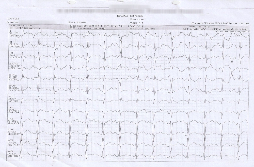
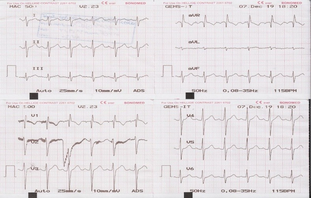
A 14-year-old Iranian boy reported experiencing shortness of breath and chest pain after exerting himself. His heart rate, rhythm and blood pressure were normal. He had non-specific changes in the ECG (Figure 1).

The ECG during the resting phase

The findings of the exercise stress test were not specific (Figure 2).

Exercise stress electrocardiography and not specific changes of ST and T change at II, III, aVF.V5, and V6 leads.

The transthoracic echocardiogram showed normal size and function of ventricles but showed abnormal origin of RCA from left sinus of Valsalva.

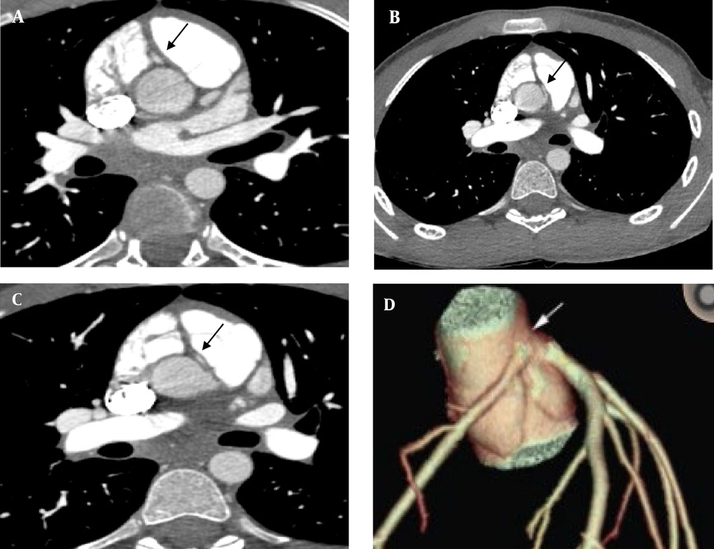
The RCA was found to originate from the left sinus of Valsalva with a distinct ostium and an intramural and inter-arterial course by computed tomographic coronary angiography (CTCA).

The RCA began adjunct to the ostium of the left main coronary artery and coursed intramural at proximal segment and then between the aorta and pulmonary artery (Figure 3).

Computed tomographic coronary angiography image with an anomalous right coronary artery (RCA) from the opposite sinus (A, B, C); three -dimensional reconstruction of coronary artery shows the course of RCA in relation to the left main and left anterior descending arteries (arrow) (D).

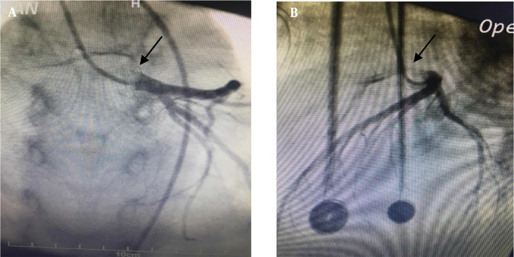
Selective coronary arteries angiography also confirmed this and RCA stenosis and slit-like coronary compressed between the pulmonary artery an aorta.

This finding is generally known as anomalous coronary artery from the opposite sinus (ACAOS) (Figure 4).

Selective left coronary artery angiography shows right coronary artery from left sinus and slit-like coronary compressed between the pulmonary artery and aorta (A, B).

The patient underwent corrective surgery. The RCA was cut from between the aorta and pulmonary artery and connected to the right coronary sinus after preparing the sinus.

The post-operative period was uneventful. The patient recovered well and he was discharged with the ability to perform sports activity. During one year follow up the patient was asymptomatic with normal clinical evaluation.

3. Discussion

The solitary ostium coronary arteries anomaly was classified by Lipton et al. based on its origin and progression (12). In 2019 and 2020 Al Jarallah et al. and Rajan et al. introduced the modified classification (13, 14). Among coronary artery anomalies, ACAOS can be considered as a high risk factor for sudden death, particularly in young patients with inter-arterial course of anomalous artery (15). According to a study, of the 387 sudden fatalities of young athletes, 53 (13.7%) were caused by coronary artery abnormalities, the majority of which were ACAOS (15).

Right ACAOS is more common and more benign than left ACAOS (where the left main coronary artery originated from right coronary sinus). This anomaly can lead to myocardial ischemia and sudden death. This is a rare congenital anomaly with an incidence of 0.5%.

When symptoms do arise, they may include chest pain, fainting, palpitations, syncope, and sudden death. This anomaly is frequently asymptomatic (10, 11). The right ACAOS was initially suspected by trans thoracic echocardiography and later confirmed by CT-Angiography in this patient who experienced chest pain and exertional dyspnea.

The mechanisms of the signs and symptoms is not fully understood in these patients, but several mechanisms have been proposed:

(1) Compression of the RCA between pulmonary artery and aorta; (2) partially orifice stenosis in RCA; (3) compression of the intramural segment of RCA by aortic wall and (4) coronary artery spasm due to endothelial injury.

The presented patients' symptoms were multifactorial: intramural course and partial hypoplasia of intramural segment, inter-arterial course, oblique and stenotic ostium (Figure 4). These risk factors dispose the patient to be symptomatic and sudden death specially during or after physical activity or emotional stress.

Treatment for asymptomatic patients include observation and limiting competitive sports. In symptomatic patients, corrective surgery is recommended. Corrective surgery procedures include coronary bypass, direct implantation of anomalous artery, un-roofing the intramural segment of the vessel within aortic wall and osteoplasty, which aims at creating a new ostium in its proper place.

3.1. Conclusions

Although the single ostium coronary arteries anomalies are rare but they are increasingly diagnosed in recent years by non-invasive methods such as trans thoracic echocardiography confirmed by CT angiography or conventional angiography. These patients are susceptible to sudden death due to above mentioned risk factors, so attention to symptoms of the patients and comprehensive approach to them especially by non-invasive methods can be lifesaving.

References

  • 1.

    Yamanaka O, Hobbs RE. Coronary artery anomalies in 126,595 patients undergoing coronary arteriography. Cathet Cardiovasc Diagn. 1990;21(1):28-40. [PubMed ID: 2208265]. https://doi.org/10.1002/ccd.1810210110.

  • 2.

    Kardos A, Babai L, Rudas L, Gaál T, Horváth T, Tálosi L, et al. Epidemiology of congenital coronary artery anomalies: a coronary arteriography study on a Central European population. Catheter Cardio Diag. 1997;42(3):270-5.

  • 3.

    Topaz O, DeMarchena EJ, Perin E, Sommer LS, Mallon SM, Chahine RA. Anomalous coronary arteries: angiographic findings in 80 patients. Int J Cardiol. 1992;34(2):129-38. [PubMed ID: 1737663]. https://doi.org/10.1016/0167-5273(92)90148-v.

  • 4.

    Garg N, Tewari S, Kapoor A, Gupta DK, Sinha N. Primary congenital anomalies of the coronary arteries: a coronary: arteriographic study. Int J Cardiol. 2000;74(1):39-46. [PubMed ID: 10854679]. https://doi.org/10.1016/s0167-5273(00)00243-6.

  • 5.

    Alexander RW, Griffith GC. Anomalies of the coronary arteries and their clinical significance. Circulation. 1956;14(5):800-5. [PubMed ID: 13374855]. https://doi.org/10.1161/01.cir.14.5.800.

  • 6.

    Pelliccia A, Spataro A, Maron BJ. Prospective echocardiographic screening for coronary artery anomalies in 1,360 elite competitive athletes. Am J Cardiol. 1993;72(12):978-9. [PubMed ID: 8213561]. https://doi.org/10.1016/0002-9149(93)91120-7.

  • 7.

    Pelliccia A. Congenital coronary artery anomalies in young patients: new perspectives for timely identification. J Am Coll Cardiol. 2001;37(2):598-600. [PubMed ID: 11216985]. https://doi.org/10.1016/s0735-1097(00)01122-0.

  • 8.

    Maron BJ, Thompson PD, Puffer JC, McGrew CA, Strong WB, Douglas PS, et al. Cardiovascular preparticipation screening of competitive athletes. A statement for health professionals from the Sudden Death Committee (clinical cardiology) and Congenital Cardiac Defects Committee (cardiovascular disease in the young), American Heart Association. Circulation. 1996;94(4):850-6. [PubMed ID: 8772711]. https://doi.org/10.1161/01.cir.94.4.850.

  • 9.

    Eckart RE, Scoville SL, Campbell CL, Shry EA, Stajduhar KC, Potter RN, et al. Sudden death in young adults: a 25-year review of autopsies in military recruits. Ann Intern Med. 2004;141(11):829-34. [PubMed ID: 15583223]. https://doi.org/10.7326/0003-4819-141-11-200412070-00005.

  • 10.

    Cheitlin MD, De Castro CM, McAllister HA. Sudden death as a complication of anomalous left coronary origin from the anterior sinus of Valsalva, A not-so-minor congenital anomaly. Circulation. 1974;50(4):780-7. [PubMed ID: 4419670]. https://doi.org/10.1161/01.cir.50.4.780.

  • 11.

    Kim CK, Park CB, Jin U, Lee BY, Song KS. Evaluation of unroofing procedure of anomalous origin of right coronary artery from left sinus of valsalva between aorta and pulmonary trunk by multidetector computed tomography. J Comput Assist Tomogr. 2005;29(6):752-5. [PubMed ID: 16272846]. https://doi.org/10.1097/01.rct.0000179242.77847.cd.

  • 12.

    Lipton MJ, Barry WH, Obrez I, Silverman JF, Wexler L. Isolated single coronary artery: diagnosis, angiographic classification, and clinical significance. Radiology. 1979;130(1):39-47. [PubMed ID: 758666]. https://doi.org/10.1148/130.1.39.

  • 13.

    Al Jarallah M, Rajan R, Kotevski V, Dashti R, Moustafa W. Multivessel percutaneous coronary intervention with bifurcation stenting in a quadfurcated single coronary artery from the right aortic sinus: a case report. Eur Heart J Case Rep. 2019;3(4):1-4. [PubMed ID: 32123787]. [PubMed Central ID: PMC7042130]. https://doi.org/10.1093/ehjcr/ytz197.

  • 14.

    Rajan R, Kotevski V, Al Jarallah M, Dashti R. Type-IV Quadfurcation of Single Coronary Artery from the Right Aortic Sinus: New Classification. Oman Med J. 2020;35(6). e207. [PubMed ID: 33335746]. [PubMed Central ID: PMC7724053]. https://doi.org/10.5001/omj.2020.101.

  • 15.

    Lilly SM, Schussler JM, Stoler RC. Anomalous origin of the right coronary artery from the left sinus of Valsalva associated with syncope in a young athlete. Proc (Bayl Univ Med Cent). 2011;24(1):13-4. [PubMed ID: 21307970]. [PubMed Central ID: PMC3012283]. https://doi.org/10.1080/08998280.2011.11928675.