1. Background
Precise aorto-ostial coronary angioplasty remains challenging due to the inability of conventional 2D angiography to accurately delineate the aorto-ostial plane. This limitation can lead to geographic miss of the ostium or protrusion of the stent into the aorta. Several strategies and techniques have been proposed to improve the accuracy of aorto-ostial stenting, each with certain limitations. For example, the aorta free floating wire technique is not very accurate for determining the ostium, the Szabo technique involves complex steps, and novel stent designs (such as the Cappella stent) and devices (such as the Ostial Pro device) face economic concerns (1, 2).
2. Objectives
We propose the "modified floating wire" technique as a novel, safe, and straightforward method for aorto-ostial coronary stenting, ensuring appropriate ostial alignment without stent protrusion into the aorta.
3. Methods
The following steps were performed:
1. A 6-French right Judkins catheter was advanced to the right coronary artery (RCA) via the right femoral approach. A 0.014-inch workhorse coronary guide wire was advanced through the lesion.
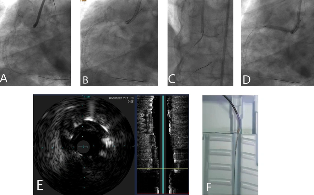
2. A second coronary workhorse guide wire was advanced to the aortic cusp as the floating wire (Figure 1A).
A, foating wire advanced to aortic cusp; B, same balloon on the floating wire; C, balloon on floating wire pulled back (video 2); D, stent is deployed when pulled-back until gets stuck by the inflated balloon (video 3); E, intracoronary ultrasound (IVUS) showed full coverage of ostium (video 5); F, bench testing of floating balloon techniques.
3. After effective predilation of the lesion with a compliant balloon, the balloon was removed.
4. The same balloon was advanced over the floating wire (Figure 1B) (Supplementary Video 1).
5. The stent was advanced over the RCA guide wire.
6. The balloon was inflated and gradually pulled back on the floating wire until it reached the guiding catheter’s tip. The guiding catheter was then moved towards the coronary ostium as much as possible (Figure 1C) (Supplementary Video 2).
7. The stent was pulled back until it was positioned by the inflated balloon, then deployed (Figure 1D) (Supplementary Video 3).
8. The stent was post-dilated and the ostium was flared, with the results shown (Supplementary Video 4).
9. Intracoronary ultrasound (IVUS) confirmed full coverage of the RCA ostium (Figure 1E) (Supplementary Video 5).
4. Results and Discussion
In the floating wire method, the second wire placed at the origin of the aorta serves as a marker for the true ostium, facilitating more accurate stent placement. However, this technique has some limitations. Since the wire does not bend at a 90-degree angle from the ostium, the true ostium is positioned a few millimeters away from the curvature of the wire, which extends from the guide to the aortic wall. The wire appears as a single line on fluoroscopic images, which can result in variable relationships between the wire, the actual ostium, and the stent in different fluoroscopic views. Any point between the tip of the guiding catheter and the actual ostium may be mistakenly considered as corresponding to the ostium.
To address this limitation, we advance a balloon catheter over the floating wire and gradually pull it back until it reaches the tip of the guiding catheter (Figure 1F). Our technique is designed to indirectly delineate the aorto-ostial plane through balloon inflation, optimizing aorto-ostial stenting while reducing the need for contrast media and preventing deep intubation of the guide catheter into the coronary artery. In fact, our technique is similar to the stent draw-back technique.

