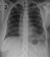

Cover image of the article: Echocardiographic and Angiographic Guided Removal of a Fractured Central Venous Access Port Using Snare Loop

Image Credit:Multidisciplinary Cardiovascular Annals

Echocardiographic and Angiographic Guided Removal of a Fractured Central Venous Access Port Using Snare Loop

Authors

Azin AlizadehaslAzin Alizadehasl ORCID1, Fatemeh ZohrianFatemeh Zohrian ORCID2,*, Seifolah Abdi3, Freidoon Noohi Bezanjani3, Bahram MohebbiBahram Mohebbi ORCID3, Arezoo Mohamadifar4, Mohsen Ziyaeifard5, Rasoul AzarfarinRasoul Azarfarin ORCID5, Solmaz Valizadeh6
1Department of Cardio-Oncology, Rajaie Cardiovascular Medical and Research Center, Tehran, Iran
2Department of Echocardiography, Rajaei Heart Center, Iran University, Tehran, Iran
3Departement of Interventional Cardiology, Rajaie Cardiovascular Medical and Research Center, Tehran, Iran
4Department of Heart Failure and Transplantation, Rajaei Cardiovascular Medical and Research Center, Tehran, Iran
5Department of Anesthesiology, Rajaie Cardiovascular Medical and Research Center, Tehran, Iran
6Department of Radiology, Shahid Beheshti University, Tehran, Iran
*Corresponding Author: Department of Echocardiography ,Rajaie Cardiovascular Medical and Research Center, Tehran, Iran. Tel: +98-9125257166, Email: [email protected]

Multidisciplinary Cardiovascular Annals:Vol. 11, issue 1; e99584
Published online:Mar 02, 2020
Article type:Case Report
Received:Nov 14, 2019
Accepted:Dec 03, 2019
How to Cite:Alizadehasl A, Zohrian F, Abdi S, Noohi Bezanjani F, Mohebbi B, et al. Echocardiographic and Angiographic Guided Removal of a Fractured Central Venous Access Port Using Snare Loop. Multidiscip Cardio Annal. 2020;11(1):e99584. doi: https://doi.org/10.5812/mca.99584

Abstract

Central venous access is commonly performed in the care of critically ill patients, for administration of repeated drug treatments. This routine procedure has multiple complications such as catheter malposition, migration, obstruction, infection, thrombosis and catheter fraction. We must recognize, consider and be able to manage these complications. We report a successful endovascular technique using a snare for retrieving broken venous port in a 36-year-old female.

1. Introduction

Fracture of a central venous catheter is very catastrophic and has been attributed to several factors such as prolonged mechanical force acting on the catheter, and forceful removal or insertion of the catheter (1). Embolization of fractured central venous catheter is a serious and rare complication. Catheter fragments may cause complications such as cardiac perforation, arrhythmias, sepsis and pulmonary embolism (2). We report a successful endovascular technique using a snare for retrieving broken venous port in a 36-year-old female.

2. Case Presentation

We report a 36-year-old female with a history of Hodgkin’s lymphoma 10 years ago and breast cancer 4 years ago who had received chemotherapy and radiotherapy. A peripherally inserted central venous port was used for chemotherapy since 10 years ago. The catheter had accidentally broken 8 months ago during an attempt to percutaneously retrieve the catheter. The patient had intermittent high grade fevers and referred to our center for extraction of the broken catheter.
Her serial chest radiographs demonstrated a long radiopaque venous port fragment from left subclavian vein extending to the junction of superior vena cava (SVC) to the right atrium (RA) (Figure 1).
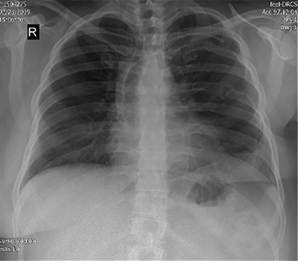
Venous port fragment from left subclavian vein extending to the junction of superior vena cava (SVC) to the right atrium (RA)
Figure 1.
Venous port fragment from left subclavian vein extending to the junction of superior vena cava (SVC) to the right atrium (RA)
Transthoracic and trans-esophageal echocardiography revealed that the distal tip of the catheter fragment was at the level of junction superior vena cava (SVC) to right atrium (RA) causing turbulences and narrowing with 3 mmHg gradient at proximal part of superior vena cava (SVC), catheter was thickened at proximal portion (0.55 cm) but more distal parts seemed to have normal thickness, there was also a 1 cm linear tissue-texture hypermobile linear density attached to the tip of the catheter which could be either vegetation or thrombosis, no other mass was visualized (Figures 2 and 3).
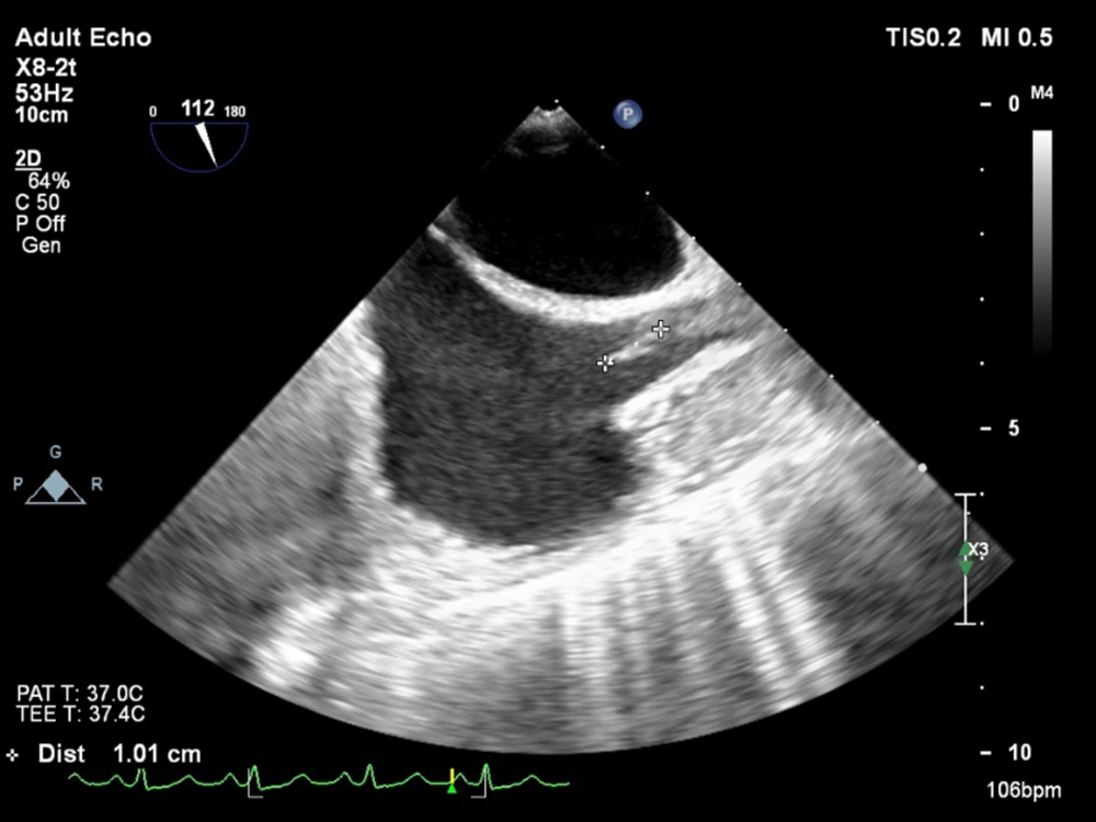
Distal tip of the catheter fragment was at the level of junction superior vena cava (SVC) to right atrium (RA)
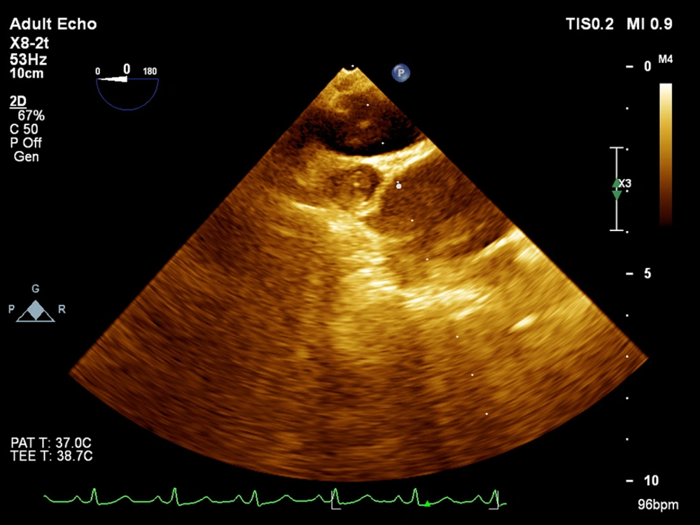
Figure 2.
Distal tip of the catheter fragment was at the level of junction superior vena cava (SVC) to right atrium (RA)
A 1 cm linear tissue-texture hypermobile linear density was attached to the tip of the catheter
Figure 3.
A 1 cm linear tissue-texture hypermobile linear density was attached to the tip of the catheter
Physical examination revealed no pathological findings, blood pressure on admission was 120/80 mmHg and oral temperature was normal. Laboratory tests including red blood cell and white blood cell count, urine culture, aerobic and anaerobic blood cultures were within normal limits except C- reactant protein (CRP) level which was 26.4 ng/mL (normal range < 6 ng/mL) and pro-calcitonin was 0.88 ng/mL with normal range (< 0.5 ng/mL).
The patient was treated with vancomycin 1 gr stat and BID and ampi sulbactam 3g IV stat and TDS, and after that percutaneous trans catheter retrieval technique via right femoral vein was chosen to be performed. The procedure was performed under mild sedation and fluoroscopic guidance. Injection in right and left subclavian, jugular and SVC revealed that the catheter was lodged at the junction of superior vena cava (SVC) to right atrium (RA). A 12 - 20 mm in diameter snare was advanced to proximal part of the catheter near the lodged site. During the procedure increasing forces released the catheter and the catheter fragment and snare were carefully pulled back via femoral sheath (Figures 4-6).
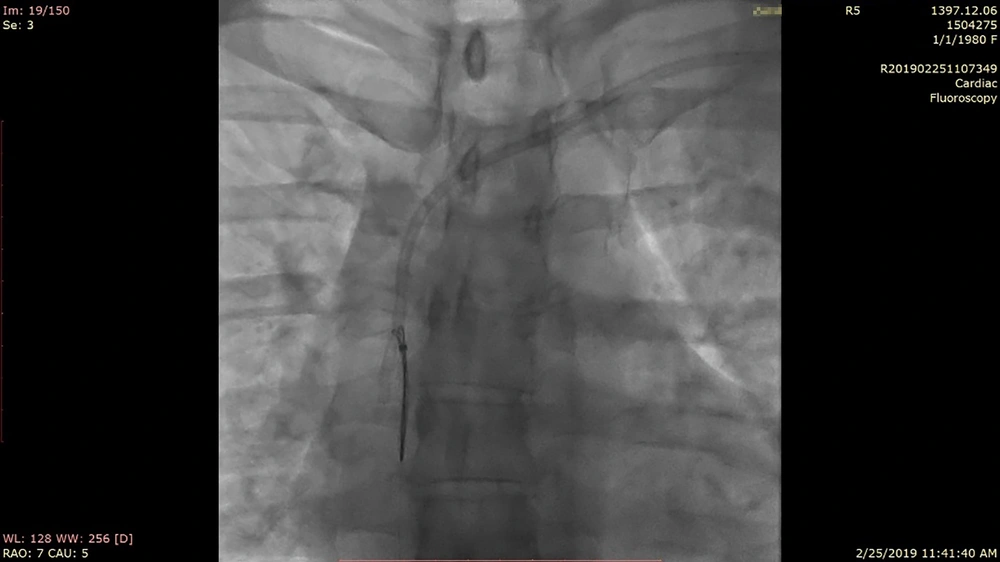
Snaring the catheter fragment
Figure 4.
Snaring the catheter fragment
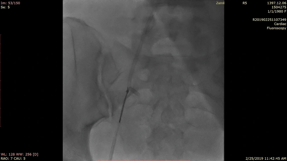
Catheter fragment and snare were carefully pulled back via femoral sheath
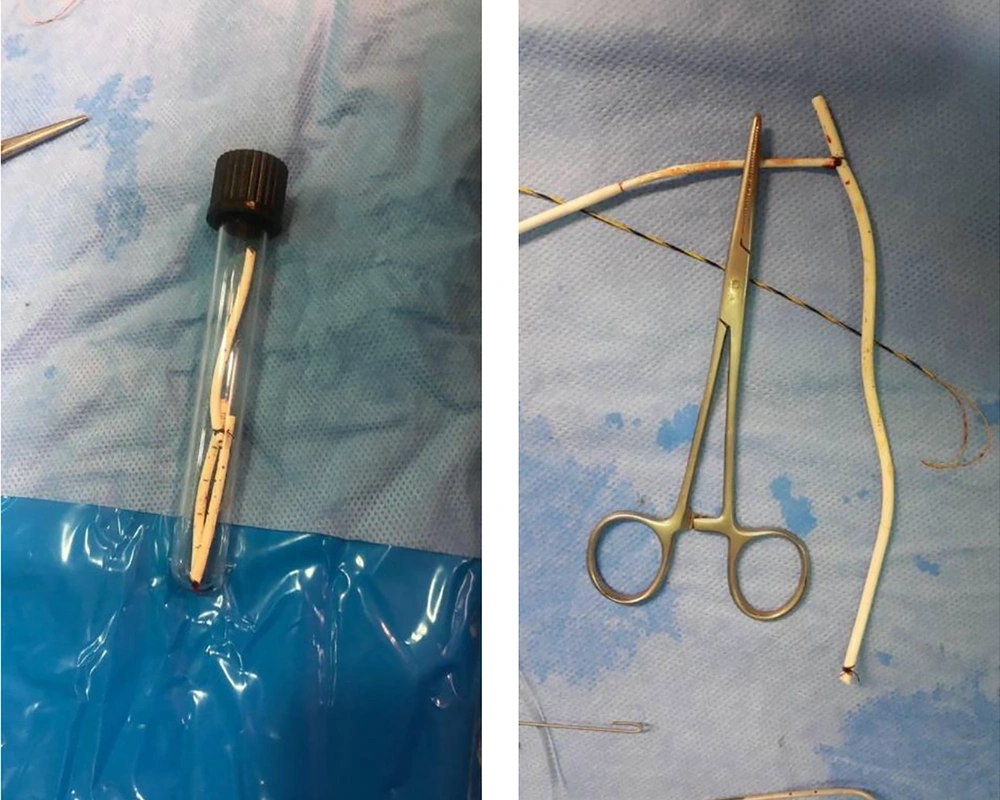
Figure 5.
Catheter fragment and snare were carefully pulled back via femoral sheath
Catheter fragment
Figure 6.
Catheter fragment
Injection in left subclavian and SVC revealed no extravasation of contrast from veins. No complication occurred during the interventional procedure.
After the procedure, the patient was monitored and control chest radiography was normal during the 24 hours after the procedure (Figure 7).
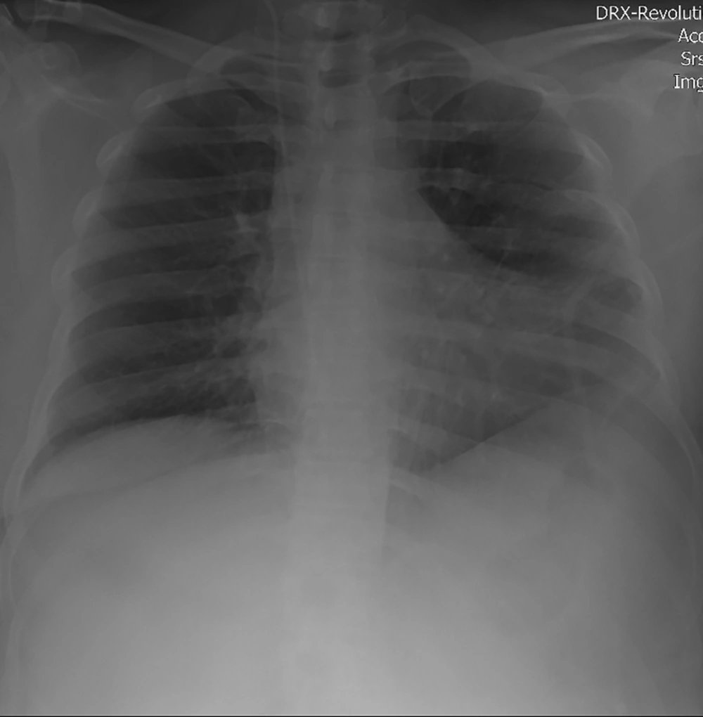
Control chest radiograph after removal of the catheter
Figure 7.
Control chest radiograph after removal of the catheter
Transthoracic and transesophageal echocardiography after removal of catheter and antibiotic therapy revealed normal findings and there were no residual mass or vegetation.
Her foreign body culture was staphylococcus aureus coagulase negative which was sensitive to linezolid and amikacin, so the patient was discharged with linezolid 600 mg BID and recommended to return to the clinic after one week.

3. Discussion

Delayed complications of venous access devices are spontaneous intravascular fracture of the catheter, catheter dislocation and embolization, and spontaneous migration and thrombosis of the internal jugular vein (1). Catheter fracture with or without embolization is a serious and rare complication in adult patients (3). If a catheter fracture occurs, the broken catheter will migrate distally along the blood stream and finally lodge in the superior vena cava, the right atrium, the right ventricle, the main pulmonary artery or its branches (1). The reported rate of intravenous catheter fracture is around 0.1% (4). Patients with a fractured catheter may remain asymptomatic or have shortness of breath, chest pain and even syncope (2). Diagnosis is often made by routine chest radiography (2). The mechanism of catheter fracture has largely been attributed to mechanical shearing forces acting on the catheter over a prolonged period (2).
In general, broken catheter management includes percutaneous trans-catheter retrieval, open thoracotomy or sternotomy and long term anticoagulation therapy. Percutaneous trans-catheter retrieval is usually performed at the first choice for the treatment of the complication (5). Regarding the patient’s request and the possibility of complications, percutaneous trans-catheter retrieval of fractured catheter was chosen.

3.1. Conclusions

Fracture of central venous port is a rare but serious condition that must be recognized and managed promptly. Early detection of the complication is crucial for the management. A chest radiography is very useful for early detection. Early removal of the catheter fragment is recommended.
Footnotes

  • Authors' Contribution: Fatemeh Zohrian did study concept and design. Azin Alizadehasl did acquisition of data. Seifolah Abdi did interventional procedure. Bahram Mohebbi did interventional procedure. Rasoul Azarfarin did critical revision of the manuscript. Freidoon Noohi Bezanjani did study supervision. Soulmaz Valizade did statistical analysis. Arezoo Mohamadifar did drafting of the manuscript.

  • Conflict of Interests: There was no conflict of interest for this case study.

  • Funding/Support: The authors have not received any funding.

  • Informed Consent: Informed consent signed by the patient.

References

  • 1.
    Biffi R, Orsi F, Pozzi S, Pace U, Bonomo G, Monfardini L, et al. Best choice of central venous insertion site for the prevention of catheter-related complications in adult patients who need cancer therapy: A randomized trial. Ann Oncol. 2009;20(5):935-40. [PubMed ID: 19179550]. https://doi.org/10.1093/annonc/mdn701.
  • 2.
    Muhm M, Sunder-Plassmann G, Apsner R, Kritzinger M, Hiesmayr M, Druml W. Supraclavicular approach to the subclavian/innominate vein for large-bore central venous catheters. Am J Kidney Dis. 1997;30(6):802-8. [PubMed ID: 9398124]. https://doi.org/10.1016/s0272-6386(97)90085-8.
  • 3.
    Cheng CC, Tsai TN, Yang CC, Han CL. Percutaneous retrieval of dislodged totally implantable central venous access system in 92 cases: Experience in a single hospital. Eur J Radiol. 2009;69(2):346-50. [PubMed ID: 17976941]. https://doi.org/10.1016/j.ejrad.2007.09.034.
  • 4.
    Gabelmann A, Kramer S, Gorich J. Percutaneous retrieval of lost or misplaced intravascular objects. AJR Am J Roentgenol. 2001;176(6):1509-13. [PubMed ID: 11373221]. https://doi.org/10.2214/ajr.176.6.1761509.
  • 5.
    Amerasekera SS, Jones CM, Patel R, Cleasby MJ. Imaging of the complications of peripherally inserted central venous catheters. Clin Radiol. 2009;64(8):832-40. [PubMed ID: 19589422]. https://doi.org/10.1016/j.crad.2009.02.021.

Copyright

Copyright © 2020, Multidisciplinary Cardiovascular Annals. This is an open-access article distributed under the terms of the Creative Commons Attribution-NonCommercial 4.0 International License (http://creativecommons.org/licenses/by-nc/4.0/) which permits copy and redistribute the material just in noncommercial usages, provided the original work is properly cited.

Similar Articles

30
Nov
2018

Suspected Short Peripheral Venous Catheter Intravascular Embolization: Identification and Management

Limael E Rodriguez,
Francisco Steven Rodriguez,
Francisco J Rivera-Pedrogo

E Rodriguez L, Steven Rodriguez F, J Rivera-Pedrogo F. Suspected Short Peripheral Venous Catheter Intravascular Embolization: Identification and Management. Int J Cardiovasc Pract. 2018;3(4):e130272. doi: https://doi.org/10.21859/ijcp-03044

27
Jul
2020
Placement of Totally Implantable Venous Access Ports Using the Single-Incision Technique: Outcomes and Complications

Placement of Totally Implantable Venous Access Ports Using the Single-Incision Technique: Outcomes and Complications

Seo Jin Jang,
Jae Hyun Kwon,
Yoon Ki Cha,
Do Yeun Kim

Jang SJ, Kwon JH, Cha YK, Kim DY. Placement of Totally Implantable Venous Access Ports Using the Single-Incision Technique: Outcomes and Complications. I J Radiol. 2020;17(3):e101804. doi: https://doi.org/10.5812/iranjradiol.101804

27
Dec
2017

Intraperitoneally Located Tip of Femoral Vein Catheter; Clinical Suspicion for Avoidance of Unnecessary Laparotomy

Hamed Shafiee,
Saeid Safari,
Reza Aminnejad

Shafiee H, Safari S, Aminnejad R. Intraperitoneally Located Tip of Femoral Vein Catheter; Clinical Suspicion for Avoidance of Unnecessary Laparotomy. Anesth Pain Med. 2017;7(6):e64557. doi: https://doi.org/10.5812/aapm.64557

5
Feb
2014

A Rare Central Venous Catheter Malposition: A Case Report

Ali Asghar Moeinipour,
Ahmad Amouzeshi,
Marjan Joudi,
Mehdi Fathi,
Saeed Jahanbakhsh,
Saeed Hafez
,et al.

Moeinipour AA, Amouzeshi A, Joudi M, Fathi M, Jahanbakhsh S, et al. A Rare Central Venous Catheter Malposition: A Case Report. Anesth Pain Med. 2014;4(1):e16049. doi: https://doi.org/10.5812/aapm.16049

7
Sep
2014

Letter to Editor on a Rare Central Venous Catheter Malposition

Jason Lie,
Manjula Yadagiri,
Catherine Aspinall

Lie J, Yadagiri M, Aspinall C. Letter to Editor on a Rare Central Venous Catheter Malposition. Anesth Pain Med. 2014;4(4):e20019. doi: https://doi.org/10.5812/aapm.20019

More by these authors

Azin AlizadehaslPubMedScholar
Fatemeh ZohrianPubMedScholar
Seifolah AbdiPubMedScholar
Freidoon Noohi BezanjaniPubMedScholar
Bahram MohebbiPubMedScholar
Arezoo MohamadifarPubMedScholar
Mohsen ZiyaeifardPubMedScholar
Rasoul AzarfarinPubMedScholar
Solmaz ValizadehPubMedScholar
Share
Cited by
Metrics