1. Background
Staphylococcus aureus is a major cause of skin infections that may present in the form of boils, folliculitis, impetigo, cellulitis, invasive soft tissue infections, foot ulcer, and furuncle. In skin infections, bacterial colonization in the skin due to skin barrier disruption leads to the expression of cytokines, which ultimately aggravates the symptoms (1-3). Infections caused by S. aureus pose a significant challenge to treating wounds and damaged tissues. Such infections are of great clinical importance because of S. aureus pathogenicity and its transmission and antibiotic resistance capacity. Therefore, timely and appropriate antibiotic therapy is essential for managing S. aureus infections.
Since the emergence and spread of multi-drug resistant (MDR) S. aureus strains, including methicillin-resistant S. aureus (MRSA) in 1961, the World Health Organization has classified this bacterium as a serious threat to the control of various infections, including skin infections. The prevalence of infection with extensively drug-resistant (XDR) S. aureus is also increasing. MDR is defined as non-susceptibility to ≥1 antimicrobial agent in ≥3 antimicrobial categories, and XDR is defined as non-susceptibility to ≥1 antimicrobial agent in all the antimicrobial categories, except in ≤2. This bacterium can acquire antibiotic resistance through several biochemical pathways, including modifying and destroying antibiotic molecules, reducing antibiotic penetration, or altering the bacterial target site (4, 5). Resistant skin infections are usually treated with beta-lactams, especially cephalosporins, such as cefazolin or glycopeptides (6). Vancomycin and teicoplanin were the first glycopeptides clinically used in 1955 and 1984, respectively. Oritavancin diphosphate (LY333328) is a semi-synthetic lipoglycopeptide introduced in 2015 with broad antibacterial activity against Gram-positive bacteria, including penicillin-resistant streptococci and coagulase-positive staphylococci. This agent acts on the peptide backbone by binding to the D-alanyl-D-alanine terminal of the peptidoglycan chain of Gram-positive bacteria, thereby blocking transglycosylation during peptidoglycan synthesis and preventing the formation of the bacterial cell wall. In addition, oritavancin can bind to the cytoplasmic membrane through its alkyl side chain, which in turn increases binding affinity to peptidoglycan residues and activity against vancomycin-resistant enterococci and vancomycin-resistant S. aureus (7-9).
2. Objectives
Considering the high prevalence of skin infections caused by S. aureus and the high rate of resistance to various antibacterial agents, this study aimed to evaluate the incidence of MDR, XDR isolates, and the minimum inhibitory concentration (MIC) of glycopeptides, particularly oritavancin, against drug-resistant S. aureus isolates from outpatients with skin infections.
3. Methods
3.1. Study Design and Patients’ Characteristics
In this prospective cross-sectional study, skin samples were taken from 250 outpatients (age range 11 – 72 years mean ± SD 43 ± 9.1) referred to seven Golestan Province (northern Iran) hospitals during 2019 - 2022. The samples were selected randomly via the convenience sampling method by dermatologists or infectious disease specialists. The inclusion criterion was clinical signs of skin infection and no antibiotic use in the last three months. The sample size was determined with a 95% confidence level using the formula below, where P1 is the number of patients referred to the infectious disease ward and P2 represents the number of patients with a positive MRSA test (α = 0.05, β = 0.10).
A questionnaire was prepared to collect characteristics of patients, including gender, age, type of skin infection, and the referral season. Patients with autoimmune diseases and those under ten (male and female) were excluded from this study.
3.2. Identification of Isolates
To identify and confirm the bacterial agents, skin swabs were first placed in tryptone soya broth (Merck, Germany) and then transferred to the medical diagnosis laboratory of the Infectious Diseases Research Center of Shahid Beheshti University. Then, the samples were inoculated onto chocolate agar, Columbia agar with 5% horse blood, and MacConkey agar. After 48 hours of incubation at 37°C, S. aureus strains were identified by examining mannitol-positive colonies and analyzing colony morphology, Gram staining, hemolysis, and catalase, coagulase (clumping factor), and DNase tests, and ultimately the Vitek-2 card system (Biometrics, France).
3.3. Determination of MRSA Isolates
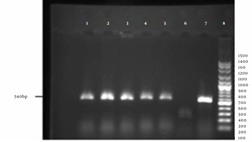
Phenotypic characterization of MRSA isolates was done by the Kirby-Bauer method using cefoxitin disks (30 µg). Detection of a growth inhibition zone with a diameter of ≤21 mm confirmed the presence of MRSA isolates. For a definite identification of MRSA strains, after DNA extraction using a commercial kit (SinaClon, Iran) according to the manufacturer's instructions, polymerase chain reaction (PCR) was done using the following primers (designed by Oligo 5 software) that are specific for the mecA gene (methicillin resistance gene): forward: 5'- AGTTCTGCAGTACCGGATTTGC -3' and reveres: 5'-AAAATCGATGGTAAAGGTTGGC-3'. Standard strains of S. aureus ATCC 33591 and S. aureus ATCC 25923 were considered positive and negative controls, respectively. The PCR reaction was carried out in a final volume of 25 µL consisting of 1 µL DNA sample, 1 µL of each primer, 12 µL of 2X Master Mix (containing 20 µM dNTP and 1.5 µM MgCl2), and 11 µL of distilled water. The reaction was performed in a thermocycler (Eppendorf, Germany) with the following cycling conditions: initial denaturation at 95°C for 5 minutes, 30 cycles of denaturation at 94°C for 15 seconds, annealing at 61°C for 1 minute, extension at 72°C for 1 minute, and final extension at 72°C for 5 minutes. The resulting PCR products were then electrophoresed on 1.5% agarose gel. The detection of 540 bp fragments confirmed the presence of MRSA isolates.
3.4. Antibacterial Susceptibility Testing
Antibiotic susceptibility test was performed on Mueller- Hinton agar (Merck, Germany) by the disk diffusion method (Kerby-Bauer method) using the following antibiotic disks: linezolid (10 µg), doxycycline (10 µg), amikacin (30 µg), daptomycin (2 µg), ciprofloxacin (5 µg), cefuroxime (30 µg), cefazolin (30 µg), clindamycin (2 µg), penicillin (10 units), and azithromycin (15 µg). All antibiotic discs were purchased from Padtan Teb Co. (Iran) except for linezolid (purchased from Mast Group, UK). After 16 - 18 hours of incubation at 37°C, the results were interpreted by measuring the diameter of the growth inhibition zone according to the Clinical & Laboratory Standards Institute (CLSI) standard guidelines (2021) (10).
3.5. Determination of Glycopeptides Minimum Inhibitory Concentration(s)
The MIC of three glycopeptides, including teicoplanin, vancomycin, and oritavancin, against the MRSA isolates was determined using the broth microdilution method according to the CLSI M100 guidelines (10). To prepare an antibacterial suspension, necessary amounts of vancomycin and teicoplanin powder (Sigma-Aldrich, USA) were inoculated into water and oritavancin powder into 0.002% polyphosphate in water. The initial concentration of each antibiotic was inoculated into wells of a 96-well microplate containing Mueller Hinton broth (Merck, Germany) and 2% salt. After preparing serial dilutions in the range of 0.06 - 64 µg/mL and inoculating the bacterial suspension at a final concentration of 1.5 × 10⁵ colony-forming units, the microplate was incubated at 37°C for 20 - 24 hours. A well containing the medium and antibiotic stock and another containing the medium with bacterial suspension were considered negative and positive controls, respectively. The MIC was determined by measuring the absorbance at 560 nm using an ELISA reader (10). Staphylococcus aureus ATCC 25923 and S. aureus ATCC 29213 were used as the control strains in the Kirby-Bauer and broth microdilution methods, respectively.
3.6. Statistical Analysis
Data were presented as frequency tables, graphs, and numerical indices. All data were analyzed using SPSS (version 23), and intergroup comparisons were done using the chi-square test at a significance level of <0.05.
4. Results
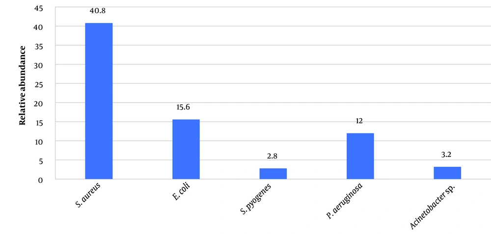
The most common and least common isolates were MRSA (102,40.80%) and Staphylococcus pyogenes (2.80%), respectively (Figure 1). Based on the phenotypic and molecular investigations (Figure 2), the frequency of MRSA isolates was significantly higher in samples collected from impetigo (53.92%) (P = 0.03) and in the summer season (44.11%) (P = 0.02) (Table 1).
| Variables | MDR (n = 91) | XDR (n = 11) | χ2 | P-Value |
|---|---|---|---|---|
| Age, y | 0.07 | |||
| 11 - 17 | 13 (14.28) | 0 (0 ) | 0.17 | |
| 18 - 44 | 63 (69.23) | 7 (63.63) | 0.21 | |
| ≥45 | 15 (16.48) | 4 (36.36) | 0.29 | |
| Gender | 0.06 | |||
| Female | 71 (78.02) | 4 (36.36) | 0.49 | |
| Male | 20 (21.97) | 7 (63.63) | 0.34 | |
| Season | 0.02 b | |||
| Spring | 33 (36.26) | 5 (45.45) | 0.37 | |
| Summer | 41 (45.05) | 4 (36.36) | 0.32 | |
| Autumn | 11 (12.08) | 2 (18.18) | 0.21 | |
| Winter | 6 (6.59) | 0 (0 ) | 0.11 | |
| Skin infection | 0.03 b | |||
| Foot ulcers | 35 (38.46) | 6 (54.54) | 0.41 | |
| Impetigo | 51 (56.04) | 4 (36.36) | 0.45 | |
| Cellulitis | 5 (5.49) | 1 (9.09) | 0.30 |
a Values are expressed as No. (%).
b Significant difference between the groups based on the chi-square test
Among MRSA isolates, 89.21% and 10.78% were MDR and XDR, respectively. In the Kirby-Bauer disk diffusion method, the highest and lowest drug resistance was related to penicillin (79.41%) and linezolid (2.94%). Cefazolin and cefuroxime were in second place in terms of antibacterial potency. Linezolid exhibited significant inhibitory effects against 98% of the isolates, especially S. aureus isolates, from foot ulcers in patients with diabetes. (Table 2). Moreover, linezolid inhibited the growth of 91% of XDR and 97% of MDR isolates (P = 0.01).
| Antibiotic | Foot ulcers (n = 41), Resistance | Impetigo (n = 55), Resistance | Cellulitis (n = 6), Resistance |
|---|---|---|---|
| Linezolid | 2 (4.87) | 0 (0 ) | 1 (16.67) |
| Daptomycin | 14 (34.14) | 9 (16.36) | 5 (83.33) |
| Clindamycin | 29 (70.73) | 33 (60 ) | 6 (100 ) |
| Ciprofloxacin | 17 (41.46) | 11 (20 ) | 3 (50 ) |
| Cefuroxime | 14 (34.14) | 7 (12.72 ) | 3 (50 ) |
| Azithromycin | 20 (48.78) | 13 (23.63) | 4 (66.67) |
| Doxycycline | 31 (75.60) | 39 (70.90) | 5 (83.33) |
| Penicillin | 35 (85.36) | 40 (72.72) | 6 (100 ) |
| Cefazolin | 15 (36.58) | 5 (9.09) | 2 (33.33) |
| Amikacin | 21 (51.21) | 12 (21.81) | 5 (83.33) |
a Values are expressed as No. (%).
At MIC ≥2 µg/mL, oritavancin inhibited the growth of 81.82% of MDR and, at MIC ≥8 µg/mL, 54.5 % of XDR isolates in a dose-dependent manner. In the case of glycopeptides, the lowest concentration of oritavancin that inhibited the growth of 90% of the MDR isolates (MIC90) was 2 µg/mL, which was eight times less than that of vancomycin (MIC90 = 16 µg/mL) and 16 times lower than that of teicoplanin (MIC90 = 32 µg/mL) (Tables 3 and 4).
| Glycopeptides | MIC | ||||||||||
|---|---|---|---|---|---|---|---|---|---|---|---|
| Concentration | 64 | 32 | 16 | 8 | 4 | 2 | 1 | 0.5 | 0.25 | 0.125 | 0.06 |
| Vancomycin | - | - | √ | - | - | - | - | - | - | - | - |
| Teicoplanin | - | √ | - | - | - | - | - | - | - | - | - |
| Oritavancin | - | - | - | - | - | √ | - | - | - | - | - |
Abbreviations: MIC (minimum inhibitory concentration): Microgram per milliliter
| Antibiotics XDR (11) | MIC90 (µg/mL) | MIC50 (µg/mL) | Range | Percentage of Resistance |
|---|---|---|---|---|
| Oritavancin | ≥8 | 2 | 1 - 8 | 45.45 |
| Vancomycin | ≥16 | 4 | 4 - 32 | 54.55 |
| Teicoplanin | >32 | 8 | 4 to 64 | 63.63 |
In the present study, the MIC average of oritavancin against MDR isolates from impetigo has been determined at 1.85 µL/mL, in which most growth fluctuations were observed in the density of 0.25 and 0.125 µL/mL. At the same time, this mean was 1.93 and 2.00 µL/mL for foot ulcer and cellulite, respectively. Most oritavancin-resistant MDR S. aureus isolates were from the specimens of cellulite (3 out of 6). In total, 15 cases (14.70%) of MDR S. aureus isolates were resistant to oritavancin, whereas (47, 85.45%) of MDR isolates from impetigo cases were sensitive to oritavancin. Oritavancin also showed favorable antibacterial activity against 60% of vancomycin-resistant XDR strains, 85.71% of all teicoplanin-resistant XDR strains, and 91% of MDR/XDR strains isolated from impetigo.
The frequency of vancomycin-resistance and teicoplanin-resistance among MDR S. aureus isolates was 26.47% and 34.31%, respectively. The average MIC of vancomycin and teicoplanin in order against MDR isolates from impetigo was determined at 14.40 and 31.62 µL/mL.
5. Discussion
It is well-documented that MRSA is one of the most important causes of skin infections in many parts of the world. However, different bacterial pathogens may cause skin infections, highlighting the role of environmental conditions, personal hygiene standards, age, infection site, and even season (11, 12). In the present study, more than half of S. aureus isolates from impetigo, and 40.8% of those from all skin infections were MRSA. These findings are in line with the findings of a previous study in Iran (13). Similarly, two other studies in Iran reported that more than half of all isolates from skin infections were MRSA (14, 15). In a recent study, 48% of S. aureus isolates from wounds, secretions, and blood samples were MRSA, of which 61% were MDR (6), which is less than the frequency of MDR strains in our study (89%), Based on our results, 10.8% of MRSA isolate were XDR. In contrast, in Pakistan (2020), 20%, and in Iran, a Middle East country (2022), 48% of the isolates were identified as XDR (16, 17).
Excessive use of antibiotics has significantly increased the prevalence of bacterial skin infections, creating a serious health challenge (18, 19).
In this study, among MDR S. aureus isolates, the highest antibiotic resistance was against penicillin (79.41%) and doxycycline (73.52%), which is consistent with previous reports (20). As an oxazolidinone, linezolid generally has a high antibacterial potential. In this study, 97% of MDR and 91% of XDR S. aureus isolates were sensitive to linezolid. Among MRSA isolates, 78.44% and 76.48% were sensitive to cephalosporines cefazolin and cefuroxime, respectively. However, it should be noted that glycopeptides are usually considered the first and second line of treatment against infections caused by Gram-positive bacteria (21, 22).
In our study, vancomycin and teicoplanin showed moderate antibacterial activity, and 26.47% and 34.31% of isolates were resistant, respectively. In a study in Iran in 2016, 23.3% of S. aureus isolates from abscesses and wounds showed resistance to vancomycin (22). However, a year later, another study in Iran reported that all S. aureus isolates were sensitive to vancomycin (23). The higher rate of vancomycin-resistance among S. aureus isolates in the present study could be attributed to the difference in the study time, the floods in 2019 in our study location, the high prevalence of impetigo in flood victims and the difference in the type of samples.
Compared to other glycopeptides, oritavancin can exert rapid and dose-dependent bactericidal activity (24). In this study, 85.30% of MDR isolates were sensitive to oritavancin, and this agent showed the highest inhibitory effect on MRSA isolates at a concentration of 1 - 2 µg/mL. It also showed an inhibitory effect on XDR S. aureus isolates at concentrations of 8 µg/mL and higher. In this regard, previous studies in Canada and the United States also reported the high efficiency of oritavancin in treating acute skin infections and in vivo pharmacokinetic models (25, 26). A study in 2021 and a meta-analysis study (2022) demonstrated the efficacy of oritavancin therapy compared with other glycopeptides for controlling skin infections in hospitalized patients, which could prevent recurrences with fewer side effects (27, 28). Various trials on animal models and patients with complex skin infections have also demonstrated that oritavancin can be the antibiotic of choice because of its shorter treatment duration, safety in children, and fewer side effects (29-31).
A limitation of this study was the small patient sample size and, most importantly, the COVID-19 pandemic that prevented us from comparing clinical features with the microbiological results. However, this study had strengths that can facilitate the development of skin infection treatment guidelines.
5.1. Conclusions
Our results highlighted the alarmingly high rate of resistance to antibiotics among S. aureus isolates from patients with skin infections. In line with previous studies, S. aureus was confirmed as the most common cause of skin infections in our study. Considering the great inhibitory properties of oritavancin against MDR S. aureus strains, especially those isolated from impetigo patients, the efficacy of this antibiotic for treating skin infections, particularly impetigo, is imperative. Moreover, oritavancin should be included in the skin sample antibiogram of medical diagnostic laboratories.


