Incidence of Carotid Atheroma in Patients Received Radiotherapy in Neck Field-A Case Series Study

authors:

avatar Maryam Jalili Sadrabad ORCID 1 , avatar Mahtab Kheirkhahi ORCID 2 , avatar Sayna Shaghayegh ORCID 3 , avatar Maryam Sami ORCID 3 , avatar Azra Mohiti ORCID 4 , *

Department of Oral and Maxillofacial Medicine, School of Dentistry, Semnan University of Medical Sciences, Semnan, Iran
Oral and Maxillofacial Radiologist, Shahid Beheshti University of Mediacal Sciences, Tehran, Iran
Student Research Committee, School of Dentistry, Semnan University of Medical Sciences, Semnan, Iran
Department of Oral and Maxillofacial Medicine, School of Dentistry, Alborz University of Medical Sciences, Karaj, Iran

How To Cite Jalili Sadrabad M, Kheirkhahi M, Shaghayegh S, Sami M, Mohiti A. Incidence of Carotid Atheroma in Patients Received Radiotherapy in Neck Field-A Case Series Study. Middle East J Rehabil Health Stud. 2025;12(2):e142401. https://doi.org/10.5812/mejrh-142401.

Abstract

Introduction:

Atheroma is a condition that affects various blood vessels, primarily forming at junctions of moderate to large vessels, such as the carotid artery, where it is known as carotid atheroma (CA). Atheroma can lead to myocardial infarction or stroke. Previous studies have shown that head and neck radiotherapy can induce CA, even in young patients. Early diagnosis of atheroma in panoramic views can help prevent ischemic attacks in the brain and potentially save lives.

Case Presentation:

Thirty-six patients were referred to Semnan Dental School for dental screening by a hematologist-oncologist. An oral medicine specialist ordered panoramic radiographs for each patient, and radiopaque lesions were detected in the para-spinal region in four patients. These lesions were diagnosed as Carotid Atheroma by an Oral and Maxillofacial Radiologist, and this case series was reported.

Conclusions:

Radiotherapy is a component of treatment for head and neck malignancies and can cause atherosclerosis in carotid junctions. Dentists should monitor panoramic views of patients receiving radiotherapy for any signs of suspicious lesions. If carotid atheroma is detected, the patient should be referred to a cardiovascular specialist for further evaluation and treatment.

1. Introduction

Atheroma is a disease affecting blood vessels, primarily forming at the junctions of moderate to large arteries, such as the carotid artery. Plaque formation causes the inner layer of the carotid vessel to thicken. This plaque consists of various cells, including monocytes, macrophages, lymphocytes, dendritic cells, smooth muscle cells, extracellular matrix, lipids, and calcium deposits (1). These plaques can rupture suddenly, leading to distant thrombosis and ischemic attacks, such as stroke and myocardial infarction (2).

Carotid atheroma can appear in panoramic views in two forms: As a round radiopaque area or as two radiopaque vertical lines with a distance of 1.5 to 4 cm, typically located at the border of the mandible or the posterior border of the mandible and anterior to the third and fourth cervical vertebrae (3).

Radiotherapy, while targeting tumor extension, also affects surrounding normal tissues. In head and neck radiotherapy, the oral mucosa, teeth, bone, and salivary glands respond differently. Acute changes include mucosal erythema, ulcers, reduced salivary secretion, damage to taste buds, and skin erythema and desquamation. Long-term effects involve reduced blood circulation in tissues leading to fibrosis (4).

Radiotherapy of the neck increases the thickness of the inner layer of the carotid artery (5), making atherosclerosis a relatively common complication following radiotherapy (6). Radiotherapy plays a crucial role in controlling head and neck cancers, with doses regulated based on the cancer's location and type. Most patients with head and neck cancer receive doses between 50 to 70 gray. Doses higher than 45 gray have been associated with carotid atheroma in previous studies (7).

Radiation-induced coronary heart disease is the second leading cause of death among patients receiving radiotherapy for breast cancer, Hodgkin lymphoma, and other mediastinal cancers (8). It has been shown that plaque formation is dose-dependent, with carotid atheroma being more severe in patients who received radiation for lymphoma (9).

Due to the severe cerebrovascular complications associated with radiotherapy in the neck region, this study investigated the incidence of carotid atheroma in patients who received radiation and were referred to the Dental Faculty of Semnan University, using panoramic views.

2. Case Presentation

Thirty-six patients were referred to the Oral Medicine Department of the Dental Faculty at Semnan University of Medical Sciences by a hematologist-oncologist. Following an evaluation of medical and dental histories and oral examinations, a panoramic radiograph was taken for each patient to screen for dental and surrounding bone diseases and to investigate carotid atheroma (CA).

All images were analyzed by an oral and maxillofacial radiologist, and four of these images showed opacities diagnosed as carotid atheroma. The patients with CA are listed below:

Case 1:

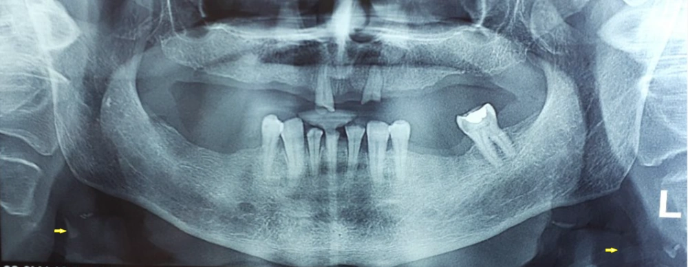
A 48-year-old man, weighing 78 kg and 175 cm tall, was diagnosed with nasopharyngeal cancer. He has no history of hyperlipidemia, diabetes, or hypertension. An X-ray taken 3 months after radiotherapy showed a single, almost round radiopaque area in front of the vertebral column between the third and fourth vertebrae, with a diameter of about 0.5 centimeters (Figure 1).

On the left side, a single, almost round radiopaque in front of the vertebral column between the third and fourth vertebrae, with a diameter of about half a centimeter. On the right side, a single radiopacity with similar characteristics is observed.

Case 2:

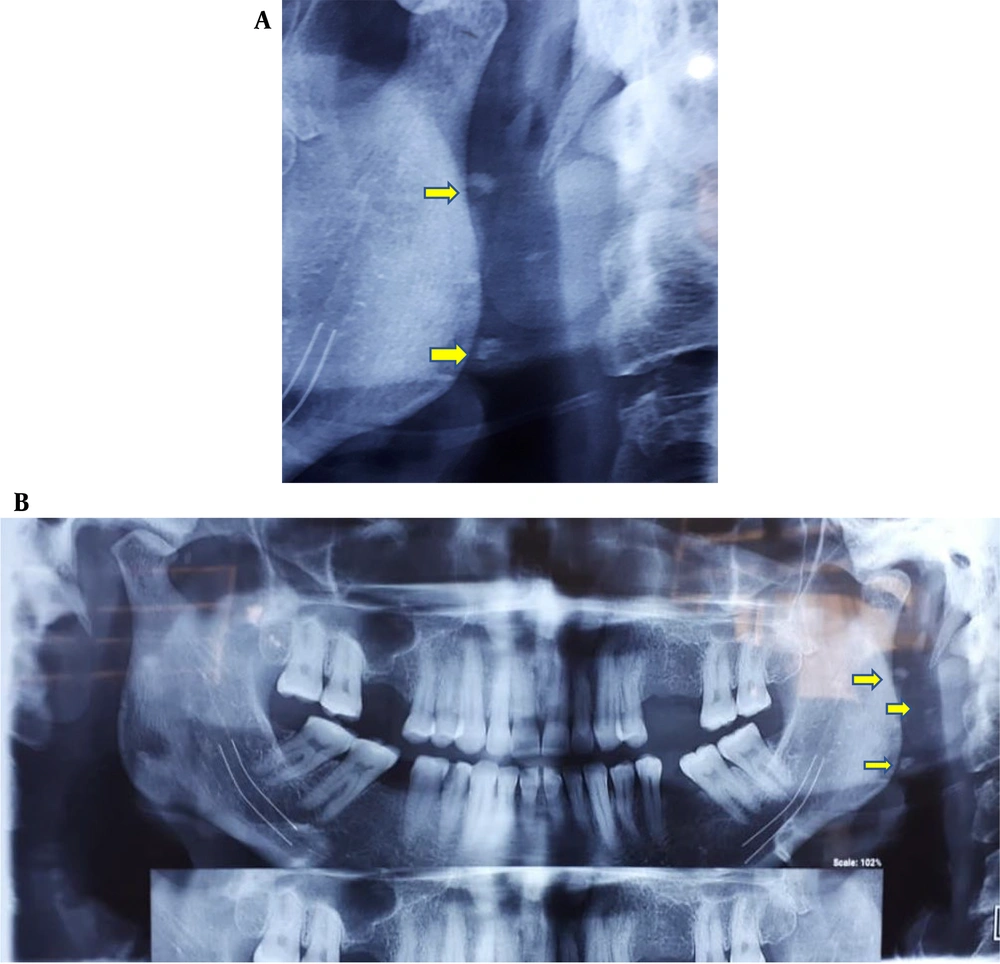
A 62-year-old woman, weighing 70 kg and 165 cm tall, with a history of hyperlipidemia, was diagnosed with breast cancer. She has no history of diabetes or hypertension. An X-ray taken 4 months after radiotherapy showed a radiopaque area (Figure 2).

This view has both sides lesions. The left side has two lesions, one about 5 × 2 mm. The second one is 1 × 1 mm, and Right side one 6 × 2 mm.

Case 3:

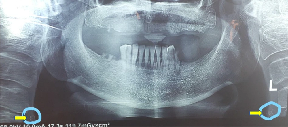
A 39-year-old woman, weighing 65 kg and 159 cm tall, with a history of breast cancer and hyperlipidemia, was referred. She has no history of diabetes or hypertension. An X-ray taken 5 months after radiotherapy revealed a radiopaque area (Figure 3).

Three rounded radiopaque lesions on the right side. The first is inclined to the left and round with a size of less than 1 cm. The second is inclined to the right and is 0.5 × 0.2 cm wide. The third is inclined to the left and round and less than 1 cm in size.

Case 4:

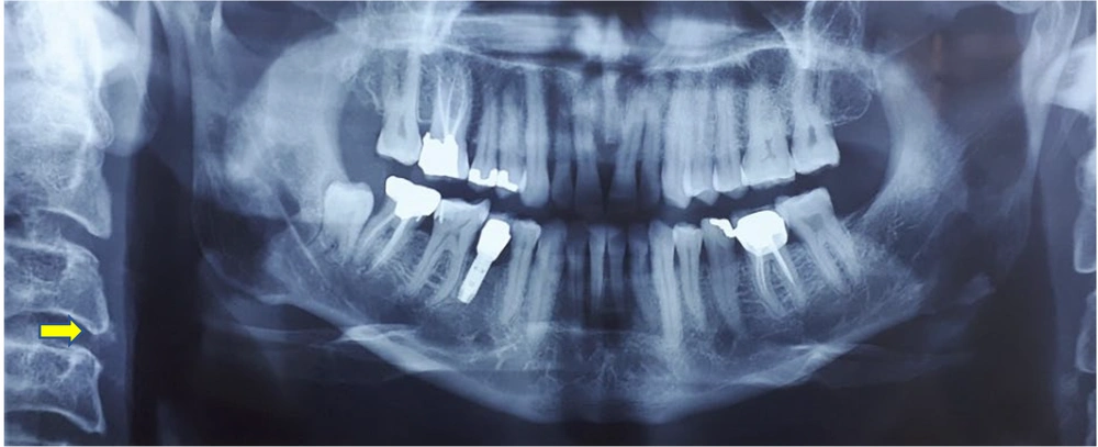
A 61-year-old woman, weighing 65 kg and 157 cm tall, with a history of breast cancer, was referred. She has no history of hyperlipidemia or diabetes. After 7 sessions of radiotherapy, an X-ray was prepared (Figure 4).

Tow round radiopaque lesions in bilateral paravertebral area are seen with around 0.5 cm.

3. Discussion

Atherosclerosis is one of the most common vascular diseases worldwide, affecting many individuals in older age. The most well-known complications of this disease include an increased risk of cardiovascular disorders, while less common but significant complications such as stroke and infarction also warrant attention.

Recent research has indicated that radiotherapy is associated with the formation of a stroke-stimulating factor known as atheroma in the cervical region of the carotid artery, which can be detected on panoramic radiographs (10).

Currently, head and neck cancers are treated with surgery, chemotherapy, and radiotherapy. The complications of radiotherapy are dose-dependent and significantly influenced by the radiation field.

One notable complication of radiotherapy is atherosclerosis in vessels such as the carotid artery, particularly when the radiation field includes regions affected by lymphoma, breast cancer, and other mediastinal cancers. Radiation doses of 45 Gy and higher are associated with this complication. The dose of radiation linked to the development of carotid atherosclerosis ranges from 40 Gy for the treatment of Hodgkin's and non-Hodgkin's lymphoma to 50-80 Gy for the treatment of squamous cell carcinoma and salivary malignancies of the head and neck (11).

Some studies, utilizing Doppler ultrasound and angiography (12), have shown that the damage caused by radiotherapy is extensive and typically involves the distal part of the common carotid artery, the bifurcation area of the artery, as well as the proximal and distal parts of the internal carotid artery. In contrast, spontaneous atherosclerosis is usually limited to the bifurcation area of the common carotid artery and the proximal internal carotid artery (12, 13).

In a study, the carotid arteries of 40 patients were examined 10 years post-radiotherapy, and 40% showed stenosis in their arteries (14). In a similar study, among 23 patients, 22% had carotid stenosis 4.9 years after completing radiotherapy (13). A recent cohort study indicated that radiotherapy either initiates new atherosclerosis or accelerates existing conditions, underscoring the importance of early diagnosis to prevent the potentially fatal outcomes of carotid atheroma (15).

Another study demonstrated that the prevalence of severe stenosis following radiotherapy in head and neck cancer patients is low. It suggests that common cardiovascular risk factors are more influential in causing these lesions. Consequently, regular and continuous examinations are essential for patients receiving radiotherapy who have underlying cardiovascular risk factors (3, 16). These lesions typically appear 1.5 to 2.5 cm below and posterior to the angle of the mandible (17). Dentists play a crucial role in early diagnosis, as patients are often referred to them before, during, or after radiotherapy for routine dental check-ups. Dentists should also consider the possibility of new or recurring diseases in radiotherapy patients and manage dry mouth while preventing osteoradionecrosis (18).

Identifying patients with carotid atheroma through panoramic radiography is vital, as radiotherapy-related atherosclerosis may be the first sign of acute ischemic stroke (19, 20). Timely diagnosis and follow-up of these lesions can significantly reduce the risk of death.

Therefore, guidelines should be developed for dentists treating patients undergoing head and neck radiotherapy. These guidelines should include educating dentists to screen for critical lesions, such as radiopaque lesions in the paravertebral area suspected of being carotid atheroma, and referring patients to appropriate physicians for further treatment if such lesions are detected. Specialist physicians must evaluate patients for high blood pressure, high cholesterol, high blood sugar, and smoking, as arteries exposed to radiation are particularly sensitive to these atherogenic factors (21). It is also recommended that doctors use ultrasound and other imaging methods to assess the extent of stenosis (22).

3.1. Conclusions

Radiotherapy, a common treatment for head and neck malignancies, can lead to carotid atheroma. Dentists should monitor panoramic views of patients receiving radiotherapy for any suspicious lesions and refer patients with detected atheroma to a cardiovascular specialist for timely treatment, thereby helping to prevent cerebral attacks and sudden death.

Acknowledgements

References

  • 1.

    Tabas I, Garcia-Cardena G, Owens GK. Recent insights into the cellular biology of atherosclerosis. J Cell Biol. 2015;209(1):13-22. [PubMed ID: 25869663]. [PubMed Central ID: PMC4395483]. https://doi.org/10.1083/jcb.201412052.

  • 2.

    Goikuria H, Vandenbroeck K, Alloza I. Inflammation in human carotid atheroma plaques. Cytokine Growth Factor Rev. 2018;39:62-70. [PubMed ID: 29396056]. https://doi.org/10.1016/j.cytogfr.2018.01.006.

  • 3.

    Markman RL, Conceicao-Vasconcelos KG, Brandao TB, Prado-Ribeiro AC, Santos-Silva AR, Lopes MA. Calcified carotid artery atheromas on panoramic radiographs of head and neck cancer patients before and after radiotherapy. Med Oral Patol Oral Cir Bucal. 2017;22(2):e153-8. [PubMed ID: 28160583]. [PubMed Central ID: PMC5359713]. https://doi.org/10.4317/medoral.21436.

  • 4.

    Vissink A, Jansma J, Spijkervet FK, Burlage FR, Coppes RP. Oral sequelae of head and neck radiotherapy. Crit Rev Oral Biol Med. 2003;14(3):199-212. [PubMed ID: 12799323]. https://doi.org/10.1177/154411130301400305.

  • 5.

    Fernandez-Alvarez V, Lopez F, Suarez C, Strojan P, Eisbruch A, Silver CE, et al. Radiation-induced carotid artery lesions. Strahlenther Onkol. 2018;194(8):699-710. [PubMed ID: 29679099]. https://doi.org/10.1007/s00066-018-1304-4.

  • 6.

    Miura M, Nakajima M, Fujimoto A, Kaku Y, Kawano T, Watanabe M, et al. High prevalence of small vessel disease long after cranial irradiation. J Clin Neurosci. 2017;46:129-35. [PubMed ID: 28974389]. https://doi.org/10.1016/j.jocn.2017.09.008.

  • 7.

    Freymiller EG, Sung EC, Friedlander AH. Detection of radiation-induced cervical atheromas by panoramic radiography. Oral Oncol. 2000;36(2):175-9. [PubMed ID: 10745169]. https://doi.org/10.1016/s1368-8375(99)00072-x.

  • 8.

    Cuomo JR, Javaheri SP, Sharma GK, Kapoor D, Berman AE, Weintraub NL. How to prevent and manage radiation-induced coronary artery disease. Heart. 2018;104(20):1647-53. [PubMed ID: 29764968]. [PubMed Central ID: PMC6381836]. https://doi.org/10.1136/heartjnl-2017-312123.

  • 9.

    Kloosterman A, Dillen TV, Bijwaard H, Heeneman S, Hoving S, Stewart FA, et al. How radiation influences atherosclerotic plaque development: A biophysical approach in ApoE(-)/(-) mice. Radiat Environ Biophys. 2017;56(4):423-31. [PubMed ID: 28866809]. [PubMed Central ID: PMC5655690]. https://doi.org/10.1007/s00411-017-0709-2.

  • 10.

    Flor N, Sardanelli F, Soldi S, Franceschelli G, Missiroli C, De Paoli F, et al. Unknown internal carotid artery atherosclerotic stenoses detected with biphasic multidetector computed tomography for head and neck cancer. Eur Radiol. 2006;16(4):866-71. [PubMed ID: 16320057]. https://doi.org/10.1007/s00330-005-0023-2.

  • 11.

    Chung TS, Yousem DM, Lexa FJ, Markiewicz DA. MRI of carotid angiopathy after therapeutic radiation. J Comput Assist Tomogr. 1994;18(4):533-8. [PubMed ID: 8040432]. https://doi.org/10.1097/00004728-199407000-00003.

  • 12.

    Dorresteijn LD, Kappelle AC, Boogerd W, Klokman WJ, Balm AJ, Keus RB, et al. Increased risk of ischemic stroke after radiotherapy on the neck in patients younger than 60 years. J Clin Oncol. 2002;20(1):282-8. [PubMed ID: 11773180]. https://doi.org/10.1200/JCO.2002.20.1.282.

  • 13.

    Tsai CF, Jeng JS, Lu CJ, Yip PK. Clinical and ultrasonographic manifestations in major causes of common carotid artery occlusion. J Neuroimaging. 2005;15(1):50-6. [PubMed ID: 15574574]. https://doi.org/10.1177/1051228404270242.

  • 14.

    Popovtzer A, Eisbruch A. Advances in radiation therapy of head and neck cancer. Expert Rev Anticancer Ther. 2008;8(4):633-44. [PubMed ID: 18402530]. https://doi.org/10.1586/14737140.8.4.633.

  • 15.

    Simonetto C, Mayinger M, Ahmed T, Borm K, Kundrat P, Pigorsch S, et al. Longitudinal atherosclerotic changes after radio(chemo)therapy of hypopharyngeal carcinoma. Radiat Oncol. 2020;15(1):102. [PubMed ID: 32381045]. [PubMed Central ID: PMC7206771]. https://doi.org/10.1186/s13014-020-01541-3.

  • 16.

    Valentin ML, Barco S, Studer G, Clemens R, Kreuzpointner R, Sebastian T, et al. Prevalence of carotid plaque stenosis after head and neck radiotherapy - an observational study of 156 survivors. Vasa. 2020;49(6):467-73. [PubMed ID: 32674693]. https://doi.org/10.1024/0301-1526/a000896.

  • 17.

    Friedlander AH, Federico M, Yueh R, Norman KM, Chin EE. Radiation-associated carotid artery atherosclerosis: Case report and review of contemporaneous literature. Spec Care Dentist. 2009;29(2):75-9. [PubMed ID: 19284506]. https://doi.org/10.1111/j.1754-4505.2008.00066.x.

  • 18.

    before Treatment OA. Oral and dental management related to radiation therapy for head and neck cancer. J Can Dent Assoc. 2003;69(9):585-90.

  • 19.

    Friedlander AH, Cohen SN. Panoramic radiographic atheromas portend adverse vascular events. Oral Surg Oral Med Oral Pathol Oral Radiol Endod. 2007;103(6):830-5. [PubMed ID: 17261373]. https://doi.org/10.1016/j.tripleo.2006.07.016.

  • 20.

    Santoro A, Bristot R, Paolini S, Di Stefano D. Radiation injury involving the internal carotid Artery: Report of two cases. Journal of Neurosurgical Sciences. 2000;44(3):159.

  • 21.

    Patel DA, Kochanski J, Suen AW, Fajardo LF, Hancock SL, Knox SJ. Clinical manifestations of noncoronary atherosclerotic vascular disease after moderate dose irradiation. Cancer. 2006;106(3):718-25. [PubMed ID: 16353211]. https://doi.org/10.1002/cncr.21636.

  • 22.

    Ecker RD, Donovan MT, Hopkins LN. Endovascular management of carotid artery disease after radiation therapy and radical neck dissection. Neurosurg Focus. 2005;18(1). e8. [PubMed ID: 15669802]. https://doi.org/10.3171/foc.2005.18.1.9.