1. Background
Football is among the world's most popular sports (1), boasting 500 million players globally. Research indicates that professional football players experience an injury incidence of 101 injuries per 1,000 hours of play (2). Hamstring muscle (HM) injuries, prevalent in sprinting-related activities like football, have an incidence rate of 6 - 29% across sports (3, 4) with football seeing 8% to 25% of all injuries resulting from hamstring strains (5). The central nervous system controls voluntary movement and muscular patterns (6), with two primary mechanisms of HM injury identified as overstretching and explosive eccentric contractions (4).
The Nordic hamstring exercise, a recent literature subject, is an eccentric strengthening exercise for the HM (7). This exercise involves individuals kneeling, their ankles secured, as they gradually lower their upper body towards the ground in a prone position (8), enhancing the eccentric strength of knee flexor muscles and reducing HM injury rates (9, 10). Studies have also shown its application as a field test for estimating HM eccentric ability (11-13), with the breakpoint angle (BPA) defined as the angle at which subjects cannot maintain contraction due to an increased gravitational moment, leading to their fall during the Nordic Hamstring test (NHT) (12).
Traditionally, Surface electromyography (sEMG) studies have focused on measuring muscle activity and timing (14), with the objective evaluation of voluntary motor control and muscular patterns proving challenging (15). Introduced in 2004, the voluntary response index (VRI) offers a novel surface EMG analysis sensitive enough to measure motor control (16) by evaluating whole muscle patterns rather than individual muscle activities, encompassing two components: Magnitude (MAG) and similarity index (SI) (15). Voluntary response index aims to reduce participation variability in EMG signal analysis by comparing voluntary motor control patterns across tasks.
Voluntary response index methodologies have been applied in diverse research areas, including incomplete spinal cord injuries (17, 18), shoulder dysfunction (19), trunk muscle fatigue (20), anterior cruciate ligament (ACL) injury (21), and trunk muscle activity during variable weight lifting (22). A higher SI value indicates superior motor control in voluntary tasks.
When selecting research tools or methods, verifying within-day reliability is essential to confirm their validity and consistency in replicating results across identical conditions and populations.
The reliability of MAG and SI was explored in individuals with incomplete spinal cord injuries (23), showing good to excellent reliability [MAG (ICC = 0.71 - 0.99), SI (ICC = 0.65 - 0.96)] in repeated tests. The reliability of BPA was also examined in football and track and field student-athletes (24), revealing perfect reliability (ICC = 0.93 - 0.98). However, the reliability of VRI in healthy professional athletic populations remains a subject of ongoing research. For example, Hajouj et al. assessed VRI reliability in athletes post-ACL reconstruction (25), discovering high to very high reliability during sit-stand-sit tasks and recommending this method for motor control assessment post-knee ligament injury. This underscores the importance of validating the VRI analysis method's reliability in participants with high physical fitness during varied functional tasks.
2. Objectives
The primary objective of our study was to evaluate the within-day reliability of the VRI, including the SI and MAG, as well as the BPA during the NHT in healthy professional football players. We hypothesized that both the VRI method and BPA would prove to be reliable tools for use in studies of motor control among professional athletes, particularly football players.
3. Methods
3.1. Study Participants
Twenty-four healthy male professional football players, aged between 21 and 34 years [mean age 26.4 ± 4.3 years, height 180 ± 6.4 cm, weight 73.6 ± 5.1 kg, BMI 22.6 ± 0.9 kg/m2 (mean ± SD)], participated in this study. The sample size was determined using online applications designed for research on reliability and measurement error (26). All participants scored 9 or higher on the Tegner activity scale and played in the second division of the Iranian football league (21). Players with a history of HM injury or other lower limb injuries, such as soft tissue damage and fractures in the past six months, hyperlordosis in the lumbar spine, or any neurological disorders, were excluded from the study (24). This research received approval from the Tabriz University of Medical Sciences Ethical Committee (IR.TBZMED.REC.1400.1089). All participants provided written informed consent and agreed to the publication of their data. They were also informed of their right to withdraw from the study at any time. The study took place in the Medical Committee laboratory of the Guilan football board in 2023.
3.2. Experimental Procedure
A single examiner, a physiotherapist with eight years of experience in sports physiotherapy, conducted all procedures. Participants were briefed on performing the NHT and completed a demographic questionnaire before the assessment. They were advised to avoid any high-intensity exercises for at least 48 hours before the test. Initially, a warm-up session consisting of muscle stretching exercises and a five-minute jog was conducted. Preparation of the skin for sEMG involved cleaning the electrode areas with alcohol and shaving any hair. Following the attachment of electrodes and verification of proper recording, participants performed three trials of the NHT.
3.3. Surface Electromyography Measurement
Muscular activities were recorded using an eight-channel portable sEMG system with 16-bit resolution (Biosignals Plux, Lisbon, Portugal). The recordings were made with preamplifier bipolar active electrodes. Each electrode had a 50 mm diameter and a 10 mm recording diameter, with a center-to-center distance of 20 mm, prepared by the examiner. The electrodes featured a built-in differential amplifier with a gain of 1000, an input impedance of over 100 GOhm, a common-mode rejection ratio of 100 dB, a bandwidth of 25 - 500 Hz, a sampling frequency of 1 kHz, and a 1 mA consumption. Reference electrodes were placed on the skin over the left proximal fibula head.
3.4. Electrode Placement
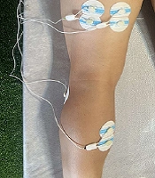
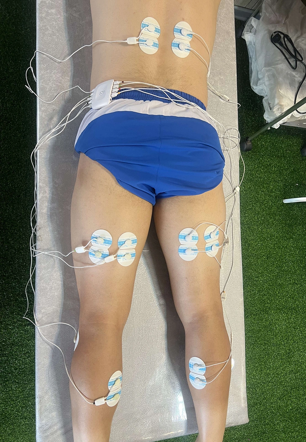
Surface EMG activity from the biceps femoris (BF), semitendinosus (ST), erector spinae (ES), and medial gastrocnemius (MG) muscles on both sides were recorded during the NHT (Figure 1). Electrode placement followed the recommendations of SENIAM (Surface EMG for Non-Invasive Assessment of Muscles) (27). The electrode for the BF was positioned at the midpoint between the lateral epicondyle of the femur and the ischial tuberosity. For the ST, the electrode was placed at the midpoint between the medial epicondyle of the femur and the ischial tuberosity. Medial gastrocnemius electrodes were positioned on the muscle's most prominent bulge, and ES electrodes were placed two fingers' width lateral to the spinous process of L1.
3.5. Nordic Hamstring Test
Participants performed three repetitions of the NHT with two-minute intervals between each repetition. Four LED markers were affixed to the lateral shoulder, hip, knee joints, and lateral malleolus by the examiner. The NHT began in a kneeling position with the trunk upright, knees flexed at 90°, and arms held by the sides. The examiner stabilized the participants' lower legs to support their body weight. Participants lowered their bodies forward towards the floor until reaching the maximum point of descent (Figure 2). They were instructed to keep the shoulder, hip, and knee joints aligned and to maintain this alignment throughout the NHT. Each trial was captured using an iPhone 7+ camera, set to record slow-motion video at 720 pixels and 240 frames per second, positioned two meters to the participant's left side. Angles were analyzed using Kinovea software (beta 0.08.27), whose validity and reliability have been established in recent research (28). This software identified LED markers in the video files, extracted joint coordinates, and plotted joint angle and speed diagrams for each trial participant. The BPA was identified as the point where the speed exceeded 10º/ second.
3.6. Data Analysis
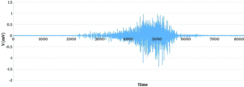
For the objective analysis of motor control during the NHT, we employed the VRI, a vector analysis method that includes two components. The MAG component quantifies the combined sEMG activity of all muscles as the length of the response vector (RV) for a given task. The SI reflects the consistency of sEMG activity distribution during a task. Initially, data files were imported into custom-developed MATLAB software (Math Works), and raw sEMG data were plotted (Figure 3). The moving average of the root mean square (RMS) values for eight muscles was calculated during each NHT attempt. The RV, composed of RMS values from all muscles of a participant, was then computed. Subsequently, a prototype response matrix (PRM) was generated using RVs from 10 professional players not included in the study. The prototype response vector (PRV) was derived from the PRM, and its MAG was determined using Equation 1. After calculating the participant's RV, the MAG of the subject's response vector (sRV) was determined using Equation 2. Finally, SI was calculated as the inner product of PRV and sRV, representing the cosine of the angle between them by Equation 3 (29). In the equations, PRV stands for prototype response vector, sRV for subject response vector, and the subsequent abbreviations denote muscles.
All statistical analyses were performed using SPSS software version 27. The Shapiro-Wilk test assessed data normality. The intra-class correlation coefficient (ICC) was used to evaluate test-retest (within-day) reliability. Intra-class correlation coefficient values were interpreted according to the Landis and Koch criteria (poor = 0 - 0.02, fair = 0.21 - 0.40, moderate = 0.41 - 0.60, substantial = 0.61 - 0.80, and almost perfect = 0.81 - 1 (30). The standard error of measurement (SEM) was calculated using the formula [SEM = Zα * SD * √(1 - ICC)]. The minimal detect change (MDC) was also determined with the formula 1.95 * √2 * SEM (31).
4. Results
Descriptive statistics, including means and standard deviations (SD) for MAG, SI, and BPA across three NHT trials, are detailed in Table 1. The reliability of VRI components (MAG, SI) and BPA is outlined in Table 2, featuring the ICC with a 95% confidence interval (CI), the standard error of measurement (SEM), and the minimal detectable change (MDC). The VRI components, MAG (ICC = 0.90) and SI (ICC = 0.81) demonstrated almost perfect reliability in the functional task. BPA exhibited substantial reliability (ICC = 0.65) during the NHT. A lower SEM indicates a reduced error in measurement across trials.
| Variables | Trial 1 | Trial 2 | Trial 3 |
|---|---|---|---|
| MAG, mV | 0.45 ± 0.14 | 0.46 ± 0.13 | 0.46 ± 0.16 |
| SI | 0.91 ± 0.04 | 0.92 ± 0.02 | 0.92 ± 0.03 |
| BPA, ° | 44.07 ± 6.14 | 46.84 ± 4.84 | 49.54 ± 8.54 |
Descriptive Analysis of the Magnitude, Similarity Index, and Breakpoint Angle in the Nordic Hamstring Test (n = 24) a
| Variables | ICC (95%CI) | SEM | MDC |
|---|---|---|---|
| MAG, mV | 0.90 | 0.25 | 1.37 |
| SI | 0.81 | 0.07 | 0.72 |
| BPA, ° | 0.65 | 17.8 | 11.6 |
Reliability of VRI Components and BPA in NHT
5. Discussion
This study aimed to evaluate the test-retest reliability of sEMG recordings using the novel VRI method and BPA in professional football players during a functional task (NHT). To our knowledge, this is the first study to explore the reliability of sEMG analysis via VRI during the NHT in professional football players. The findings indicated that VRI achieved almost perfect reliability, and BPA showed substantial reliability during the NHT among professional football players.
Surface EMG technology has traditionally been employed for assessing muscle activity for medical purposes. Literature suggests that surface electrode recordings may offer more reliability than needle EMG (32). This greater reliability and ease of use of sEMG make it a preferred choice for researchers conducting EMG analysis studies (32).
Recently, the VRI analysis method has been applied to evaluate motor control and muscular patterns by assessing the overall activity of muscles involved in specific motor tasks (15). Voluntary response index analysis can identify motor control deficits and compensatory strategies by examining the collective muscle activity involved in a task (16). In one study, VRI analysis was utilized to investigate differences in motor control patterns during a specific voluntary task in patients with incomplete spinal cord injuries (33).
The VRI method has also been applied in studying the motor control of shoulder muscles in patients with shoulder disorders across four functional tasks. It was discovered that lower SI values could indicate abnormal muscular patterns during these tasks (19).
In another study, VRI was used to assess the reciprocal coactivation of knee muscles in patients with patellofemoral pain syndrome, revealing ICC values ranging from 0.85 to 0.99 for VRI components (34).
The findings of the present study demonstrated nearly perfect reliability for the MAG (ICC = 0.90) and SI (ICC = 0.81). In a similar vein, Ghofrani et al. explored the reliability of trunk muscle VRI during the lifting of variable loads in healthy individuals (22), discovering that VRI was a reliable and repeatable method (ICC = 0.73 - 0.97) for assessing trunk muscle recruitment patterns during load lifting. Hajouj et al. assessed the reliability of the VRI approach in individuals with knee ligament injuries engaging in a specific voluntary motor task (35). Their findings indicated that VRI is a reliable technique (ICC = 0.75 - 0.92) for individuals who have undergone knee ligament surgery when performing functional tasks.
This study also revealed that the BPA could effectively illustrate hamstring eccentric strength in football players (12). The ICC for BPA was moderate (0.65) during the NHT. Sadri-Aghdam et al. recently assessed the reliability of BPA among recreational football players and track and field student-athletes during the NHT (24), showing high reliability (ICC = 0.93 - 0.98) for BPA in both groups.
The NHT's Similarity Index exhibited the lowest SEM and MDC, indicating a sensitive level of measurement error. The comparatively lower ICC for BPA in our study might be attributed to the higher physical activity levels of the athletes, who likely adopt varied strategies to prevent falling during the NHT compared to those in the study by Sadri-Aghdam et al. (24). The implications of this study are specifically relevant to professional football players; hence, further research is suggested to explore VRI in various sports and among athletes with hamstring injuries.
In our study, reliability was assessed among male professional football players. However, further research involving more participants and various sports disciplines is necessary to understand the differences between genders and sports.
In EMG analysis studies, skin preparation, electrode placement, and motion artifacts can influence sEMG data. The authors utilized best practices and appropriate filtering to minimize sEMG signal noise, recommending these procedures for future measurements.
Additionally, it is recommended that researchers design studies to assess the reliability of the VRI method during different competition seasons to account for variations in athletes' physical condition. Evaluating the VRI among athletes with and without Hamstring injuries is also suggested.
5.1. Conclusions
The results of these studies demonstrated almost perfect reliability for the VRI. Thus, utilizing VRI to assess motor control for specific tasks could be a beneficial and reliable method to evaluate the impact of eccentric exercise interventions on professional football players.


